Leukocoria: Difference between revisions
Andrew.R.Lee (talk | contribs) (added citation for differential, and image) |
Andrew.R.Lee (talk | contribs) No edit summary |
||
| (20 intermediate revisions by 2 users not shown) | |||
| Line 1: | Line 1: | ||
{{Article | {{Article | ||
|Authors=Sarah.g.chaudhry | |Authors=Sarah.g.chaudhry | ||
|Category=Articles, Pediatric Ophthalmology/Strabismus | |Category=Articles,Pediatric Ophthalmology/Strabismus | ||
|Assigned editor=Andrew.R.Lee | |Assigned editor=Andrew.R.Lee | ||
|Date reviewed= | |Reviewer=Andrew.R.Lee | ||
|Article status= | |Date reviewed=August 12, 2024 | ||
|Article status=Up to Date | |||
}} | }} | ||
= Definition and Terminology = | |||
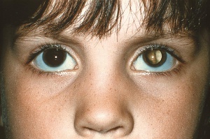
Leukocoria, meaning “white pupil,” originates from the Greek words “leukos” (white) and “kore” (pupil). It refers to the reflection of white light seen upon direct illumination of the fundus through the pupil, in contrast to the usual red glow.[[File:Retinoblastoma White Reflex.jpeg|thumb|Leukocoria of the left eye due to retinoblastoma]] | |||
== Etiopathogenesis == | |||
Direct interference of the normal red reflex from opacities and abnormalities occurring anywhere from the cornea through to the posterior pole can create the leukocoric reflex. For example, the white retinal mass seen in [[retinoblastoma]] creates leukocoria. | |||
[[ | |||
= Diagnosis = | |||
==History== | |||
Comprehensive history should include age of onset, duration, and other ocular symptoms. Past ocular history and past medical history including prematurity, [[Retinopathy of Prematurity|retinopathy of prematurity]], trauma, arthritis, prenatal infection, and other systemic diseases should be assessed. Family history of ocular diseases including [[retinoblastoma]], [[Familial Exudative Vitreoretinopathy (FEVR)|familial exudative vitreoretinopathy]], and [[coloboma]] should be obtained as well.<ref name=":0" /> | |||
==Clinical Examination== | |||
=== Red Reflex === | |||
Red reflex testing recommendations from the American Academy of Pediatrics in 2008 are as follows: | |||
"All neonates, infants, and children should have an examination of the red reflex of the eyes performed by a pediatrician or other primary care clinician trained in this examination technique before discharge from the neonatal nursery and during all subsequent routine health supervision visits." | |||
' | "The red reflex test is properly performed by holding a direct ophthalmoscope close to the examiner's eye with the ophthalmoscope lens power set at “0” (see Fig 1). In a darkened room, the ophthalmoscope light should then be projected onto both eyes of the child simultaneously from approximately 18 inches away. To be considered normal, a red reflex should emanate from both eyes and be symmetric in character. Dark spots in the red reflex, a markedly diminished reflex, the presence of a white reflex, or asymmetry of the reflexes (Bruckner reflex) are all indications for referral to an ophthalmologist who is experienced in the examination of children. The exception to this rule is a transient opacity from mucus in the tear film that is mobile and completely disappears with blinking."<ref>AMERICAN ACADEMY OF PEDIATRICS, Section on Ophthalmology, AMERICAN ASSOCIATION FOR PEDIATRIC OPHTHALMOLOGY AND STRABISMUS, AMERICAN ACADEMY OF OPHTHALMOLOGY, AMERICAN ASSOCIATION OF CERTIFIED ORTHOPTISTS; Red Reflex Examination in Neonates, Infants, and Children. ''Pediatrics'' December 2008; 122 (6): 1401–1404. 10.1542/peds.2008-2624</ref> | ||
=== Comprehensive Ophthalmic Examination === | |||
In cases of abnormal red reflex, a comprehensive ophthalmic examination is indicated. Anterior segment abnormalities can include [[Microphthalmos|microphthalmia]], microcornea, [[Persistent Pupillary Membrane|persistent pupillary membrane]], iris [[coloboma]], [[cataract]], or neovascularization or inflammation. Fundus abnormalities can include vitreous opacities, optic nerve anomalies, retinal vascular abnormalities (in [[Retinopathy of Prematurity|ROP]], [[Coats Disease|Coats disease]], [[retinoblastoma]], and [[Familial Exudative Vitreoretinopathy (FEVR)|FEVR]]), and retinal masses, exudation, [[Retinal Detachment|detachment]], stalk, or fibrosis. | |||
== Ancillary Testing == | |||
Ancillary testing should be performed as indicated by other findings on the clinical examination. Ultrasonography can help detect calcification in [[retinoblastoma]] and can also diagnose findings in other diseases in the above differential diagnosis including [[Coats Disease|Coats disease]], [[Persistent Hyperplastic Primary Vitreous|PFV]], and [[Retinal Detachment|retinal detachment]].<ref name=":0" /> Ultrasonography is particularly valuable in cases with media opacities which may prevent direct visualization of the posterior segment. | |||
Other ancillary testing may include [[Fluorescein Angiography|fluorescein angiography]], [[Optical Coherence Tomography|optical coherence tomography]], or neuroimaging such as computerized tomography or magnetic resonance imaging. | |||
== Differential Diagnosis<ref name=":0">[https://www.aao.org/eyenet/article/stepwise-approach-to-leukocoria Syed R, Ramasubramanian A. Ophthalmic Pearls: A Stepwise Approach to Leukocoria. EyeNet Magazine. July 2016.]</ref><ref>[https://www.ncbi.nlm.nih.gov/pmc/articles/PMC2704541/ Balmer A, Munier F. Differential diagnosis of leukocoria and strabismus, first presenting signs of retinoblastoma. ''Clin Ophthalmol''. 2007;1(4):431-439.]</ref><ref>American Academy of Ophthalmology. Pediatric Ophthalmology and Strabismus. Basic and Clinical Science Course 2021-2022.</ref>== | |||
* Tumors | |||
**[[Retinoblastoma]] | |||
**[[Medulloepithelioma]] | |||
**[[Combined Hamartoma of Retina and Retinal Pigment Epithelium]] | |||
**[[Retinal astrocytic hamartoma]] ([[Ocular Manifestations of Phakomatoses (Neurocutaneous Syndromes)|tuberous sclerosis]]) | |||
* Congenital malformations | |||
**[[Persistent Hyperplastic Primary Vitreous|Persistent Fetal Vasculature]] | |||
**[[Coloboma|Chorioretinal or optic nerve coloboma]] | |||
** Retinal fold | |||
**[[Myelinated Retinal Nerve Fiber Layer|Myelinated retinal nerve fiber layer]] | |||
**[[Morning Glory Anomaly]] | |||
**[[Norrie Disease|Norrie disease]] | |||
** [[Incontinentia Pigmenti|Incontinentia pigmenti]] | |||
** Retinal dysplasia | |||
* Media opacities | |||
** [[Cataract]] | |||
** Corneal opacity | |||
** Organizing [[Vitreous Hemorrhage|vitreous hemorrhage]] | |||
* [[Retinal Detachment|Retinal detachment]] | |||
* Vascular diseases | |||
** [[Retinopathy of Prematurity|Retinopathy of prematurity]] | |||
** [[Coats Disease|Coats disease]] | |||
** [[Familial Exudative Vitreoretinopathy (FEVR)|Familial exudative vitreoretinopathy]] | |||
* Inflammatory diseases | |||
** [[Toxocariasis|Ocular toxocariasis]] | |||
** [[Toxoplasmosis|Congenital toxoplasmosis]] | |||
** [[CMV Retinitis|Cytomegalovirus retinitis]] | |||
** Herpes simplex retinitis | |||
** [[Endophthalmitis]] | |||
** Uveitis | |||
* Trauma | |||
**[[Commotio Retinae|Commotio retinae]] | |||
* Refractive | |||
** High [[myopia]] or anisometropia | |||
* Miscellaneous | |||
** Strabismus (Brückner’s phenomenon) | |||
** Photographic artifact | |||
** Pseudoleukocoria due to optic nerve reflex | |||
= References = | |||
<references /> | |||
Latest revision as of 18:38, August 12, 2024
All content on Eyewiki is protected by copyright law and the Terms of Service. This content may not be reproduced, copied, or put into any artificial intelligence program, including large language and generative AI models, without permission from the Academy.
Definition and Terminology
Leukocoria, meaning “white pupil,” originates from the Greek words “leukos” (white) and “kore” (pupil). It refers to the reflection of white light seen upon direct illumination of the fundus through the pupil, in contrast to the usual red glow.
Etiopathogenesis
Direct interference of the normal red reflex from opacities and abnormalities occurring anywhere from the cornea through to the posterior pole can create the leukocoric reflex. For example, the white retinal mass seen in retinoblastoma creates leukocoria.
Diagnosis
History
Comprehensive history should include age of onset, duration, and other ocular symptoms. Past ocular history and past medical history including prematurity, retinopathy of prematurity, trauma, arthritis, prenatal infection, and other systemic diseases should be assessed. Family history of ocular diseases including retinoblastoma, familial exudative vitreoretinopathy, and coloboma should be obtained as well.[1]
Clinical Examination
Red Reflex
Red reflex testing recommendations from the American Academy of Pediatrics in 2008 are as follows:
"All neonates, infants, and children should have an examination of the red reflex of the eyes performed by a pediatrician or other primary care clinician trained in this examination technique before discharge from the neonatal nursery and during all subsequent routine health supervision visits."
"The red reflex test is properly performed by holding a direct ophthalmoscope close to the examiner's eye with the ophthalmoscope lens power set at “0” (see Fig 1). In a darkened room, the ophthalmoscope light should then be projected onto both eyes of the child simultaneously from approximately 18 inches away. To be considered normal, a red reflex should emanate from both eyes and be symmetric in character. Dark spots in the red reflex, a markedly diminished reflex, the presence of a white reflex, or asymmetry of the reflexes (Bruckner reflex) are all indications for referral to an ophthalmologist who is experienced in the examination of children. The exception to this rule is a transient opacity from mucus in the tear film that is mobile and completely disappears with blinking."[2]
Comprehensive Ophthalmic Examination
In cases of abnormal red reflex, a comprehensive ophthalmic examination is indicated. Anterior segment abnormalities can include microphthalmia, microcornea, persistent pupillary membrane, iris coloboma, cataract, or neovascularization or inflammation. Fundus abnormalities can include vitreous opacities, optic nerve anomalies, retinal vascular abnormalities (in ROP, Coats disease, retinoblastoma, and FEVR), and retinal masses, exudation, detachment, stalk, or fibrosis.
Ancillary Testing
Ancillary testing should be performed as indicated by other findings on the clinical examination. Ultrasonography can help detect calcification in retinoblastoma and can also diagnose findings in other diseases in the above differential diagnosis including Coats disease, PFV, and retinal detachment.[1] Ultrasonography is particularly valuable in cases with media opacities which may prevent direct visualization of the posterior segment.
Other ancillary testing may include fluorescein angiography, optical coherence tomography, or neuroimaging such as computerized tomography or magnetic resonance imaging.
Differential Diagnosis[1][3][4]
- Tumors
- Congenital malformations
- Media opacities
- Cataract
- Corneal opacity
- Organizing vitreous hemorrhage
- Retinal detachment
- Vascular diseases
- Inflammatory diseases
- Ocular toxocariasis
- Congenital toxoplasmosis
- Cytomegalovirus retinitis
- Herpes simplex retinitis
- Endophthalmitis
- Uveitis
- Trauma
- Refractive
- High myopia or anisometropia
- Miscellaneous
- Strabismus (Brückner’s phenomenon)
- Photographic artifact
- Pseudoleukocoria due to optic nerve reflex
References
- ↑ Jump up to: 1.0 1.1 1.2 Syed R, Ramasubramanian A. Ophthalmic Pearls: A Stepwise Approach to Leukocoria. EyeNet Magazine. July 2016.
- ↑ AMERICAN ACADEMY OF PEDIATRICS, Section on Ophthalmology, AMERICAN ASSOCIATION FOR PEDIATRIC OPHTHALMOLOGY AND STRABISMUS, AMERICAN ACADEMY OF OPHTHALMOLOGY, AMERICAN ASSOCIATION OF CERTIFIED ORTHOPTISTS; Red Reflex Examination in Neonates, Infants, and Children. Pediatrics December 2008; 122 (6): 1401–1404. 10.1542/peds.2008-2624
- ↑ Balmer A, Munier F. Differential diagnosis of leukocoria and strabismus, first presenting signs of retinoblastoma. Clin Ophthalmol. 2007;1(4):431-439.
- ↑ American Academy of Ophthalmology. Pediatric Ophthalmology and Strabismus. Basic and Clinical Science Course 2021-2022.


