Combined Hamartoma of Retina and Retinal Pigment Epithelium
All content on Eyewiki is protected by copyright law and the Terms of Service. This content may not be reproduced, copied, or put into any artificial intelligence program, including large language and generative AI models, without permission from the Academy.
Disease Entity
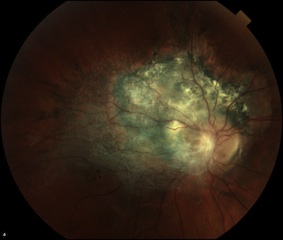
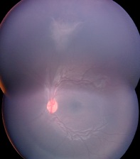
Combined hamartoma of the retina and the retinal pigment epithelium is a rare benign lesion found in the macula, juxtapapillary, or periphery that is commonly diagnosed in children and consists of glial cells, vascular tissue, and sheets of pigment epithelial cells.
Disease
Although histopathologic descriptions were reported earlier, Gass was the first to use the term combined hamartoma of the retina and the retinal pigment epithelium (RPE) in 1973.[1] It was described in 7 patients as (1) slightly elevated, charcoal grey mass involving the RPE, retina, and overlying vitreous; (2) extending in a fanlike projection toward the periphery; (3) blending imperceptibly with surrounding RPE; (4) covered by thickened grey-white retinal and preretinal tissue; (5) showing contraction of the inner surface; (6) absence of RPE or choroidal atrophy at the margin; and (7) with absence of retinal detachment, hemorrhage, exudation, and vitreous inflammation.[2] In 1984, Schachat et al in conjunction with Macula Society Research Committee reviewed 60 cases of combined hamartoma of retina and RPE.[3] More recently, Shields et al performed a review of 77 consecutive patients with the diagnosis.[4] Combined hamartoma of the retina and RPE is important to recognize as it be confused with such serious entities as uveal melanoma, rpe adenocarcinoma and retinoblastoma.
Etiology
The etiology of combined hamartomas of the retina and RPE is not known but they are believed to be congenital lesions even thought the lesion has not been reported at the time of birth. .[3] A recent study by Pujari et al. suggest the tumors may arise from undifferentiated ectopic progenitor cells destined for the RPE that fail to complete fail to complete differentiation due to lack of necessary homeostatic factors leading to hyperplasia and accumulation within the neurosensory retina.[5]
Risk Factors
There are no known risk factors. In terms of gender, in the study by Schachat et al males and females were equally affected (31 males and 29 females).[3] However, in the reports by Font et al and Shields et al most patients were male (70% and 68%, respectively).[6][4] Caucasian race appears to predominate.[3][4] The combined hamartomas of the retina and RPE has been most strongly associated with neurofibromatosis type II.[7][8][9][10] The diagnosis of neurofibromatosis should be especially considered in patients with bilateral combined hamartomas. There have also been case reports in the literature of association of combined hamartoma with neurofibromatosis type I,[11] Gorlin Goltz syndrome,[12] Poland anomaly,[13] branchio-oculo-facial syndrome,[14] brachio-oto-renal syndrome,[15] and juvenile nasopharyngeal angiofibroma.[16]
Classification
Recently, Dedania and colleagues[17] developed a classification system for these tumors which would guide the follow-up frequency determination. The classification scheme was based on the tumor's location (posterior, mid-periphery, or far periphery), characteristics (presence of traction, retinoschisis, or retinal detachment), and optical coherence tomography findings (epiretinal, partial retinal, or full-thickness retina/RPE involvement). Depending on the stages, the authors suggested management options ranging from complete ophthalmologic evaluation at least every 6 months to surgical intervention.
General Pathology
Given that a combined hamartoma, especially when there is a recorded growth of the lesion, can be mistaken for a melanoma or retinoblastoma, there are several reports in literature of enucleation performed due to a concern of a malignant process. Font et al reports 8 enucleations among patients with combined hamartoma in his review of 54 eyes from 1952 to 1988.[6] Histopathologically, the tumor is described as disorganized glial tissue intermixed with numerous blood vessels and cords and tubules of proliferating RPE.[6]
Pathophysiology
Hamartomas are benign, focal overgrowths of mature tissue elements normally found at that site, but which are growing in a disorganized manner. Combined hamartoma of retina and RPE is characterized by variable pigmentation, slight elevation, retinal vascular tortuosity, and epiretinal membrane (ERM) formation. They occur most commonly in a peripapillary location, but can also be found in the macula and periphery.
Diagnosis
History, Signs, and Symptoms
Combined hamartoma of the retina and retinal pigment epithelium is usually found in young children with symptoms of decreased visual acuity (VA) and strabismus. The patients commonly present with painless decrease in VA, strabismus, asymptomatic, or with nonspecific ocular irritation. Less common presenting symptoms include floaters, ocular pain, and leukocoria. The mean age of presentation differs between the studies from 1 year of age in Shields et al study to 15 years in Schachat et al study, possibly reflecting practice patterns. Patients with tumors involving the macula were found to be younger with strabismus and worse presenting VA. The percent of the correct referring diagnosis ranged from 3% to 31% depending on a study. The most common incorrect referring diagnoses include possible choroidal melanoma, choroidal nevus, retinoblastoma, toxocariassis, astrocytoma, and hemangioma.[3][4]
Physical Examination
The presenting VA depends on the location of the lesion: macular, peripapillary, or peripheral. In Schachat et al the initial VA was 20/15–20/40 in 42%, 20/50–20/100 in 17%, and ≤ 20/200 in 42%. Shields et al found presenting VA ≤ 20/200 in 47% of all cases, in 69% of macular, and in 25% of extramacular tumors. The tumor usually presents as a unilateral solitary juxtapapillary, macular, or peripheral lesion; peripheral lesions are the least common. Bilateral cases have been reported. The tumor can be dusky brown, green, yellow, grey, or orange. Retinal vascular changes are prominent: the feeder and distal vessels are straight from traction and intrinsic vessels are tortuous and corkscrewed from contraction. The lesions are usually pigmented and elevated. The vitreoretinal interface changes, traction, fibrosis, gliosis, and ERM formation are common and foveal dragging is seen in 100% of macular tumors and in 42% of extramacular tumors. Less common presentations include mild retinal exudation, detachment, macular edema, choroidal neovascularization (CNV), retinoschisis, retinal holes, vitreous hemorrhage, and pre-retinal neovascularization.[3][4][18][19][20]
Clinical Diagnosis
Clinical diagnosis of the combined hamartoma of retina and RPE depends on thorough history and review of the presenting signs, symptoms, and fundoscopic exam. In case of infants and young children the exam under anesthesia is warranted. A combined hamartoma should be excluded in all young patients with epiretinal membrane or vitreomacular adhesion.
Diagnostic procedures
The fluorescein angiography and OCT are of use in establishing the diagnosis of combined hamartoma of retina and RPE and also in its management. The fluorescein angiography shows early hypoflourescence due to blockage in the region of hyperpigmentation. In the arterial-venous phase, microaneurysms and a fine network of abnormal dilated capillaries with leakage may be observed. In the late phase, leakage from dilated tortuous vessels might be visible.[3][21] The peripheral lesions are characterized on fluorescein angiography by straightening of the vessels and relative avascularity. A report by Helbig et al discusses a case with significant areas of capillary nonperfusion that resulted in preretinal neovascularization peripheral to the hamartoma, suggesting that significant retinal ischemia can occur.[22] OCT is a helpful tool when evaluating the combined hamartoma of the retina and RPE as it allows one to visualize epiretinal membrane (ERM) commonly associated with the disease. The study by Shields et al examined 11 patients and showed that a distinct ERM with secondary retinal folds and striae was present in almost all patients. Horizontal traction induced by membrane was more common than vertical traction. The membrane was preretinal with no evidence of intertwining into the tumor, which has been previously hypothesized.[3] Arepalli and colleagues[15] from the Shields' Oncology have described 'mini-peaks' as saw tooth appearance of the inner retina without deep retinal disturbance (maxi-peaks).
Additional findings included retinal disorganization. However, the adjacent retina was normal in architecture and appeared to gradually thicken into the disorganized tissue.[23] At this point there are no studies assessing the progression of ERM formation as the patient ages. Ultrasonography might be performed to rule out other similar appearing pathology. There is usually a tumor with plaque-like configuration with no choroidal excavation or extrascleral extension. If neurofibromatosis type II is suspected, appropriate work-up is warranted.
Laboratory test
There are no laboratory testing indicated for patients with combined hamartoma of retina and RPE.
Differential diagnosis
- Choroidal melanoma
- Choroidal nevus
- Adenoma or adenocarcinoma of RPE
- Melanocytoma
- Morning glory anomaly
- Retinoblastoma
Management
General treatment
Since combined hamartoma of retina and RPE presents at a young age, amblyopia prevention is paramount. Surgery for associated ERMs is still a subject of debate since visual acuity may not improve despite membrane removal Also, it may not be possible to remove the whole ERM without damaging the retina. There are case reports of treatment of complications and non-classic presentations of combined hamartoma such as CNV and vitreous hemorrhage.
Non-surgical management therapy
Schachat et al reports that of 4 patients that improved by two or more lines, 3 had amblyopia therapy, highlighting its importance.[3] Theodossiadis et al reports laser treatment for juxtapapillary lesion with CNV.[24]
Non-surgical follow-up
The patients should be monitored for decrease in vision, CNV lesions, ERM formation, vitreous hemorrhage, and neovascularization.
Surgery
Surgical intervention consisting of vitrectomy for ERM associated with combined hamartoma of the retina and RPE has been a subject of a debate. Out 60 patients reviewed by Schachat et al, three underwent surgery and only one improving in VA from 20/200 to 20/40.[3] In a study by McDonald et al two patients age 44 and 26 had no improvement.[25] Subsequently, there have been case reports of a 27-year-old patient improving from count fingers to 20/400, 20-year-old improving from 20/400 to 20/60, and a 10-year-old child improving from 2/200 to 20/40.[26][27][28] A study by Cohn et al looked at surgical outcomes for pediatric patients aged 1 to 14 years who underwent pars plana vitrectomy with membrane peeling with or without autologous plasmin enzyme.[29] The results showed that 8 of 11 showed improved VA postoperatively, and 3 of 11 showed stabilized vision. Four eyes had recurrences of ERM, and three of these eyes required additional surgery. They concluded that in the pediatric population, pars plana vitrectomy with membrane peeling with or without the use of autologous plasmin enzyme for ERM associated with combined hamartomas of the retina and RPE can result in improved retinal architecture and VA. There are also reports of surgical intervention for vitreous hemorrhage and subfoveal CVN membrane.[30][31]
Surgical follow-up
ERM can recur after the surgery and sometimes additional surgery is necessary. It has been observed, however, that visual acuity may improve despite recurrence of the ERM.[29]
Complications
Complications of combined hamartoma of the retina and the RPE include reduced visual acuity due to amblyopia, formation of ERM, retinal holes, retinoschisis, CNV, retinal neovascularization, retinal heme, and retinal detachment.
Prognosis
Patients with combined hamartoma of the retina and the RPE can suffer from progressive visual loss due to tumor growth or complications associated with the tumor. In the study by Schachat et al, at 4 years, 66% had final VA within two lines of the original VA, 24% lost two or more lines, and 10% improved by two or more lines (with amblyopia therapy in 3 patients and vitreous surgery in 1).[3] Shields at a compared the macular versus extramacular tumors, and found that vision loss of ≥3 Snellen lines at 4 years was seen in 60% of eyes with macular tumors and 13% of those with extramacular tumors.[4]
References
- ↑ Gass JD. An unusual hamartoma of the pigment epithelium and retina simulating choroidal melanoma and retinoblastoma. Trans Am Ophthalmol Soc. 1973;71:171,83; discussions 184-5.
- ↑ Gass JD. An unusual hamartoma of the pigment epithelium and retina simulating choroidal melanoma and retinoblastoma. Trans Am Ophthalmol Soc. 1973;71:171,83; discussions 184-5.
- ↑ Jump up to: 3.00 3.01 3.02 3.03 3.04 3.05 3.06 3.07 3.08 3.09 3.10 Schachat AP, Shields JA, Fine SL, et al. Combined hamartomas of the retina and retinal pigment epithelium. Ophthalmology. 1984 Dec;91(12):1609-15.
- ↑ Jump up to: 4.0 4.1 4.2 4.3 4.4 4.5 Shields CL, Thangappan A, Hartzell K, Valente P, Pirondini C, Shields JA. Combined hamartoma of the retina and retinal pigment epithelium in 77 consecutive patients visual outcome based on macular versus extramacular tumor location. Ophthalmology. 2008 Dec;115(12):2246,2252.e3.
- ↑ Pujari A, Agarwal A, Chawla R et al. Congenital simple hamartoma of the retinal pigment epithelium: What is the probable cause? Med Hypotheses 2019; 123:79-80. doi: 10.106/j.mehy2018.2018.12.019.
- ↑ Jump up to: 6.0 6.1 6.2 Font RL, Moura RA, Shetlar DJ, Martinez JA, McPherson AR. Combined hamartoma of sensory retina and retinal pigment epithelium. Retina. 1989;9(4):302-11.
- ↑ Meyers SM, Gutman FA, Kaye LD, Rothner AD. Retinal changes associated with neurofibromatosis 2. Trans Am Ophthalmol Soc. 1995;93:245,52; discussion 252-7.
- ↑ Kaye LD, Rothner AD, Beauchamp GR, Meyers SM, Estes ML. Ocular findings associated with neurofibromatosis type II. Ophthalmology. 1992 Sep;99(9):1424-9.
- ↑ Destro M, D'Amico DJ, Gragoudas ES, et al. Retinal manifestations of neurofibromatosis. diagnosis and management. Arch Ophthalmol. 1991 May;109(5):662-6.
- ↑ Grant EA, Trzupek KM, Reiss J, Crow K, Messiaen L, Weleber RG. Combined retinal hamartomas leading to the diagnosis of neurofibromatosis type 2. Ophthalmic Genet. 2008 Sep;29(3):133-8.
- ↑ Vianna RN, Pacheco DF, Vasconcelos MM, de Laey JJ. Combined hamartoma of the retina and retinal pigment epithelium associated with neurofibromatosis type-1. Int Ophthalmol. 2001;24(2):63-6.
- ↑ De Potter P, Stanescu D, Caspers-Velu L, Hofmans A. Photo essay: Combined hamartoma of the retina and retinal pigment epithelium in gorlin syndrome. Arch Ophthalmol. 2000 Jul;118(7):1004-5.
- ↑ Stupp T, Pavlidis M, Bochner T, Thanos S. Poland anomaly associated with ipsilateral combined hamartoma of retina and retinal pigment epithelium. Eye (Lond). 2004 May;18(5):550-2.
- ↑ Demirci H, Shields CL, Shields JA. New ophthalmic manifestations of branchio-oculo-facial syndrome. Am J Ophthalmol. 2005 Feb;139(2):362-4.
- ↑ Jump up to: 15.0 15.1 Arepalli S, Pellegrini M, Ferenczy SR, Shields CL. Combined hamartoma of the retina and retinal pigment epithelium: findings on
enhanced depth imaging optical coherence tomography in eight eyes. Retina. 2014;34(11):2202-7
- ↑ Fonseca RA, Dantas MA, Kaga T, Spaide RF. Combined hamartoma of the retina and retinal pigment epithelium associated with juvenile nasopharyngeal angiofibroma. Am J Ophthalmol. 2001 Jul;132(1):131-2.
- ↑ Dedania VS1, Ozgonul C, Zacks DN, Besirli. NOVEL CLASSIFICATION SYSTEM FOR COMBINED HAMARTOMA OF THE RETINA AND RETINAL PIGMENT EPITHELIUM. Retina. 2018 Jan;38(1):12-19.
- ↑ Kahn D, Goldberg MF, Jednock N. Combined retinal-retina pigment epithelial hamartoma presenting as a vitreous hemorrhage. Retina. 1984 Winter-Spring;4(1):40-3.
- ↑ Inoue M, Noda K, Ishida S, et al. Successful treatment of subfoveal choroidal neovascularization associated with combined hamartoma of the retina and retinal pigment epithelium. Am J Ophthalmol. 2004 Jul;138(1):155-6.
- ↑ Helbig H, Niederberger H. Presumed combined hamartoma of the retina and retinal pigment epithelium with preretinal neovascularization. Am J Ophthalmol. 2003 Dec;136(6):1157-9.
- ↑ Heimann H, Kellner U, Foerster MH. Atlas of fundus angiography. Stuttgart ; New York: Thieme; 2006.
- ↑ Helbig H, Niederberger H. Presumed combined hamartoma of the retina and retinal pigment epithelium with preretinal neovascularization. Am J Ophthalmol. 2003 Dec;136(6):1157-9.
- ↑ Shields CL, Mashayekhi A, Dai VV, Materin MA, Shields JA. Optical coherence tomographic findings of combined hamartoma of the retina and retinal pigment epithelium in 11 patients. Arch Ophthalmol. 2005 Dec;123(12):1746-50.
- ↑ Theodossiadis PG, Panagiotidis DN, Baltatzis SG, Georgopoulos GT, Moschos MN. Combined hamartoma of the sensory retina and retinal pigment epithelium involving the optic disk associated with choroidal neovascularization. Retina. 2001;21(3):267-70.
- ↑ McDonald HR, Abrams GW, Burke JM, Neuwirth J. Clinicopathologic results of vitreous surgery for epiretinal membranes in patients with combined retinal and retinal pigment epithelial hamartomas. Am J Ophthalmol. 1985 Dec 15;100(6):806-13.
- ↑ Sappenfield DL, Gitter KA. Surgical intervention for combined retinal-retinal pigment epithelial hamartoma. Retina. 1990;10(2):119-24.
- ↑ Stallman JB. Visual improvement after pars plana vitrectomy and membrane peeling for vitreoretinal traction associated with combined hamartoma of the retina and retinal pigment epithelium. Retina. 2002 Feb;22(1):101-4.
- ↑ Mason JO,3rd. Visual improvement after pars plana vitrectomy and membrane peeling for vitreoretinal traction associated with combined hamartoma of the retina and retinal pigment epithelium. Retina. 2002 Dec;22(6):824,5; author reply 825-6.
- ↑ Jump up to: 29.0 29.1 Cohn AD, Quiram PA, Drenser KA, Trese MT, Capone A,Jr. Surgical outcomes of epiretinal membranes associated with combined hamartoma of the retina and retinal pigment epithelium. Retina. 2009 Jun;29(6):825-30.
- ↑ Inoue M, Noda K, Ishida S, et al. Successful treatment of subfoveal choroidal neovascularization associated with combined hamartoma of the retina and retinal pigment epithelium. Am J Ophthalmol. 2004 Jul;138(1):155-6.
- ↑ Kahn D, Goldberg MF, Jednock N. Combined retinal-retina pigment epithelial hamartoma presenting as a vitreous hemorrhage. Retina. 1984 Winter-Spring;4(1):40-3.


