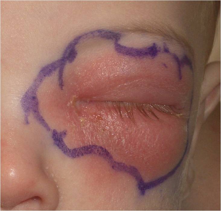
Orbital Cellulitis
All content on Eyewiki is protected by copyright law and the Terms of Service. This content may not be reproduced, copied, or put into any artificial intelligence program, including large language and generative AI models, without permission from the Academy.
Disease Entity
Orbital cellulitis is an infection of the soft tissues of the eye socket behind the orbital septum, a thin tissue that divides the eyelid from the eye socket. Infection isolated anterior to the orbital septum is considered to be preseptal cellulitis. Orbital cellulitis most commonly refers to an acute spread of infection into the eye socket due to extension from periorbital structures (most commonly the adjacent ethmoid or frontal sinuses [90%], skin, dacryocystitis, dental infection, intracranial infection), exogenous causes (trauma, foreign bodies, postsurgical), intraorbital infection (endophthalmitis, dacryoadenitis), or spread through the blood (bacteremia with septic emboli).
Disease
Orbital Cellulitis (ICD-9 #376.01)
Etiology
Orbital cellulitis most commonly occurs when a bacterial infection spreads from the paranasal sinuses into the orbit. In children younger than 10 years, paranasal sinusitis most often involves the ethmoid sinus and spreads through the thin lamina papyracea of the medial orbital wall into the orbit. It can also occur when an eyelid skin infection, an infection in an adjacent area, or an infection in the bloodstream spreads into the orbit. The drainage of the eyelids and sinuses occurs largely through the orbital venous system: more specifically, through the superior and inferior orbital veins that drain into the cavernous sinus. This venous system is devoid of valves, and for this reason infection might spread in preseptal and orbital cellulitis into the cavernous sinus, causing a sight-threatening complication such as cavernous sinus thrombosis.
Risk Factors
Risk factors include recent upper respiratory illness, acute or chronic bacterial sinusitis, trauma, ocular or periocular infection, or systemic infection.
General Pathology
Orbital cellulitis can be caused by an extension from periorbital structures (paranasal sinuses, face and eyelids, lacrimal sac, dental structures). Its cause can also be exogenous (trauma, foreign body, surgery), endogenous (bacteremia with septic embolization), or intraorbital (endophthalmitis, dacryoadenitis).[1] The orbital tissues are infiltrated by acute and chronic inflammatory cells and the infectious organisms may be identified on the tissue sections. The organisms are best identified by microbiologic culture. The most common infectious pathogens include gram-positive streptococcal and staphylococcal species. In a landmark article by Harris et al, it was noted that in children younger than 9 years, the infections are typically from 1 aerobic organism; in children older than 9 years and in adults, the infections may be polymicrobial with both aerobic and anaerobic bacteria.[2]
Streptococcal infections are identified on culture by their formation of pairs or chains. Streptococcus pyogenes (group A strep) requires blood agar to grow and exhibits clear (beta) hemolysis on blood agar. Streptococcal bacteria such as Streptococcus pneumoniae produce green (alpha) hemolysis, or partial reduction of red blood cell hemoglobin. Staphylococcal species appear as a cluster arrangement on Gram stain. Staphylococcus aureus forms a large, yellow colony on rich medium in distinction to Staphylococcus epidermidis, which forms white colonies. Gram-negative rods can be seen in both orbital cellulitis related to trauma and in neonates and immunosuppressed patients. Anaerobic bacteria such as Peptococcus, Peptostreptococcus, and Bacteroides can be involved in infections extending from sinusitis in adults and older children. Fungal infections with either Mucor or Aspergillus need to be considered in immunocompromised or diabetic patients; immunocompetent patients may also have fungal infections in rare cases.
Pathophysiology
Orbital cellulitis most commonly occurs in the setting of an upper respiratory or sinus infection. The human upper respiratory tract is normally colonized with S pneumoniae, and infection can occur through several mechanisms. S pyogenes infections also occur primarily in the respiratory tract. The complex cell surface of this gram-positive organism determines its virulence and ability to invade the surrounding tissue and incite inflammation. S aureus infections occur commonly in the skin and spread to the orbit from the skin. Staphylococcal organisms also produce toxins that help to promote their virulence and lead to the inflammatory response seen in these infections. The inflammatory response elicited by all of these pathogens plays a major role in tissue destruction in the orbit.
Primary Prevention
Identifying patients and effectively treating upper respiratory or sinus infections before they evolve into orbital cellulitis is an important aspect of preventing preseptal cellulitis from progressing to orbital cellulitis. Equally important in preventing orbital cellulitis is prompt and appropriate treatment of preseptal skin infections as well as infections of the teeth, middle ear, or face before they spread into the orbit.
Diagnosis
The diagnosis of orbital cellulitis is based on clinical examination. The presence of the following signs is suggestive of orbital involvement: proptosis, chemosis, pain with eye movements, ophthalmoplegia, and optic nerve involvement as well as fever, leukocytosis (75% of cases), and lethargy. Other signs and symptoms include rhinorrhea, headache, tenderness on palpation, and eyelid edema. Intraocular pressure (IOP) may be elevated if there is increased venous congestion. In addition to clinical suspicion, diagnostic imaging using computed tomography (CT) may help distinguish between preseptal and orbital cellulitis while also looking for complications of orbital cellulitis (see below).
History
The presence of a painful red eye with lid edema in a child with a recent upper respiratory infection is the typical presentation of orbital cellulitis. Patient history should also include the presence of headache, orbital pain, double vision, progression of symptoms, recent upper respiratory symptoms (e.g., nasal discharge or stuffiness), pain over sinuses, fever, or lethargy. In addition, recent periocular trauma or injury; family or health care contacts with methicillin-resistant Staphylococcus aureus (MRSA); history of sinus, ear, dental, or facial infections or surgery; recent ocular surgery; pertinent medical conditions; and current medications should be noted, as well as the presence of diabetes mellitus and the immune status of the patient. Specific questions regarding any change in vision, mental status, pain with neck movement, or nausea or vomiting should also be asked.
Physical Examination
The physical examination should include the following:
- Best-corrected visual acuity (BCVA). Decreased vision might be indicative of optic nerve involvement or could be secondary to severe exposure keratopathy or retinal vein occlusion.
- Color vision assessment to assess the presence of optic nerve involvement.
- Proptosis measurements using Hertel exophthalmometry.
- Visual field assessment via confrontation.
- Assessment of pupillary function with particular attention paid to the presence of a relative afferent pupillary defect (RAPD).
- Ocular motility and presence of pain with eye movements. Also, there might be involvement of the III, IV, and V1/V2 cranial nerve in cases of cavernous sinus involvement.
- Orbit examination should include documentation of the direction of displacement of the globe (e.g., a superior subperiosteal abscess will displace the globe inferiorly), resistance to retropulsion on palpation, and unilateral or bilateral involvement (bilateral involvement is virtually diagnostic of cavernous sinus thrombosis[3]).
- Measurement of intraocular pressure. Increased venous congestion may result in increased IOP.
- Slit-lamp biomicroscopy of the anterior segment, if possible, to look for signs of exposure keratopathy in cases of severe proptosis.
- Dilated fundus examination will exclude or confirm the presence of optic neuropathy or retinal vascular occlusion.
Signs
As a preseptal infection progresses into the orbit, the inflammatory signs typically increase with increasing redness and swelling of the eyelid with a secondary ptosis. As the infection worsens, proptosis develops and extraocular motility becomes compromised. When the optic nerve is involved, loss of visual acuity is noted and an afferent pupillary defect can be appreciated. The IOP often increases and the orbit becomes resistant to retropulsion. The skin can feel warm to the touch and pain can be elicited with either touch or eye movements. Examination of the nose and mouth is also warranted to look for any black eschar which would suggest a fungal infection. Purulent nasal discharge with hyperemic nasal mucosa may be present.
Symptoms
Systemic symptoms including fever and lethargy may or may not be present. Change in the appearance of the eyelids with redness and swelling is frequently a presenting symptom. Pain, particularly with eye movement, is commonly noted. Double vision may also occur.
Diagnostic Procedures
Computed tomography of the orbit is the imaging modality of choice for patients with orbital cellulitis. Most of the time, CT is readily available and will give the clinician information regarding the presence of sinusitis, subperiosteal abscess, stranding of orbital fat, or intracranial involvement. Nevertheless, in cases of mild to moderate orbital cellulitis with no optic nerve involvement, the initial management of the patient remains medical. Imaging is warranted in children and in cases of poor response to intravenous antibiotics with progression of orbital signs in order to confirm the presence of complications such as subperiosteal abscess or intracranial involvement. Although a magnetic resonance imaging (MRI) scan is safer in children because there is no risk of radiation exposure, the long acquisition time and the need for prolonged sedation make CT the imaging modality of choice. However, if there is suspicion of a concomitant cavernous sinus thrombosis, MRI may be a useful adjunct to a CT scan.
Laboratory Test
Admission to the hospital is warranted in all cases of orbital cellulitis. A complete blood count with differential, blood cultures, and nasal and throat swabs should be ordered.
Differential Diagnosis
The differential diagnosis includes the following:
- Idiopathic inflammation/specific inflammation (e.g., orbital pseudotumor, granulomatosis with polyangiitis, sarcoidosis)
- Neoplasia (e.g., lymphangioma, hemangioma, leukemia, rhabdomyosarcoma, lymphoma, retinoblastoma, metastatic carcinoma)
- Trauma (e.g., retrobulbar hemorrhage, orbital emphysema)
- Systemic diseases (e.g., sickle cell disease with bony infarcts and subperiosteal hematomas)
- Endocrine disorders (e.g., thyroid eye disease)
Management
General Treatment
The management of orbital cellulitis requires admission to the hospital and initiation of broad-spectrum intravenous antibiotics that address the most common pathogens. Blood cultures and nasal/throat swabs may be undertaken, and the antibiotics should be modified based on the results. In infants with orbital cellulitis, a third-generation cephalosporin is usually initiated such as cefotaxime, ceftriaxone, or ceftazidime, along with a penicillinase-resistant penicillin. In older children, because sinusitis is most commonly associated with aerobic and anaerobic organisms, clindamycin might be another option. Metronidazole is also being increasingly used in children. If there is concern for MRSA infection, vancomycin may be added as well.[4] As mentioned before, the antibiotic regimen should be modified based on the results of the cultures if needed. Intravenous corticosteroids may also be of benefit in the management of pediatric orbital cellulitis.[5] [6][7] The patient should be followed closely in the hospital setting for progression of orbital signs and development of complications such as cavernous sinus thrombosis or intracranial extension, which can be lethal.[3] Once improvement has been documented with 48 hours of intravenous antibiotics, consideration for switching to oral antibiotics may be appropriate.
Medical Follow-up
A multidisciplinary approach is usually warranted for patients with orbital cellulitis under the care of pediatricians, ENT surgeons, ophthalmologists, and infectious disease specialists.
Surgery
The prevalence of subperiosteal or orbital abscess complicating an orbital cellulitis approaches 10%. The clinician should suspect the presence of such an entity if there is progression of orbital signs and/or systemic compromise despite the initiation of appropriate intravenous antibiotics for at least 24-48 hours. In these cases, a contrast-enhanced CT scan should be ordered to evaluate the orbit, paranasal sinuses, and/or brain. If there is associated sinusitis, ENT should be consulted. If an orbital abscess is present, it should be drained. The management of subperiosteal abscess remains controversial because there are cases of resolution with the use of intravenous antibiotics only. As a general recommendation (as initially described by Garcia and Harris), observation with intravenous antibiotics only (i.e., no drainage of the subperiosteal abscess) is indicated as follows:
- Child is younger than 9 years
- No intracranial involvement
- Medial wall abscess is of moderate or smaller size
- No vision loss or afferent pupillary defect
- No frontal sinus involvement
- No dental abscess
If there is evidence of intracranial extension of the infection, evidence of optic nerve compromise, clinical deterioration despite 48 hours of intravenous antibiotics, suspicion of anaerobic infection (e.g., presence of gas in abscess on CT), or recurrence of subperiosteal abscess after prior drainage, surgery is almost always indicated.[1] At the time of surgery, cultures and susceptibility testing may be obtained from samples to help tailor therapy.
Complications
The complications of orbital cellulitis are ominous and include severe exposure keratopathy with secondary ulcerative keratitis, neurotrophic keratitis, secondary glaucoma, septic uveitis or retinitis, exudative retinal detachment, inflammatory or infectious neuritis, optic neuropathy, panophthalmitis, cranial nerve palsies, optic nerve edema, subperiosteal abscess, orbital abscess, central retinal artery occlusion, retinal vein occlusion, blindness, orbital apex syndrome, cavernous sinus thrombosis, meningitis, subdural or brain abscess, and death.
Prognosis
With prompt recognition and aggressive medical and/or surgical treatment, the prognosis is excellent.
Additional Resources
- Boyd K. What is cellulitis? American Academy of Ophthalmology. EyeSmart/Eye Health. September 10, 2024. Accessed April 3, 2025. https://www.aao.org/eye-health/diseases/what-is-cellulitis
- Mawn LA, Jordan DR, Donahue SP. Preseptal and orbital cellulitis. Ophthalmol Clin North Am. 2000;13(4):633-641.
References
- ↑ Jump up to: 1.0 1.1 Basic and Clinical Science Course, Section 7. Oculofacial Plastic and Orbital Surgery. American Academy of Ophthalmology; 2019.
- ↑ Harris GJ. Subperiosteal abscess of the orbit. Age as a factor in the bacteriology and response to treatment. Ophthalmology. 1994;101(3):585-595.
- ↑ Jump up to: 3.0 3.1 Basic and Clinical Science Course, Section 6. Pediatric Ophthalmology and Strabismus. American Academy of Ophthalmology; 2019.
- ↑ Liao S, Durand ML, Cunningham MJ. Sinogenic orbital and subperiosteal abscesses: microbiology and methicillin-resistant Staphylococcus aureus incidence. Otolaryngol Head Neck Surg. 2010;143(3):392-396.
- ↑ Yen MT, Yen KG. Effect of corticosteroids in the acute management of pediatric orbital cellulitis with subperiosteal abscess. Ophthalmic Plast Reconstr Surg. 2005;21(5):363-366; discussion 366-367. doi:10.1097/01.iop.0000179973.44003.f7
- ↑ Chen L, Silverman N, Wu A, Shinder R. Intravenous steroids with antibiotics on admission for children with orbital cellulitis. Ophthalmic Plast Reconstr Surg. 2018;34(3):205-208. doi:10.1097/IOP.0000000000000910
- ↑ Davies BW, Smith JM, Hink EM, Durairaj VD. C-reactive protein as a marker for initiating steroid treatment in children with orbital cellulitis. Ophthalmic Plast Reconstr Surg. 2015;31(5):364-368. doi:10.1097/IOP.0000000000000349
- Brook I. Role of methicillin-resistant Staphylococcus aureus in head and neck infections. J Laryngol Otol. 2009;123(12):1301-1307.
- Cannon PS, Mc Keag D, Radford R, Ataullah S, Leatherbarrow B. Our experience using primary oral antibiotics in the management of orbital cellulitis in a tertiary referral centre. Eye (Lond). 2009;23(3):612-615.
- Fu SY, Su GW, McKinley SH, Yen MT. Cytokine expression in pediatric subperiosteal orbital abscesses. Can J Ophthalmol. 2007;42(6):865-869.
- Garcia GH, Harris GJ. Criteria for nonsurgical management of subperiosteal abscess of the orbit: analysis of outcomes 1988-1998. Ophthalmology. 2000;107(8):1454-1456; discussion 1457-1458.
- Goldstein SM, Shelsta HN. Community-acquired methicillin-resistant Staphylococcus aureus periorbital cellulitis: a problem here to stay. Ophthalmic Plast Reconstr Surg. 2009;25(1):77.
- Harris GJ. Subperiosteal abscess of the orbit: computed tomography and the clinical course. Ophthalmic Plast Reconstr Surg. 1996;12(1):1-8.
- Kornelsen E, Mahant S, Parkin P, et al. Corticosteroids for periorbital and orbital cellulitis. Cochrane Database Syst Rev. 2021;2021(4):CD013535.
- McKinley SH, Yen MT, Miller AM, Yen KG. Microbiology of pediatric orbital cellulitis. Am J Ophthalmol. 2007;144(4):497-501.
- Miller A, Castanes M, Yen M, Coats D, Yen K. Infantile orbital cellulitis. Ophthalmology. 2008;115(3):594.
- Nageswaran S, Woods CR, Benjamin DK Jr, Givner LB, Shetty AK. Orbital cellulitis in children. Pediatr Infect Dis J. 2006;25(8):695-699.


