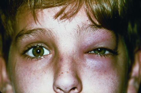
Preseptal Cellulitis
All content on Eyewiki is protected by copyright law and the Terms of Service. This content may not be reproduced, copied, or put into any artificial intelligence program, including large language and generative AI models, without permission from the Academy.
Disease Entity
Preseptal cellulitis is an inflammation of the tissues localized anterior to the orbital septum. The orbital septum is a fibrous tissue that divides the orbit contents into 2 compartments: preseptal (anterior to the septum) and postseptal (posterior to the septum). The inflammation that develops posterior to the septum is known as “orbital cellulitis.” Both entities are caused by an infectious process.
Pathophysiology
There are 3 main routes for pathogen inoculation in the periorbital tissues:
- Direct inoculation: after eyelid trauma and infected insect bites
- Spread from contiguous structures: paranasal sinuses are the most common (specially the ethmoids, since nerves and vessels traverse the lamina papyracea that divides the ethmoid sinuses from the orbit), chalazia/hordeolum, dacryocystitis, dacryoadenitis, canaliculitis, impetigo, erysipelas, herpes simplex and herpes zoster skin lesions, endophthalmitis
- Hematogenous: via blood vessels from an upper respiratory tract or a middle ear infection
The venous drainage of the orbit, eyelids, and sinuses goes primarily to the superior and inferior orbital veins, which drain to the cavernous sinus. Because these veins are devoid of valves, infection can easily spread to preseptal and postseptal space, and it can also lead to cavernous sinus thrombosis.
Classification
A modification of Chandler’s classification of periorbital infections is still in use by many clinicians.:
- Preseptal cellulitis
- Orbital cellulitis
- Subperiosteal abscess
- Orbital abscess
- Cavernous sinus thrombosis
Etiology
The majority of these infections are caused by bacteria. Adenovirus, herpes simplex, and varicella zoster are also associated with cellulitis. In the immunocompromised patient, the clinician must suspect fungi as a possible etiology. Gram-positive cocci are the most prevalent microorganisms identified in preseptal cellulitis—typically Staphylococcus and Streptococcus species (pyogenes and pneumonia). Staphylococcus aureus and epidermidis are commonly found after a penetrating eyelid trauma. Streptococcus pneumoniae is a common etiology in preseptal cellulitis secondary to sinusitis. In the era before the establishment of the universal vaccination against Haemophilus influenzae type b, this was a frequent etiology, especially in children under 5 years of age. It is still common in unvaccinated patients. In preseptal cellulitis secondary to a human bite, it is frequent to isolate anaerobic bacteria such as Clostridium.
Diagnosis
Patients complain of eyelid swelling and redness. But general malaise and low-grade fever are also commonly reported. Among the classic signs of preseptal cellulitis are eyelid edema/erythema/warmth and fever. There are clinical keys that help us distinguish between preseptal and orbital cellulitis.
- Preseptal cellulitis: eyelid edema and erythema, normal visual acuity, absence of proptosis, pupil with normal reaction to light, normal color saturation, normal conjunctiva, and normal ocular movements
- Orbital cellulitis: eyelid edema and erythema, diminished visual acuity, proptosis is present, relative afferent pupillary defect may be present, reduced color saturation, chemotic conjunctiva, and reduced extraocular movements with pain elicited by these movements
Cellulitis may extend to the cheek and forehead. Also, it is common to see an eyelid abscess associated with preseptal cellulitis, which may require incision and drainage.
Work up
It is useful to delineate the area of the face affected with cellulitis using a skin marker, in order to monitor progression along time. Photographs are also an invaluable tool.
Tests
- Complete blood count to document leukocytosis.
- Computed tomography (CT) scan: Sometimes the eyelid edema is so severe that it precludes eye examination, thus making the distinction between preseptal and orbital cellulitis impossible. In these cases, it is useful to order a CT scan of the orbit and sinuses (to diagnose an associated sinusitis).
- Cultures of the eyelid wound (if evident), conjunctiva, blood (if febrile), abscess contents (if present and drained), or paranasal sinus secretion. These are important in order to prescribe the most appropriate antibiotic according to bacteria sensitivity.
- Lymph nodes of the head and neck to assess for lymphadenomegaly
- Check for signs of meningeal irritation to evaluate the presence of intracranial complications.
Differential diagnosis
- Orbital cellulitis
- Adenoviral keratoconjunctivitis
- Allergic conjunctivitis
- Contact dermatitis
- Kawasaki’s disease (children)
- Idiopathic orbital inflammation
- Thyroid eye disease
- Dacryocystitis
- Dacryoadenitis
Management
General treatment
Once diagnosed, preseptal cellulitis can be treated in an outpatient or inpatient basis, depending on the characteristics of the patient.
- If the patient is afebrile, with a mild preseptal cellulitis, he can be followed as an outpatient with oral antibiotics and daily visits to monitor the progress of the disease. However, if the patient does not respond to oral antibiotics in 48 hours or if extension of the infectious process into the orbit is suspected, he or she should be admitted to the hospital: A CT scan must be performed to evaluate for orbital extension, and intravenous antibiotics must be indicated.
- Usually, children under 2 years of age or febrile patients with a severe cellulitis are managed with intravenous antibiotics during hospitalization, with close follow-up. Hospitalization is also recommended in patients who cannot be followed up as outpatients. Intravenous antibiotics are usually indicated for 2 or 3 days, depending on improvement. If the condition improves, treatment can be switched to the appropriate oral antibiotics based on cultures.
These patients should be treated by a multidisciplinary team: ophthalmologist, pediatrician/primary care physician, and otolaryngologist (in case of an associated sinusitis).
Empiric antibiotic therapy
Broad-spectrum antibiotics must be prescribed to cover gram-positive and gram-negative bacteria.
Oral
- Against gram-positive and gram-negative bacteria: ampicillin, amoxicillin/clavulanate, fluoroquinolones (levofloxacin), azithromycin (also covers some anaerobic bacteria), clindamycin.
- Against gram-positive (Staphylococcus) in case of an evident eyelid trauma: dicloxacillin, flucloxacillin, first-generation cephalosporins (cefalexin, cefazolin).
Intravenous
These antibiotics provide coverage to gram-positive and gram-negative bacteria.:
- Third-generation cephalosporins (these medications are less sensitive to β-lactamase producing bacteria such as S. aureus): ceftriaxone, cefotaxime, ceftazidime
- Ampicillin/sulbactam
The results of antibiotic sensitivities should guide the treatment whenever possible. When the cultures reveal a methicillin-resistant Staphylococcus aureus (MRSA) the therapy choice must be reevaluated. Community-associated MRSA is susceptible to these antibiotics administered in an oral route:
- Trimethoprim-sulfamethoxazole
- Rifampin
- Clindamycin
- Fluoroquinolones
Hospital-associated MRSA is susceptible only to:
- Intravenous vancomycin
- PO linezolid
If there was a penetrating eyelid injury with organic material or a human bite, antibiotics should also cover anaerobic organisms: metronidazole, clindamycin, piperacillin-tazobactam.
If an abscess localized in the preseptal space develops, it should be incised and drained. The surgeon must not open the orbital septum during the procedure, since this may spread the infection to the postseptal space and aggravate the infection. As mentioned in the work up section, the contents of the abscess should be cultured to determine appropriate antibiotic therapy.
Prognosis and Complications
Prognosis is usually good when this entity is promptly diagnosed and treated. However, complications can develop even with prompt treatment.
- Orbital extension and complications: orbital cellulitis, subperiosteal abscess, orbital abscess, cavernous sinus thrombosis
- Central nervous system involvement (after orbital extension): meningitis, abscesses (brain, extradural or subdural).
- Necrotizing fasciitis: it is a rare complication caused by β-hemolytic Streptococcus. It presents as a rapidly progressive cellulitis with poorly demarcated borders and violaceous skin discoloration, which can lead to necrosis and toxic shock syndrome. The patient must be admitted to the hospital, intravenous fluids should be replenished, IV broad-spectrum antibiotics must be prescribed, and surgical debridement could be necessary.
References
- ↑ American Academy of Ophthalmology. Preseptal cellulitis. https://www.aao.org/image/preseptal-cellulitis-4 Accessed July 17, 2019.
- Chandler JR, Langenbrunner DJ, Stevens ER. The pathogenesis of orbital complications in acute sinusitis. Laryngoscope 1970; 80:1414-28.
- Ambati BK, Ambati J, Azar N, et al. Periorbital and orbital cellulitis before and after the advent of Haemophilus influenzae type B vaccination. Ophthalmology 2000; 107: 1450–3.
- Watts P. Preseptal and orbital cellulitis in children: a review. J Paediatr Child Health. 2012; 22(1): 1-8
- Pelton RW, Klapper SR. Focal Points Clinical Modules for Ophthalmologists: Preseptal and Orbital Cellulitis. American Academy of Ophthalmology. 2008; 26(11).
- Chapter 4: Orbital inflammatory and infectious diseases. Orbit, Eyelids, and Lacrimal System, Section 7. Basic and Clinical Science Course 2011-2012. American Academy of Ophthalmology. San Francisco, CA. 2011.
- Uddin JM, Scawn RL. Chapter 13: Preseptal and orbital cellulitis. Hoyt CS and Taylor D. Pediatric Ophthalmology and Strabismus. Elsevier Saunders. 4th edition; China, 2013. Pp 89-99.
- Rose GE, Howard DJ, Watts MR. Periorbital necrotising fasciitis. Eye 1991; 5: 736–40.
- Sweeney A.R., Yen M.T. (2020) Eyelid Infections. In: Albert D., Miller J., Azar D., Young L. (eds) Albert and Jakobiec's Principles and Practice of Ophthalmology. Springer, Cham. https://doi.org/10.1007/978-3-319-90495-5_75-1
- Brook I. Treatment of anaerobic infection. Expert Rev Anti Infect Ther. 2007;5(6):991-1006. doi:10.1586/14787210.5.6.991


