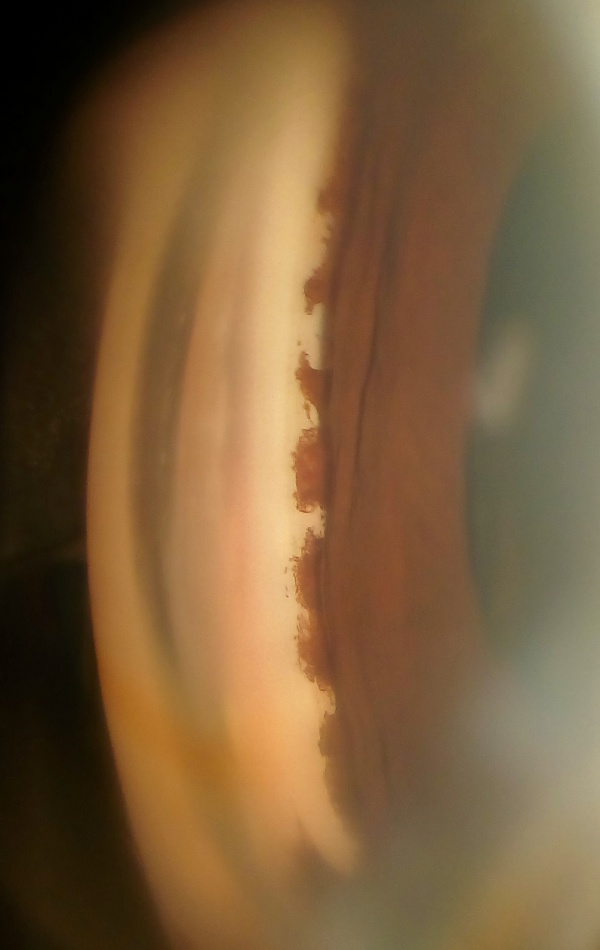
Sarcoid Uveitis
All content on Eyewiki is protected by copyright law and the Terms of Service. This content may not be reproduced, copied, or put into any artificial intelligence program, including large language and generative AI models, without permission from the Academy.
![(A) Exterior photograph showing a Koeppe nodule with granulomatous changes on the pupil margin (yellow arrow).[1] (B) Fundus image revealing vitritis with snowball opacities (green arrows), characteristic of intermediate uveitis in sarcoidosis. (C) Fundus photograph demonstrating chorioretinitis, vascular sheathing, candle wax dripping phlebitis, granulomas as yellowish lesions, and diffuse vitreous haze in the posterior pole. (D) Wide-field fundus image illustrating peripheral multifocal chorioretinal scars, described as punch-out lesions, which are not pathognomonic of sarcoidosis and can also be seen in other conditions such as Histoplasmosis (Presumed Ocular Histoplasmosis Syndrome - POHS), Fuchs' Heterochromic Iridocyclitis (FHI), toxoplasmosis, syphilis, or tuberculosis (red arrows) in sarcoid-associated posterior uveitis. (Courtesy of J. Khadamy)](/w/images/thumb/7/7b/Ocular_manifestations_of_sarcoidosis.jpg/500px-Ocular_manifestations_of_sarcoidosis.jpg)
Disease Entity
Sarcoid Uveitis
Disease
Sarcoidosis is a systemic inflammatory disease characterized by the formation of noncaseating granulomas in affected organs, most commonly the lungs, lymph nodes, skin, and eyes. The disease was first described in 1878 by noted surgeon Sir Jonathan Hutchinson as a dermatologic disorder[2]. It was not until 1909 that a Danish ophthalmologist, Heerfordt Christian Frederik[3], described uveitis as part of the disease process. His eponymous “Heerfordt Syndrome” consisted of uveitis, parotitis and fever (uveoparotid fever) with or without facial palsy.
Etiology
While the etiology of sarcoidosis is unknown, several hypothesis regarding genetic and environmental factors have been studied. Some hypothesize that it is a clinical syndrome consisting of different diseases with different underlying etiologies. Associations between sarcoid and HLA-DRB1[4] and M. tuberculosis DNA[5] are currently being evaluated.
Risk Factors
In the United States, the incidence of systemic sarcoidosis ranges from 5-40 per 100,000 population[6]. This prevalence is 10 times greater for African Americans when compared to white Americans. African Americans are also more likely to have an acute presentation of the disease and a more severe clinical course than Caucasian patients.
A large retrospective chart review from the University of Illinois uveitis service indicated that in biopsy-proved sarcoidosis, African-American patients were more likely to be diagnosed as having uveitis than whites. African-Americans younger than 50 years were more likely to present with uveitis.[7]
Several studies have noted a slight female preponderance, with most symptomatic patients being between 20-50 years old.
Pathology
Histologically, sarcoid nodules are composed of collections of noncaseating epithelioid histiocytes and lymphocytes. Multinucleated giant cells, asteroid bodies and Schaumann bodies may be seen within the giant cells.
Pathophysiology
The exact etiology of sarcoidosis is unknown, making it largely a diagnosis of exclusion. It is characterized by the appearance of noncaseating granulomas in affected tissue, most frequently seen in the lungs as bilateral hilar lymphadenopathy or pulmonary infiltration. Granulomas can also be seen on the skin, in the eyes, liver, spleen, salivary glands, heart, bones and nervous system. Ocular involvement occurs in approximately 25% of patients with sarcoidosis. The uveal tract is the most common site of ocular involvement by sarcoidosis. In most large series, sarcoidosis accounts for between 3-10% of all cases of uveitis.
Diagnosis
Sarcoid is a systemic inflammatory disease and can manifest itself in multiple ways. Therefore, clinical suspicion is often the driving force behind correct diagnosis. A thorough review of systems if necessary in all patients with recurrent uveitis. An individual with pulmonary or dermatologic complaints consistent with sarcoid should be further evaluated. Uveitis can also precede pulmonary symptoms by several years. Orbital and eyelid granulomas are also common, as well as lacrimal gland infiltration.
History
Clinical presentation of sarcoidosis depends on the severity and organ involved. A history of pulmonary disease in an individual with granulomatous uveitis should raise the suspicion of sarcoidosis.
Physical Examination
Granulomatous anterior uveitis, either acute or chronic, is the most common ocular manifestation of sarcoidosis. Less than 1/3rd of patients present with posterior uveitis without anterior involvement.
In 2009, an international group of uveitis specialists met for the International Workshop On Ocular Sarcoidosis (IWOS)[8]. Subsequently in 2017, the group met again in Nusa Dua, Bali, Indonesia, to discuss and revise the IWOS criteria for the diagnosis of OS. [9]
Seven Signs of Intraocular Sarcoidosis[9]
- Mutton-fat keratic precipitates (large and small) and/or iris nodules at pupillary margin (Koeppe) or in stroma (Busacca).
- Trabecular meshwork nodules and/or tent-shaped peripheral anterior synechia.
- Snowballs/string of pearls vitreous opacities.
- Multiple chorioretinal peripheral lesions (active and atrophic).
- Nodular and/or segmental periphlebitis (±candle wax drippings) and/or macroaneurysm in an inflamed eye.
- Optic disc nodule(s)/granuloma(s) and/or solitary choroidal nodule.
- Bilaterality (assessed by ophthalmological examination including ocular imaging showing subclinical inflammation).
Posterior segment lesions occur in 14-20% of patients with ocular sarcoidosis. Cystoid macular edema is frequently present in the setting of posterior segment involvement.
Symptoms
Ocular sarcoid can manifest itself with blurred vision, photophobia, floaters, redness, and pain from uveitis. Cranial nerve palsies causing diplopia and decreased vision from optic nerve infiltration have also been reported. Lacrimal gland involvement may cause proptosis and diplopia secondary to mass affect.
Systemic symptoms of sarcoidosis include febrile illness with cough and dyspnea, fatigue, erythema nodosum, acute polymyositis, arthritis, musculoskeletal anomalies, or sarcoid nodules of the skin.
Diagnostic Criteria
The revised IWOS criteria state: (1) other causes of granulomatous uveitis must be ruled out; (2) seven intraocular clinical signs suggestive of OS; (3) eight results of systemic investigations in suspected OS and (4) three categories of diagnostic criteria depending on biopsy results and combination of intraocular signs and results of systemic investigations.[9].
In the revised IWOS criteria, the classification of OS has become simpler, with only three categories (ie, definite OS, presumed OS and probable OS)[9]. The IWOS refers to definitive ocular sarcoidosis as biopsy-supported diagnosis with a compatible uveitis. Presumed ocular sarcoidosis was defined by having bilateral hilar lymphadenopathy with two intraocular signs and with a biopsy that was not supportive. If a biopsy was not done and the chest x-ray did not show BHL but there were 3 of the above intraocular signs and 2 positive laboratory tests, the condition was labeled as probable ocular sarcoidosis. The category of "possible ocular sarcoidosis" has been removed from the revised IWOS criteria.
Diagnostic procedures
When the diagnosis of sarcoid uveitis is suspected, a chest x-ray remains the single best screening test, it is abnormal in 90% of patients with sarcoidosis. In cases of a high clinical suspicion and normal chest radiograph, a thin-cut spiral CT scan of the chest may be valuable. In addition, serum angiotensin converting enzyme (ACE)and gallium scan are noninvasive diagnostic tools.
According to the IWOS, individuals with clinical signs of ocular sarcoidosis may show the following results on testing:
- Bilateral hilar lymphadenopathy (BHL) by chest X-ray and/or chest computed CT scan.
- Negative tuberculin test or interferon-gamma releasing assays.
- Elevated serum ACE.
- Elevated serum lysozyme.
- Elevated CD4/CD8 ratio (>3.5) in bronchoalveolar lavage fluid
- Abnormal accumulation of gallium-67 scintigraphy or 18F-fluorodeoxyglucose positron emission tomography imaging.
- Lymphopenia.
- Parenchymal lung changes consistent with sarcoidosis, as determined by pulmonologists or radiologists.
The sensitivity of abnormal liver enzyme tests was reported to be extremely low and has been removed from the results of systemic investigations.
Definitive diagnosis of sarcoidosis requires a biopsy. Typically, the most accessible site with the lowest associated morbidity is chosen, such as a palpable lymph node. Conjunctival biopsies can also be performed. Bilateral blind conjunctival biopsies are more likely to be positive in biopsy-proven sarcoidosis from other sites. Conjunctival biopsies are particularly useful in the presence of conjunctival follicles, ocular abnormalities consistent with sarcoidosis, and in patients with pulmonary infiltrates on chest x-ray. If lacrimal gland involvement is noted on physical exam or gallium scan, a lacrimal gland biopsy can also be performed. In cases of active pulmonary disease, fiberoptic bronchoscopy can be biopsy positive in 63-80%, depending on the stage of disease[10].
Laboratory test
Serum level of ACE is often elevated in sarcoidosis and can be used to monitor disease activity. Higher serum ACE levels indicate more active disease. Elevated serum ACE is 73% sensitive and 83% specific for sarcoid when used alone. The specificity increases to almost 100% when used in combination with whole-body gallium (67GA) scanning.[11] This has also been shown in studies of ocular sarcoidosis with normal or equivocal chest radiographs. Serum lysozyme also plays a role in the diagnosis of ocular sarcoid. Serum lysozyme greater than 8mg/l was found to be 60% sensitive and 76% specific for sarcoid uveitis[12].
Differential diagnosis
The differential includes autoimmune and infectious causes of granulomatous uveitis, including syphilis, Lyme disease, tuberculosis, Behcet Disease, Vogt-Koyanagi-Harada disease, and sympathetic ophthalmoplegia.
Management
Management should be based on the ocular signs and symptoms. Careful attention should be paid to the amount of anterior segment inflammation and the presence of retinal involvement. Topical, periocular, and systemic corticosteroids are the mainstays of therapy. Systemic disease should be managed by a primary care physician.
Medical therapy
The 7th IWOS was held in June 2019 and the group established consensus guidelines for the management of sarcoidosis associated anterior, intermediate, and posterior uveitis.[13] First and second-line therapies were discussed and consensus recommendations were formulated.
Aggressive management of uveitis should be initiated with topical steroids such as prednisolone acetate 1% and topical cycloplegics (e.g. homatropine 5%, scopolamine 0.25%, or atropine 1%). Cycloplegia is useful for comfort and for prevention of synechiae. Topical steroids can be tapered as long as clinical signs continue to resolve. If topical steroids are unsuccessful, or intermediate uveitis is noticed, consider periocular subtenon steroid injections of Kenalog 20- 40 mg (triamcinolone). However, this carries the risk of increased intraocular pressure and glaucoma.
Oral prednisone may be indicated in anterior uveitis not responding to topical steroids. Individuals with posterior uveitis, neovascularization, or orbital disease with visual symptoms or optic nerve compromise can also benefit from oral steroid or steroid sparing immunosuppressive medications including methotrexate, mycophenolate mofetil, azathioprine and ciclosporin. IWOS consensus guidelines also recommended the use of TNF-alpha inhibitor, adalimumab, if necessary.
Regular monitoring of intraocular pressure for steroid-response glaucoma should be performed and treated appropriately with topical aqueous suppressants.
Cystoid macular edema can be treated with topical nonsteroidal anti-inflammatory agents such as Ketorolac tromethamine and diclofenac sodium. Intravitreal injections of triamcinolone may be necessary depending on the degree of edema. Other options for cystoid macular edema include steroid implants (dexamethasone or fluocinolone), posterior subtenon triamcinolone or peribulbar triamcinolone. Retinal neovascularization with evidence of ischemia on angiography responds well to panretinal photocoagulation.
Medical follow up
Uveitis patients require close follow up during acute episodes and regular follow-up. Reevaluation of the inflammation and intraocular pressures at 1- to 2-week intervals for the first month after onset of treatment is routine. If inflammation is not improving at any given taper interval, maintain that dosing and taper only after the cells and flare decrease.
In an individual with uveitis, recurrent episodes warrant a work-up including: complete blood count (CBC with differential), fluorescent treponemal antibody absorption test (FTA-Abs), reactive plasma reagin (RPR), purified protein derivative with anergy panel (PPD with anergy panel), anti-nuclear antibody (ANA), angiotensin converting enzyme (ACE), Lyme titre, rheumatoid factor (RF), sickle preparation, chest x-ray (CXR) and sacroiliac joint films.
Systemic involvement should be monitored by a primary care physician.
Surgical Therapy
Cataract surgery may be performed when the intraocular inflammation is absolutely controlled, typically for 3 months on or off medication. Vitrectomy may be required for severe vitreous opacification. Steroid-induced glaucoma that is unresponsive to maximal medical therapy, may require either trabeculectomy or glaucoma drainage device implant procedures.
Complications
Complications vary based on the involved organ system. Ocular complications from sarcoid uveitis are similar to complications seen from other types of uveitis: band keratopathy, cataracts, glaucoma, cystoid macular edema (CME), vitreous hemorrhage, retinal detachment, and blindness from macular lesions.
Nuero-ophthalmologic involvement can result in optic atrophy and diplopia.
Systemic complications include pulmonary fibrosis, cardiac dysrhythmias, seizures, hydrocephalus, deafness, and motor/sensory deficits from spinal lesions.
Prognosis
Prognosis of sarcoid uveitis is highly variable. In general, close to two-thirds experience a benign self-limiting course with spontaneous remission. Fundus lesions have an increased incidence of associated neurosarcoidosis.
Adverse prognostic factors include lupus pernio, chronic uveitis, glaucoma, CME, age older than 40 years at onset, chronic hypercalcemia, nephrocalcinosis, black race, progressive pulmonary sarcoidosis, nasal mucosal involvement, cystic bone lesions, neurosarcoidosis, myocardial involvement, and chronic respiratory insufficiency.
Additional Resources
- The Ocular Immunology and Uveitis Foundation: MERSI
- The Uveitis Resource Center for Eye Care Professionals
- Ophthalmic Pathology and Intraocular Tumors, Section 4. Basic and Clinical Science Course, AAO, 2009: 185.
- Intraocular Inlammation and Uveitis, Section 9. Basic and Clinical Science Course, AAO, 2009: 199-203.
References
- ↑ Khadamy J (January 08, 2024) Anterior-Segment Optical Coherence Tomography Unlocks Novel Perspectives: Lacking Iris Anterior Limiting Layer Signal in Uveitis. Cureus 16(1): e51872. doi:10.7759/cureus.51872
- ↑ Young, Jr, Roscoe C MD; Rachal, Raylinda E, MD; Cowan Jr, Claude L., MD. Sarcoidosis – The Beginning: Historical Highlights of Personalities and Their Accomplishments During the Early Years. Journal of the National Medical Association. Vol 76. No 9, 1984. 887-896.
- ↑ Young, Jr, Roscoe C MD; Rachal, Raylinda E, MD; Cowan Jr, Claude L., MD. Sarcoidosis – The Beginning: Historical Highlights of Personalities and Their Accomplishments During the Early Years. Journal of the National Medical Association. Vol 76. No 9, 1984. 887-896.
- ↑ Grosser M, Luther T, Guessel M, et al. Clinical course of sarcoidosis in dependence on HLA-DRB1 allele frequencies, inflammatory markers, and the presence of M. tuberculosis DNA fragments. Sarcoidosis Vasc Diffuse Lung Dis. Mar 2005 22(1): 66-74
- ↑ Grosser M, Luther T, Muller L, et al. Detection of M. tuberculosis DNA in sarcoidosis: correlation with T-cell response. Lab Invest. Jul 1999 79(7):775-84
- ↑ Crystal RG. Sarcoidosis. In: Fauci AS, Isselbacher KJ, Braunwald E, et al, eds. Harrison's Principles of Internal Medicine. 14th ed. McGraw-Hill; 1998:1922-8.
- ↑ Birnbaum AD, Oh FS, Chakrabarti A, Tessler HH, Goldstein DA. Clinical features and diagnostic evaluation of biopsy-proven ocular sarcoidosis. Arch Ophthalmol. Apr 2011;129(4):409-13
- ↑ Herbort CP, Rao NA, Mochizuki M, et al. International criteria for the diagnosis of ocular sarcoidosis: results of the first International Workshop On Ocular Sarcoidosis (IWOS). Ocul Immunol Inflamm. 2009 May-Jun;17(3):160-9.
- ↑ Jump up to: 9.0 9.1 9.2 9.3 Br J Ophthalmol. 2019 Oct;103(10):1418-1422. Mochizuki M, Smith JR, Takase H, Kaburaki T, Acharya NR, Rao NA; International Workshop on Ocular Sarcoidosis Study Group. Revised criteria of International Workshop on Ocular Sarcoidosis (IWOS) for the diagnosis of ocular sarcoidosis.
- ↑ Koonitz CH, Joyner LR, Nelson RA. Transbronchial lung biopsy via the fiberoptic bronchoscope in sarcoidosis. Annals of Internal Medicine. 1976 Jul;85(1):64-6.
- ↑ Nosal A, Schleissner LA, Mishkin FS, Lieberman J. Angiotensin-I-converting enzyme and gallium scan in noninvasive evaluation of sarcoidosis. Annals of Internal Medicine. 1979 Mar; 90(3): 328-31.
- ↑ Baarsma GS, La Hey E, Glasius E, de Vries J, Kijlstra A. The predictive value of serum angiotensin converting enzyme and lysozyme levels in the diagnosis of ocular sarcoidosis. American Journal of Ophthalmology. 1987 Sep 15;104(3):211-7
- ↑ Takase H, Acharya NR, Babu K, Bodaghi B, Khairallah M, McCluskey PJ, Tesavibul N, Thorne JE, Tugal-Tutkun I, Yamamoto JH, Rao NA, Smith JR, Mochizuki M; 7th IWOS Study Group. Recommendations for the management of ocular sarcoidosis from the International Workshop on Ocular Sarcoidosis. Br J Ophthalmol . 2020 Sep 15;bjophthalmol-2020-317354


