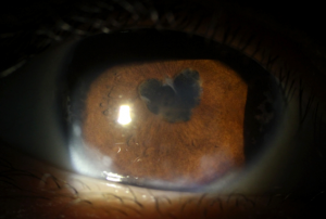
Synechiae
All content on Eyewiki is protected by copyright law and the Terms of Service. This content may not be reproduced, copied, or put into any artificial intelligence program, including large language and generative AI models, without permission from the Academy.
Disease Entity
Synechiae are adhesions that are formed between adjacent structures within the eye usually as a result of inflammation.
Disease
The term synechiae comes from the Greek synekhes, which means “hold together.” Synechiae are adhesions that typically attach the anterior iris to the trabecular meshwork in the iridocorneal angle (peripheral anterior synechiae) or the posterior iris adheres to the anterior lens capsule (posterior synechiae).
Etiology and Pathophysiology
Synechiae are most commonly formed during states of inflammation and cellular proliferation. Patients presenting with synechiae typically have an underlying inflammatory disease process such as uveitis and will present with related symptoms. The pathophysiology is thought to be related to inflammatory cells, fibrin and protein deposition, which stimulates the formation of adhesions between structures. However, peripheral anterior synechiae (PAS) can also be formed in a non-proliferative state. A posterior pushing mechanism can cause apposition of the iris on the trabecular meshwork, which may result in continuous PAS and angle closure. Other causes include trauma and increased intraocular pressure.
Diagnosis
Diagnosis is made with slit lamp exam and with gonioscopy of angle structures. Special attention should be paid to the pupillary margin.
Physical Examination
Posterior synechiae are visualized on standard slit lamp exam. Adhesions noted between posterior portion of iris and anterior capsule of lens.
Peripheral anterior synechiae are visualized on gonioscopic examination. Peripheral iris attachments noted anteriorly in the angle, which may extend anywhere from ciliary body to Schwalbe's line and corneal endothelium are important to differentiate from normally occurring iris processes.
Complications
Posterior synechiae, if substantial, may affect the movement of aqueous from the posterior to the anterior chamber, a condition known as iris bombe. As pressure builds up posteriorly, the iris may bow forward, resulting in secondary angle closure. Seclusio pupillae occurs when the synechiae extend 360 degrees around pupillary border.
Peripheral anterior synechiae may lead to secondary angle-closure glaucoma if they fuse circumferentially and are typically found inferiorly. PAS are more often found superiorly when associated with primary angle closure. Chronic angle-closure may develop following secondary shallowing of anterior chamber due to traction from PAS, which results in blockage of outflow through trabecular meshwork.
Management
- Treat the underlying cause
- Cycloplegics may prevent and also break adhesions
- Anti-inflammatory medications often prevent further formation of synechiae
- IOP lowering agents may be employed as needed (although, prostaglandin-analogues are often avoided if inflammatory causes are identified)
- Peripheral laser iridotomy may be indicated if patient develops angle closure
- Surgical synechiolysis and iridotomy are, at times, required
References
1. Moorthy RS, Mermoud A, Baerveldt G, et al. Glaucoma associated with uveitis. Surv Ophthalmol 1997; 41:361-394.
2. Ritch R. Pathophysiology of glaucoma in uveitis. Trans Ophthalmol Soc UK. 1981; 101:321-324.
3. "Glaucoma: Angle Closure" Digital Reference of Ophthalmology. Columbia University. http://dro.hs.columbia.edu/
4. Lee JY, Kim YY, Jung HR. Distribution and characteristics of peripheral anterior synechiae in primary angle-closure glaucoma. Korean J Ophthalmol. 2006 Jun;20(2):104-8.


