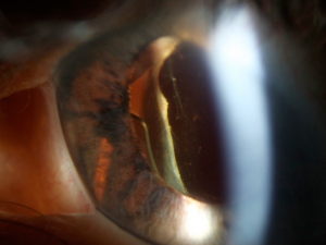
Pseudophakic and Aphakic Glaucoma
All content on Eyewiki is protected by copyright law and the Terms of Service. This content may not be reproduced, copied, or put into any artificial intelligence program, including large language and generative AI models, without permission from the Academy.
Disease Entity
Increase in intraocular pressure is a phenomenon that could present in both aphakic and pseudophakic eyes. In general, the terms are usually applied to the eye post cataract surgery. Given that there are over two million[1] cataract surgeries performed in the US each year, it is important to anticipate a possibility of postoperative glaucoma. However, there are multiple mechanisms, as well as combination of mechanisms, that can lead to increase in the eye pressure and thus a through evaluation of an eye is necessary.
Etiology
Aphakic glaucoma is referred to a condition that is a known complication which follow congenital cataract surgery in children[2]. Pseudophakic glaucoma refers to the glaucoma following implantation of the lens with cataract surgery. Aphakia or pseudophakia themselves are not the direct causative conditions in the aphakic or pseudophakic patient presenting with glaucoma[3]. There are multiple mechanisms that could be working alone or in synergy, leading to glaucoma in patients with aphakia or pseudophakia. Refer to the sections below for potential mechanisms.
Distortion of Anterior Chamber Angle
Kirsch characterized the existence of an internal white ridge protruding into the anterior chamber as an inverted snowbank along the ridge of the corneal incision as seen by gonioscopy during and after cataract surgery. The mechanism of the ridge formation is still unclear with some suggesting distortion of the chamber by sutures and others suggesting edema of the corneal stroma to be the cause. The ridge formation is associated with peripheral anterior synechiae (PAS), vitreous adhesions and hyphema[4]. It appears that the presence of the ridge has a transient effect on the intraocular pressure, affecting the trabecular meshwork and the aqueous outflow in the eyes[5]. Distortion of anterior chamber angle applies to previous cataract surgery techniques that required much larger incision than today’s modern, small incision cataract surgery. If anything, modern cataract surgery deepens the anterior chamber angle and reduces intraocular pressure[6].
Influence of Viscoelastic Substances
Viscoelastic Substances are used in cataract surgeries primarily to protect the endothelial cells of the cornea and maintain anterior chamber depth for the duration of the surgery. The use of these substances has been associated with increases in intraocular pressure (IOP) post surgically. The most likely mechanism to be implicated in the pressure elevation is a transient obstruction of the trabecular meshwork. There are many different viscoelastic substances that are used, such as sodium hyaluronate (Healon, AMO, Abbott Park, Illinois), combination of sodium hyaluronate and chondroitin sulfate (Provisc and Viscoat, Alcon Laboratories, Inc, Fort Worth, Texas), methylcellulose and others[5]. Dispersive viscoelastics (i.e. Viscoat) have lower viscosity and are harder to remove, and hence are more likely to increase IOP than cohesive ones such as Provisc and Healon.
Inflammation and Hemorrhage
Some level of inflammation is expected in eyes after cataract surgery. Depending on the extent of inflammation, excessive inflammatory cells may lead to obstruction and fibrosis of the trabecular meshwork and promote PAS formation. Uveitis-Glaucoma-Hyphema (UGH) syndrome can rarely arise from the rubbing of the lens implant on the iris and the ciliary body, leading to inflammation and hemorrhage[5]. Improperly sized and malpositioned anterior chamber lens implant is usually associated with UGH syndrome although UGH with posterior chamber lens implant placed in sulcus has been infrequently reported as well[7]. Rarely, in the presence of zonular laxity or plateau iris, in-the- bag IOLs can also cause UGH[8]. In addition, spontaneous, recurrent hyphema has been reported in eyes with neovascularization of internal corneal wound after cataract surgery (Swan syndrome).
Pigment Dispersion
It is common for some amount of pigment granules from the iris epithelium to be released during cataract surgery. This pigment can lead to obstruction of the trabecular meshwork, thus precipitating IOP elevation. Improperly inserted posterior chamber lenses are most commonly associated with pigment release that blocks the trabecular meshwork resulting in IOP elevation in a variant of UGH syndrome[5]. There are a number of the published cases describing eyes with increased pigment dispersion, inflammation, and elevated IOP due to insertion of single piece acrylic into the sulcus. Single piece lens, which is designed to be placed entirely in a capsular bag has unpolished and thicker haptics that can easily cause pigment dispersion, transillumination defects, hyphema and elevated IOP if placed into the sulcus[9].
Vitreous Filling the Anterior Chamber
Rarely, anteriorly prolapsing vitreous humor can fill the anterior chamber after a cataract surgery resulting in an acute angle closure glaucoma. While cases can resolve on their own, others require a surgical procedure such as iridotomy or anterior vitrectomy and laser vitreolysis[5].
Pupillary Block
Pupillary block does not commonly present in aphakic patients. Rarely, it occurs if there is no surgical iridectomy following congenital cataract removal. The mechanism of glaucoma, in this case, is attributed to an adherence between the vitreous humor and the iris following removal of the lens and the capsule that inhibits the flow of aqueous humor into the anterior chamber. The accumulating aqueous humor then pushes the iris forward, closing the anterior chamber angle.This mechanism may depend on the presence of an intact anterior hyaloid. Deep anterior chamber centrally and forward bowing of peripheral bowing can differentiate this condition from malignant glaucoma in aphakia[5]. Pseudophakic pupillary block glaucoma can be seen with anterior chamber, iris supported and posterior chamber lenses. Anterior chamber lenses and iris supported lenses require peripheral iridectomy to prevent pupillary block glaucoma. Generally, glaucoma appears soon after the surgery, but can also occur in a matter of months or years. The mechanisms underlying glaucoma include the bulging of the iris over the sides of the lens for the anterior chamber lenses, pupillary block and inflammation with peripheral anterior synechiae. Pupillary capture of posterior chamber lens implant can be seen after improperly placed implant in the sulcus, particularly when the lens is in reversed position.
Peripheral Anterior Synechiae and/or Trabecular Damage
Shallowing of the anterior chamber and increased post-operative inflammation can contribute to the formation of the peripheral anterior synechiae in pseudophakic and aphakic glaucoma. The anterior chamber can also shallow following cataract surgery due to wound leak, resulting in hypotony and choroidal detachment. Decrease aqueous production and a forward shift of iris and vitreous can result, contributing to chronic angle closure glaucoma[5].
Influence of alpha-chymotrypsin
In the past, alpha-chymotrypsin enzymatic zonulolysis was used for intracapsular cataract removal, and has been associated with acute IOP elevation due to obstruction of the trabecular meshwork by lysed zonular fragments. Alpha-chymotrypsin is not longer used in cataract surgery[5].
Lens-Particle Glaucoma
Small nuclear or cortical pieces of natural lens left in the eye following a routine cataract surgery or larger pieces dropped into vitreous after broken posterior capsule during complicated cataract surgery may induce glaucoma. These protein structures are too large to go through trabecular meshwork and they block the outflow and increase the pressure. There is also an inflammatory component contributing to the process.
Neodymium: YAG Laser posterior capsulotomy
Use of Neodymium: YAG Laser to remove posterior capsular opacities after cataract surgery has been shown to cause an intraocular pressure increase. The increase in IOP is usually transient, but in rare cases persists beyond the immediate post-procedural period.. There are multiple risk factors that can contribute to the increase in the eye pressure after the use of the Nd:YAG laser, such as presence of glaucoma or high eye pressure before the surgery, absence of posterior chamber lens, myopia, laser energy used, etc.[5].
Ghost cell glaucoma
Ghost cells are red blood cells without hemoglobin most often from chronic vitreous hemorrhage, and can enter the anterior chamber in aphakic eyes. The ghost cells can obstruct aqueous outflow through the trabecular meshwork leading to the increase in IOP[3].
Epidemiology
Depending on the studies, the estimate for incidence of glaucoma post cataract surgery varies widely. In patients with aphakia, chronic glaucoma post cataract surgery was found to be less prevalent at 3% than a transient increase in pressure with varying percentages of pressure rise post cataract surgery[5]. Other sources report the incidence between 5-41%[2].
In patients with pseudophakia, the incidence of IOP rise went down with the advent of the extracapsular cataract extraction (ECCE) and posterior chamber intraocular lens (PCIOL) implantation. Transient increases in IOP are seen at rates of 29-50% on the first postoperative day in the eyes without pre-existing glaucoma. Chronic glaucoma prevalence in pseudophakic eyes post-surgery was noted between 2.1-4% after the standard extracapsular extraction[5].
Diagnosis
Presentation of glaucoma in aphakic/pseudophakic eyes may be very similar to those in phakic eye, and a thorough patient history complete with disease onset in relation to ocular surgeries is necessary. Some of the ways to evaluate a patient presenting with glaucoma include utilizing gonioscopy to visualize angle morphology and presence of lens fragments, ultrasound biomicroscopy (UBM) and Scheimpflug video imaging to evaluate the location and stability of the lens behind the iris[7][8]. In a published series of UBM in UGH patients, 8/9 study patients had posterior chamber intraocular lens. In 5 cases, UBM visualized at least one of lens haptics was in contact with posterior iris pigment epithelium and in remaining, one of the haptics was embedded into ciliary body[7].
Differential Diagnosis for IOP Elevation after Cataract Surgery
- At 1-7 days post-op: consider pre-existing open angle glaucoma, retained viscoelastic, trabecular edema or angle distortion, surgical hyphema, pigment Dispersion, inflammation, pupillary block, aqueous misdirection, choroidal hemorrhage or effusion.
- At 1-7 weeks post-op: consider pre-existing open angle glaucoma, vitreous in the anterior chamber, steroid-induced glaucoma, ghost cell glaucoma, lens particle glaucoma, neovascular glaucoma.
- After 2 months: pre-existing open angle glaucoma, ghost cell glaucoma, lens particle glaucoma, UGH syndrome, pigment dispersion, chronic uveitis, epithelial downgrowth or fibrous ingrowth, pupillary block[10].
Management
Preoperative
Surgeons can choose to reduce the pressure in the eye as part of the pre-operative care. Digital pressure can be applied to the globe. Intravenous hyperosmotic agents such as mannitol can be utilized to reduce the risk of complications.
Intraoperative
Caution in the manipulation of the tissues during the surgery as well as meticulous removal of the viscoelastic substances should minimize the risks of post-op IOP elevation. An intraocular lens can also have an effect on the outcome, with PCIOL being less commonly associated with increases in IOP than ACIOL. Use of adjacent intraoperative anticholinesterase agent Miostat® Alcon laboratories (carbachol) provides pressure reduction in addition to constriction of the pupil (miosis) up to 24 hours after surgery.
Postoperative
Management of postoperative glaucoma will depend on the mechanism causing the increase in IOP. Transient pressure increases in eyes with no pre-existing glaucoma require no treatment other than immediate IOP control. In eyes with pre-existing glaucoma, transient increases in pressure early in post-surgical period can cause damage and thus should be thoroughly managed with drops such as dorzolamide-timolol, brimonidine or oral acetazolamide. Use of prostaglandin analogs in early post-operative period varies among practitioners due to their inflammatory properties. If IOP is causing significant corneal edema, discomfort, and there is pre-existing glaucoma, anterior chamber tap could be done successfully at the slit lamp by releasing aqueous through existing paracentesis from cataract surgery. The aim should be to lower the IOP to high 10s or low 20s rather then returning IOP to single digits, which might induce hypotony-associated complications. When managing IOP-elevated related to UGH syndrome, mydriatics or miotic drops in different sequence has been tried to capture the IOL properly. Usually, these measurements are not useful. Chronic or recurrent inflammation can cause cystoid macular edema and chronic use of steroid drops is associated with elevated IOP, further complicating the course of treatment. Therefore, in the presence of chronic pigment dispersion and inflammation in a pseudophakic eye, lens removal/exchange is definitely.
Pupillary block in an aphakic eye commonly requires iridotomy. For pupillary block in pseudophakic eyes, a variety of therapies can be considered ranging from mydriatic therapy to release the block as initial treatment to immediate iridotomy[5]. Malignant glaucoma post cataract surgery will likely require vitrectomy.
Preferred treatment of glaucoma in the late postoperative period is medical including the use of carbonic anhydrase inhibitors, prostaglandin analogs, beta-blockers, alpha2-antagonists or miotic therapy. Surgery for glaucoma should be performed in cases that fail medical management.
The role for newer microinvasive glaucoma surgery in the treatment of pseudophakic glaucoma has not been adequately evaluated.
Pseudophakia has been associated with higher risk for trabeculectomy failure. Conjunctival scarring from earlier techniques of cataract surgery, which involved incision of conjunctiva was presumed for the failure of consequent glaucoma surgery, however, clear corneal incision phacoemulsification also has been contributed to surgical failure of trabeculectomy with anti-metabolite. Takihara et al. reported 4.6 times higher risk of failure in 51 pseudophakic patients who underwent trabeculectomy with mitomycin than risk in 175 phakic patients with mean follow up of about 3 years[11]. Glaucoma drainage devices may also be utilized to treat aphakic and pseudophakic glaucoma[12]. There may be some advantage inserting tube into the sulcus behind the iris and in front of the IOL in pseudophakic glaucoma to decrease the corneal compromise associated with the tube.
Diode cyclophotocoagulation are typically resolved for those with advanced glaucoma and poor visual potential.
References
- ↑ http://www.preventblindness.org/cataract-surgery
- ↑ 2.0 2.1 Yi K, Chen T. Aphakic glaucoma following congenital cataract surgery. Indian J Ophthalmol. 2004 Sep;52(3):185-98. Review.
- ↑ 3.0 3.1 Tomey KF, Traverso CE. The glaucomas in aphakia and pseudophakia. Surv Ophthalmol. 1991 Sep-Oct;36(2):79-112. Review.
- ↑ Kirsch RE, Levine O, Singer JA. Ridge at internal edge of cataract incision. Arch Ophthalmol. Dec 1976;94(12):2098-2104
- ↑ 5.00 5.01 5.02 5.03 5.04 5.05 5.06 5.07 5.08 5.09 5.10 5.11 Allingham, R. Rand, and M. Bruce Shields. 2005. Shields' textbook of glaucoma. Philadelphia: Lippincott Williams & Wilkins. Chapter 26.
- ↑ Kurimoto Y, Park M, Sakaue H, Kondo T. Changes in the anterior chamber configuration after small-incision cataract surgery with posterior chamber intraocular lens implantation. Am J Ophthalmol. 1997 Dec;124(6):775-80.
- ↑ 7.0 7.1 7.2 Piette S, Canlas OAQ, Tran HV et al. Ultrasound biomicroscopy in uveitis-glaucoma-hyphema syndrome. Am J Ophthalmol 2002;133:839-841.
- ↑ 8.0 8.1 Zhang L,Hood CT, Vrabec JP et al. Mechanisms for in-the-bag uveitis-glaucoma-hyphema syndrome. J Cataract Refract Surg 2014;40:490-492.
- ↑ Uy HS, Chan PST. Pigment release and secondary glaucoma after implantation of single-piece acrylic intraocular lenses in the ciliary sulcus. Am J Ophthalmol 2006;142:330-332.
- ↑ Johnson, S. 2009. Cataract Surgery in the Glaucoma Patient. Springer.
- ↑ Takihara Y, Inatani M, Seto T, et al. Trabeculectomy with mitomycin for open-angle glaucoma in phakic vs pseudophakic eyes after phacoemulsification. Arch Ophthalmol. 2011; 29 (2):152-7.
- ↑ GeddeSJ, Schiffman JC, Feuer WJ et al. Treatment outcomes in the Tube versus Trabeculectomy (TVT) study after five years of follow-up. Am J Ophthalmol 2012;153(5):789-803.


