Pupil Expansion Devices and Mechanical Stretching of the Pupil
All content on Eyewiki is protected by copyright law and the Terms of Service. This content may not be reproduced, copied, or put into any artificial intelligence program, including large language and generative AI models, without permission from the Academy.
Introduction
Nondilating or poorly-dilating pupils pose significant challenges during and after cataract surgeries. The challenges include-
- Reduced surgical space that may lead to small capsulorrhexis.
- This causes difficulty in maneuvering the lens during surgery (reduced surgical access) and
- There is a higher chance of trauma to the anterior capsular rim/capsulorrhexis edge with instruments. An anterior capsular tear may extend beyond the equator causing a posterior capsular rent (PCR).
- Less visibility of the peripheral lens and the placement of the tip of certain instruments. This.may increase the risk of complications including iris trauma (including chafing, sphincter tear, iridodialysis), hyphema, PCR
- Iris prolapse
- Intraoperative further reduction in pupillary dilation likely related to prostaglandins release or iris touch with instruments,
- Retained cortical matter in the peripheral capsular bag which may swell up in immediate postoperative period and may need resurgery for removal
- Inadvertent placement of one haptic of intraocular lens (IOL) in the capsular bag and the other in sulcus which predisposes to iris chafing, inflammation after surgery, and concentration of the IOL after capsular healing
- Increased postoperative inflammation, corneal edema, and cystoid macular edema
Intraoperative floppy iris syndrome (IFIS) is an important clinical entity which was described in 2005 by Chang and Campbell, This may cause poor pupillary dilation.[1] The iris loses tone and is flaccid in IFIS. IFIS is usually associated with the use of oral Tamsulosin ( alpha-1A antagonist), a drug used for benign prostatic hypertrophy. Multiple other drugs can also cause a similar problem. IFIS is characterized by a triad[2][3] of
- Floppy and flaccid iris that billows in the fluid currents during the cataract surgery
- Chances of iris prolapse out of the main and side wounds
- Progressive intraoperative miosis.
Surgical strategies in small pupils
Various strategies have been employed in such patients to overcome the challenges and to dilate the pupil.
These strategies include
- ensuring maximal pupillary dilation with the use of mydriatic drops including tropicamide and phenylephrine,
- preoperative nonsteroidal anti-inflammatory drug (NSAID) drop to possibly prevent intraoperative miosis,
- placing a cotton pledget soaked in a mydriatic agent in the inferior fornix just before surgery,
- bolus intracameral mydriatic and local anesthetic [including 'epi-Shugarcaine'[4] (epinephrine 1:1000 and lidocaine 1%) described by Joel K Shugar[5], phenylephrine 1.5%, Mydrane (Thea Pharmaceuticals Limited, Newcastle-under-Lyme, UK)- lidocaine (1%), tropicamide (0.02%), and phenylephrine (0.31%)]
- mydriatic with or without NSAID in the irrigation fluid to maintain the dilation and prevent miosis- adrenaline, Omidria® (Omeros Corp., Washington, USA)-phenylephrine 1% & ketorolac 0.3%. Omidria® has been approved by the FDA (Food and Drug Administration, USA) for use in the irrigating solution during cataract surgery to maintain the pupillary dilation (which has been already achieved) by preventing miosis and to prevent pain after surgery.
- viscodilation/viscomydriasis of the pupil,
- mechanical stretching,
- pupillary expansion devices, and
- iridectomy/sphincterotomy/pupilloplasty.
This article discusses various available methods for pupillary stretching and mechanical pupil expansion devices/pupil expanders/ pupil enlarging devices.
Prerequisites before pupillary stretching and using pupil expansion devices
Maximal preoperative pharmacological dilation of the pupil should be achieved in such cases of poorly dilating or non-dilating pupils. Ophthalmic viscosurgical devices (OVDs) must be used in the anterior chamber to dilate the pupil to the maximum and to maintain the anterior chamber. Care should be taken to avoid overfilling the anterior chamber which may push iris backward and may cause difficulty in fixing the pupil expansion devices. Also, some OVD causing a mild separation of the pupil and the anterior capsule of the lens helps in the proper engagement of the expansion device. Any pupillary membrane should be removed using Utrata forceps or a similar instrument. Posterior synechia and all anterior capsular adhesions with the iris should be lysed using a blunt/smooth instrument. Care should be taken to avoid rupturing the anterior capsule and to avoid traumatizing the iris root (which can cause severe pain, hyphema, and iridodialysis in severe cases). As pupillary stretching may cause pain, it is prudent to augment the anesthesia using topical, intracameral, sub-Tenon, or peribulbar anesthesia. Only after this, pupillary stretching methods or pupil expansion devices can be used. Very rigid and small pupils may need to be stretched bimanually with other instruments before pupil expansion rings can be engaged to the pupil.
Pupillary stretching
Bimanual stretching of the pupil with 2 instruments
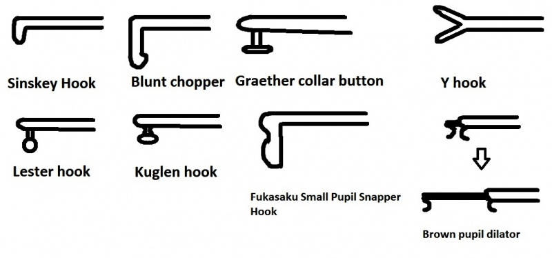
Luther L Fry, MD from Garden City, Kansas, USA popularized this technique originally described by Dr. Keener (Indianapolis, Indiana). This can be performed using 2 different instruments which may be
- Sinskey hooks
- Blunt choppers
- Graether collar buttons
- Y hooks
- Lester hook
- Kuglen hooks
- Fukasaku Small Pupil Snapper Hook
These 2 instruments are inserted from 2 different wounds at least 30° apart. The anterior chamber should be filled with OVD and should not collapse. The instrument tip is used to engage the pupillary margin 180° apart. Then the 2 instruments are carefully moved in the opposite direction toward the iris root. This stretch is maintained for some time (around 3-5 seconds). Some minor bleeding points at the pupillary margin may be noted during this maneuver. If reasonable pupillary dilation is not achieved, similar pupillary stretching is performed at a line perpendicular to the previous location. Peters pupillary dilator (Rhein Medical Inc., Tampa, Florida, USA) has a J-like loop at the tip which helps in engaging the pupillary margin. Pupillary stretching and sphincterotomies are effective for rigid/fibrotic pupils and not for IFIS in which the inherent tone of the iris is lost and intraoperative miosis is the norm even after pupillary stretching.
[Video credit- Uday Devgan MD - CataractCoach.com]
Brown pupillary dilator (Rhein Medical Inc., Tampa, Florida, USA)
The tip has a curve to engage the pupillary margin. Both side port and main incision versions are available. Side port version has one c loop facing distally. Two such instruments can be used for pupillary stretching. The main incision (phaco incision) version has 2 C loops facing away from each other and these engage the sub-incisional and distal pupillary margin and the difference between C loops can be increased leading to pupillary dilation.
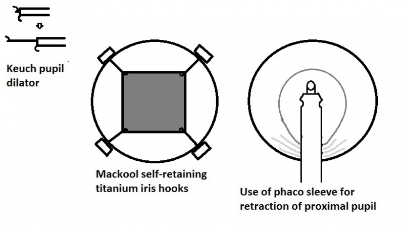
Keuch Pupil dilator (Katena Products, Inc., Parsippany, New Jersey, USA)
It is similar to the phaco-incision Brown pupillary dilator. The pupillary margin is caught at 2 places 180° apart, and then the 2 catches are separated maximally to dilate the pupil.
Beehler pupil dilator
This is used for stretching the pupil at 4 points. Initially, the dilator is introduced inside the anterior chamber through the single plane main wound (around 2.5 mm). The primary microhook engages the sub-incisional pupillary margin. The other 3 microhooks are also engaged at the pupillary margin and slowly simultaneous and symmetrical dilation at 4 points is achieved using this dilator. The pupil is held at maximal dilation for a few seconds and then the dilator is disengaged and removed. This creates multiple microsphincterotomies and makes the pupil to about 6-7 mm in diameter. Another version of the instrument with 2 microhooks is also available.
[Video credit- Dr. Tarek Youssef]
Use of phaco sleeve
The phaco sleeve may be used to stretch the sub-incisional iris peripherally, thereby increasing the field of view. However, this maneuver may cause iris prolapse later in the surgery and may be associated with pigment loss/iris chafing/iris injury.
Using the lens or lens nucleus to keep the pupil dilated/ semi-dilated
This is a type of flip and chop. In very small pupils, after hydrodissection, a part of the lens is brought up to the anterior chamber while part of it remains in the bag. A sufficiently large capsulorrhexis is mandatory for safe manipulation of the lens in the pupil and for bringing it out of the capsular bag. This lens now mechanically keeps the pupil dilated, The corneal endothelium is coated with cohesive OVD and the lens is now chopped. The energy delivery in the eye may be reduced with phaco power modulations, and the phaco is performed at the iris plane, not at the anterior chamber. Such maneuvers may protect from posterior capsular damage, but care should be taken to protect the corneal endothelium also.
[Video credit- Uday Devgan MD - CataractCoach.com]
Iris hooks/retractors
Mackool self-retaining titanium mechanical hooks
Usually, 3-4 hooks are used via self-sealing peripheral corneal/limbal paracentesis (1.5mm) incisions leading to a triangular or square-shaped pupil. The pupil is stretched with another instrument (iris pusher) and then the pupillary margin is engaged in the hooks. The hooks are removed after the surgical procedure. Challenges with these hooks include increased surgical time and fluid loss through the incisions which may accumulate over the cornea in deep-seated eyes causing increased reflections and compromised visibility.
Nylon/Polypropylene iris hooks with silicone sleeve
These are flexible J shaped iris retractors made of nylon (usually 5-0, or 6-0) which can be inserted through smaller paracenteses compared to Mackool retractors. A silicone sleeve is present to act as a stop or anchor. The design was attributed to De Juan. The plane of the sleeves usually has a defined relationship with the plane of the hook so that the sleeve may give a clue to the orientation of the tip of the hook when the tip is not visible. The entry point for the iris hooks should be at the posterior limbus at the level of iris and anterior corneal wounds (which cause tenting of the iris) should be avoided. Most surgeons use at least 4 iris hooks which gives a quadrangular opening. Four wounds are made 90° apart. All iris hooks are inserted and the pupillary margin is engaged. Then, the iris hooks are retracted slowly causing pupillary dilation and the silicone sleeve is slid forwards. The sleeve helps in anchoring the hook so that it does not slide inside the anterior chamber making the pupil small. When tightening the hooks, the maneuvers should be gentle, otherwise, hyphema, iris injury, or anterior capsular injury may occur. It is suggested that the pupil should not be stretched to more than 5mm using iris hooks to avoid tissue overstretch and resultant iris trauma, hyphema, and postoperative atonic and irregular pupil.[4][6][7] Some surgeons make wounds for the iris hooks just behind the primary wound and side wounds. Iris hooks can be inserted at any stage of the cataract surgery if the pupil becomes small. However, since the ends of some iris hooks are quite sharp, care should be taken not to include the capsulorrhexis margin in the iris hook which can cause radial tear of the anterior capsule and even posterior extension to cause a posterior capsular tear. Adequate OVD should be injected inside the anterior chamber and also between the iris and lens capsule especially when the iris hook is inserted after the capsulorrhexis has been made.
The iris hooks are removed after the implantation of the intraocular lens (IOL). For removal, the anterior chamber should be filled with OVD. The iris hook is first pushed inside to disengage the pupil. then it is drawn outwards.
The affordable cost is an important advantage of iris hooks. The hooks may be placed at the axis of intended toric IOL placement, thereby marking the axis. As the exposure at these areas is maximum, the toric marks of the IOL are easily seen. Putting an iris hook just behind the phaco wound avoids tenting up of iris in front of the phaco probe, reduces the chance of iris prolapse, and increases visibility. When 4 iris hooks are used at 90° to each other, a diamond or quadrangular opening or pupil is made and the surgical exposure can be excellent. With experience, the time needed for insertion and removal of the iris hook can be reduced considerably.
Reusable polypropylene iris hooks (4-0) are also available. The ability to reuse reduces the cost further.
Challenges of iris hooks include the need for multiple paracenteses which may be disadvantageous in pterygium, bleb, and radial keratotomy incisions. Because the entries are often made behind the limbus or at the posterior limbus, there may be subconjunctival hemorrhage, conjunctival ballooning with the irrigation fluid, and the irrigation fluid may accumulate around and in front of the cornea in deep-set eyes causing poor visibility during surgery. Another problem is that the outward end of the hook may be pushed by the eye speculum or eyelid in patients with tight palpebral fissure. The sharp tip of the iris hook may rupture the anterior capsular rim or even the posterior capsule.
[Video credit- Uday Devgan MD - CataractCoach.com]
[Video credit- Dr. Sourabh Patwardhan]
Pupil expansion devices
Hydroview Iris Protector Ring (Grieshaber & Co. AG Schaffhausen, Schaffhausen, Switzerland)
This is a hydrogel device which in its dehydrated oval form can be placed in the anterior chamber through a 3 mm incision. The device engages the pupillary margin via flanges and enlarges in size with hydration. It can be manipulated to aid in its expansion. It is removed after the placement of IOL through the same incision.
Siepser iDeal iris ring (Eagle Vision, Memphis, Tennessee, USA)
This ring has been devised by Dr. Steven B Siepser from Philadelphia, USA. This is similar to the Hydroview iris protector ring. It met FDA guidelines in 2016 and was clinically launched in 2017. This is an irregular oval hydrogel ring which expands on hydration.
[Video credit- Siepser Laser Eyecare]
Graether 2000 pupil expander system (Eagle vision, Memphis, Tennessee, USA/Katena Products, Inc., Denville, New Jersey, USA)
It is described by John Graether from Iowa, USA.[8] This is a clear soft silicone ring. It has a circumferential groove for engaging the pupillary margin. It is inserted inside the eye using a preloaded sterile device. It dilates the pupil to 6.3 mm. The ring is incomplete and a strap connects the ends of the ring. This is inserted inside the eye using an insertion instrument and iris glide-retractor.
[Video credit- The Fladen Eye Center]
Morcher Pupil dilator Type 5S (FCI Ophthalmics, Pembroke, Massachusetts, USA)
This is an incomplete elastic ring made of solid polymethylmethacrylate (PMMA) which can be injected into the anterior chamber with Geuder pupil dilator injector (2.5mm incision, #G-32970, FCI Ophthalmics, Pembroke, MA) or manually. The maximal overall diameter of this device is 7.5mm and the thickness minimum thickness of the ring is 0.15 mm. At the sides, to engage the pupil the external area has a 0.6mm notch. The thickness of the device at the notch is 0.9mm. This device provides pupillary dilation of around 5-6mm. It creates even tension at around 300° of the supported pupil and the chances of sphincter damage and postoperatively deformed pupil may be less. After insertion in the anterior chamber, the central segment of the device is manipulated to engage the distal pupillary margin. Then, the ends of the device are engaged in the pupillary margin with the help of the eyelets in the ring. For the removal of the device, first, the ends are disengaged. Then, the ring is removed with the use of forceps.
[Video credit- FCI Ophthalmics]
Malyugin ring (MicroSurgical Technology/MST Inc., Redmond, Washington, USA)
This single-use disposable ring is devised by Dr. Boris Malyugin from Moscow, Russia. Of all the pupil expansion rings, this ring is most commonly used by Ophthalmic surgeons worldwide. The initial version was marketed in 2007. It is a square-shaped device made of 4-0 polypropylene with coils/scrolls at its 4 corners which engage the iris. However, it has 8 points of fixation with the pupillary margin (at 4 coils and points located at the middle between the coils) and creates a round and dilated pupil. The device is injected inside the anterior chamber (via a 2.5mm incision) with a special disposable injector that is supplied with the ring. The coils may be engaged to the pupillary margin with Osher/Malyugin manipulator, Sinskey hook, Kuglen hook, Lester hook, or similar instruments.
The current version is called Malyugin ring 2.0 can be inserted through a 2 mm incision using a modified injector. Other modifications from the previous model include thinner material (5-0 polypropylene), wider coil gap, and more soft and elastic material. This model was released in 2016. Because of the thin profile, chances of damage to delicate ocular structures may be less. Also, the device typically does not hinder the surgical maneuvers, and inadvertent disengagement from the iris during surgery is very rare. Insertion and removal of this ring may be easier and faster than other options.[9]
Two sizes are available 7 mm and 6.25 mm.
The inventor recommends that high viscosity OVD like Healon 5 is used to create a space between the anterior lens capsule and the pupil, as the material of the device is elastic. Three coils (distal and 2 side coils) may be used to engage the pupillary margin automatically during insertion, leaving the proximal-most coil the only coil to be manipulated to hold the iris margin. This ring costs around 135 US dollars/ring which may hinder its access to the areas with a lack of resources.
[Video credit- Microsurgical Technology]
Modifications/Special situations
Malyugin ring has been successfully used in femtosecond laser-assisted cataract surgery.[10]
Agarwal's modification- A suture is passed through the leading coil so that the device can be retrieved in cases with suspected posterior capsular rupture.[11]
For ectopic pupil- The ring can be used to dilate the pupil and an iris hook may be used to center the ring. [12]
In patients with ectopia lentis- The pupil can be dilated using a Malyugin ring. The ring is then drawn (using a 10-0 polypropylene suture via a scroll) toward the displaced lens to center the pupillary opening over the lens so that a femtolaser anterior capsulotomy could be performed.[13]
In cases with zonular instability and small pupil, the side coils of the ring may be used to hold both the iris margin and the capsulorrhexis edge. Thus, a single device is used for both pupillary dilation and stabilization of the capsular bag [14]
Malyugin ring has been successfully used in the triple procedure in patients with positive vitreous pressure where safe cataract surgery could be performed as an open-sky procedure with the Malyugin ring supporting the iris just beneath the cut margin of the host cornea.[15]
Challenges with Malyugin ring
Some increase in surgical time, loss of iris pigment, and minor areas of sphincter trauma are common with the device. The side clips may entangle and manual separation with the use of other devices may be needed to separate the clips. Hyphema, anterior capsular injury, iris trauma/iridodialysis, and minor irregularity at the pupillary margin after surgery are other complications. A patient of intraoperative suprachoroidal hemorrhage has been reported who was operated after 1 week of initial surgery and the Malyugin ring was removed in the second surgery with no apparent harm to the eye.
[Video credit- Uday Devgan MD - CataractCoach.com]
[Video credit- Uday Devgan MD - CataractCoach.com]
Milvella Perfect Pupil (Becton-Dickinson, Franklin Lakes, New Jersey, USA/Milvella, Savage, Minnesota, USA)
This is described by Dr. John Milverton, of Sydney, Australia. This is a device with a 7 mm internal diameter made from polyurethane. The groove at the outer surface of the device engages the pupillary margin. It may be inserted inside the anterior chamber with a forceps or an injector through the main port which has to be mildly enlarged to accommodate both the phaco/irrigation aspiration handpiece and the portion of the ring which protrudes outside the phaco wound. The device is first inserted into the anterior chamber which has been filled with OVD. The proximal-most area of the ring is first engaged, then adjacent areas are progressively engaged in the pupillary margin. The protruding portion of the ring helps in the removal of the ring after its use. The pupillary margin may get caught in the device resulting in iris injury, but excellent pupillary dilation is usually achieved.
[Video credit- user johnrlee4171]
B-HEX Pupil Expander (Med Invent Devices, Kolkata, India)
The B-HEX Pupil Expander, invented by Dr Suven Bhattacharjee from India is the third generation Bhattacharjee Ring.[16]
It is a flexible hexagonal plastic (Polyimide) ring with a 0.075 mm (75 microns) profile having notches at corners and flanges at sides, all disposed in a single plane. The disposable 6.5 mm B-HEX provides a 5.5 mm expanded pupil. It is preloaded in a transparent single-use carrier with an ergonomic handle that delivers the device sterile at the incision.
The notches at the corners and flanges at the sides hold the pupillary margin. Alternate flanges are tucked under the iris to engage the notches to the margin of the pupil to provide a 5.5 mm expanded pupil. The thin profile and uniplanar design allow it to glide through much smaller incisions without an injector. Unlike other devices with scrolls or pockets which require an injector to avoid snagging the incision, the preloaded B-HEX is inserted and removed through a 1 mm or larger incision using the B-HEX-23 gauge micro-forceps. The thin profile and uniplanar design allow unhindered instrument movement during phacoemulsification, cortical cleaning, and IOL implantation. To engage to the iris B-HEX-23 G forceps is needed. The B-HEX is safely used even after capsulorhexis since the thin uniplanar notches are directly visualized to avoid the capsule margin. It is useful in coaxial phacoemulsification, coaxial sub-2.00 mm microincision cataract surgeries (MICS), biaxial 1.5 mm MICS, femtosecond laser-assisted cataract surgery(FLACS), small pupil pars plana vitrectomy and shallow anterior chamber eyes.[17]
In the first series describing the use of B-HEX for combined Phacoemulsification and Vitrectomy due to myriad vitreoretinal causes, the B-HEX remained well engaged and maintained excellent mydriasis throughout the surgery despite wide fluctuations in intraocular pressure and anterior chamber fluidics. The application of pupil expansion rings for vitreoretinal surgery is much different from their use in cataract surgery due to very different fluidic dynamics and pressure fluctuations during retinal surgeries. Other advantages of the B-HEX in vitreoretinal surgery are the ease of insertion and removal even via the 0.9 mm side port incision with an intraocular forceps without any collapse of the anterior chamber despite the presence of tamponading agents behind the intraocular lens (IOL). [18]
[Video credit- Dr. Suven Bhattacharjee]
[Video credit- Uday Devgan MD - CataractCoach.com]
Assia pupil expander (APX Ophthalmology, Haifa, Israel)
This scissor-like device with a spring mechanism has been developed by Dr. Ehud Assia from Israel. The initial version (introduced in 2013), APX100 was made of metal and was reusable. APX200 is the second generation of the device which was introduced in 2015. It is made of plastic with a blue color. Two devices are used via opposite 19G paracenteses placed at 90° to the main wound. Exactly opposite paracenteses create a quadrangular opening, whereas if the angle between the paracenteses is not exactly 180°, it creates a trapezoidal opening. The corneal incision is made parallel to the iris. The closed device is inserted via the paracentesis. The device is opened slowly only when the pivot pin is inside the anterior chamber, or at least within the incision. The pupillary margins are engaged on to the tip of the device one by one in the half-opened stage of the device. Then it is opened fully and the special forceps are released (see video). The pupillary opening is usually 6x6mm and intraocular manipulations are not required. Also, only 2 paracenteses suffice compared to 4 or 5 in cases with iris hooks. One end of the APX hook in each side is bent downwards for a good hold of the pupillary margin.
[Video credit- APX Ophthalmology]
I-Ring pupil expander (Beaver-Visitec International, Waltham, MA, USA)
This is a circular soft ring made of polyurethane which causes a circular dilation of the pupil to 6.3 mm. There are 4 outward projecting triangular portions with holes in each which help in positioning the device properly. The device may give a more circular appearance of postoperative pupil compared to the Malyugin ring.[19]
[Video credit- Dr. Sourabh Patwardhan]
Xpand NT iris speculum-X1 (Diamatrix Ltd, Woodlands, Texas, USA)
It is prepared from thin nitinol wire and dilates the pupil to around 6.75mm. To position the device, manipulation with other instruments is needed. This device creates an octagonal pupillary opening. It does not pinch the iris margin. The speculum is inserted inside the eye using an inserter.
[Video credit- Dr. Alan Malouf]
Oasis Iris Expander (Oasis Medical Inc. Glendora, California, USA)
This single-use square-shaped device is made of one piece blue polypropylene and comes in 2 sizes 6.25 mm and 7 mm. There are 4 pockets at corners to lodge the iris margin and may reduce the trauma to the pupillary margin. The expander is provided with an inserter. Initially, the expander needs to be loaded into the inserter, and then it is delivered within an OVD filled anterior chamber. Usually, the distal pocket, followed by proximal pocket and side pockets are engaged in the pupillary margin. However, the engagement sequence may vary according to the surgeon's preference.
[Video credit- Dr. Arup Chakrabarti]
Canabrava ring (AJL Ophthalmic S.A, Araba, Spain)
This was developed by Dr. Sergio Canabrava from Brazil.[20] In cases with iris defect/coloboma, this ring can be used whereas other rings including the Malyugin ring may be difficult to use as support for one clip is not present. Canabrava ring is made of PMMA, and the internal diameter is 6.3mm. The indents are alternating (one is above the iris, surrounded by 2 indents which are below iris) and hence the vertical thickness of this ring is only 0.4mm and the chances of endothelial injury may be less. There are 7 indents and the ring is open on one side. The device makes a 300° of a circle. No special instrument is needed for its insertion which reduces the cost. At the 2 ends of the device, there are hooks that go under the iris. Each indent has a hole for help in positioning the device.
[Video credit- Dr. Sergio Canabrava]
Results
A study[21] on 40 eyes noted that among iris hooks, Morcher ring, Beehler hooks, and pupil stretching; iris hooks, followed by Morcher ring had longest surgical times, though these achieved stable pupillary dilation throughout the surgery. Morcher ring caused the least iris trauma. Beehler pupil dilator and pupil stretching were associated with fast surgery.[21]
Plastic devices with high vertical thickness and high rigidity (including Morcher 5S) may have a higher chance of endothelial touch and damage.[9] A study noted that stress over the sphincter was highest in APX pupil expander.[22] Iris hooks caused less stress than APX device but more stress than Malyugin ring.[22] Uniform expansion caused less stress. Overall, all the devices induced higher stress than is needed for pupillary expansion.[22] A study using computer simulations showed that pupil expansion devices inhibit iris billowing in a model simulating IFIS.Lockington D, Wang Z, Qi N, et al. Modelling floppy iris syndrome and the impact of pupil size and ring devices on iris displacement [published online ahead of print, 2020 Feb 4]. Eye (Lond). 2020;10.1038/s41433-020-0782-7. doi:10.1038/s41433-020-0782-7
Malyugin ring may offer easier and faster insertion and removal, and when carefully used iris trauma is minimal.[23] A study[9] on 30 eyes using Malyugin ring noted minor hyphema in 1 eye, spontaneous disengagement of the device in 1 eye (one scroll was not engaged properly to the iris during its positioning), minor sphincter tears visible in slit-lamp in 5 eyes (pupil was round and reactive), iris chafing and transillumination defect in 8 eyes, and high intraocular pressure in 3 eyes. Other challenges noted in the study included temporary deformation of the device during loading or injection which might have been related to insufficient cooling (immediate use within 5 minutes of flash sterilization). Entanglement of the side scrolls was noted in one case, which needed removal of the device and insertion of a new one.[9]
A study comparing Malyugin ring with manual iris stretching showed that Malyugin ring was associated with better visual outcome and less endothelial cell loss.[24] Another large study noted that though both iris hooks and Malyugin ring were safe and effective in cases with small pupil, surgery with Malyugin ring was faster.[25] Complete retrieval of the Malyugin ring into the inserter after its use during surgery may be unpredictable and can cause trauma to the delicate ocular structures. To prevent this, the ring can be removed with forceps, or only partial retraction of the ring into the inserter after the disengagement of the distal and proximal scrolls can be safely done.[26] After this inserter along with the ring is removed from the eye.
In another study,[27] both Malyugin ring and I ring was noted to achieve 'smooth and atraumatic pupillary dilatation'. However, a case report noted that circular I ring device caused less distortion of the pupil (11%) compared to diamond-shaped Malyugin ring (49%).[28]
Initial results of Bhattacharjee ring in 42 eyes showed that there was adequate dilation, no incidence of spontaneous device engagement, intraoperative hyphema in 2 eyes, iridodialysis in one eye, postoperative round pupils in all but 2 eyes, microscopic sphincter tear in 10 eyes, and iris chafing in 6 eyes.[29] Bhattacherjee ring has a very low vertical height of 0.1 mm which may offer an advantage in very shallow anterior chambers.
The specific gravity of the material of Bhattacharjee ring (nylon 6.6 or nylon 6, specific gravity 1.14-1.35) is higher than water (specific gravity 1 ) or balanced salt solution (1.006) and Malyugin ring (polypropylene, specific gravity 0.9-0.91) is lighter. Thus, there is a theoretical possibility of inadvertent touch to the endothelium if Malyugin ring inadvertently disengages during cataract surgery.[29]
In a study on 30 eyes, Canabrava ring was noted to disengage intraoperatively in only one and the postoperative pupil size was significantly larger than preoperative size.[20]
Postoperative care
After using these hooks/methods, multiple sphincterotomies may be noted and the pupil may appear dilated. The pupil in such cases may be constricted intraoperatively using gentle centripetal strokes over iris with blunt instruments including Lester hook and intraoperative miotic agents (including pilocarpine). However, in most cases, the pupillary sphincter is not damaged till the full radial extent, and pupils retain a good cosmetic appearance and good pupillary response to light and accommodation. In the immediate postoperative period, miotic agents may be considered to prevent iridocapsular adhesion/synechia. In cases with severe iris manipulation, frequent topical steroids and close observation are must in the immediate postoperative period and oral/intravenous steroids may be added in a case-based approach.
References
- ↑ Chang DF, Campbell JR. Intraoperative floppy iris syndrome associated with tamsulosin. J Cataract Refract Surg. 2005;31(4):664‐673. doi:10.1016/j.jcrs.2005.02.027
- ↑ Chang DF, Braga-Mele R, Mamalis N, et al. ASCRS White Paper: clinical review of intraoperative floppy-iris syndrome. J Cataract Refract Surg. 2008;34(12):2153‐2162. doi:10.1016/j.jcrs.2008.08.031
- ↑ Tripathy K, Sharma YR, Chawla R, Basu K, Vohra R, Venkatesh P. Triads in Ophthalmology: A Comprehensive Review. Semin Ophthalmol. 2017;32(2):237‐250. doi:10.3109/08820538.2015.1045150
- ↑ Jump up to: 4.0 4.1 Malyugin BE. Recent advances in small pupil cataract surgery. Curr Opin Ophthalmol. 2018;29(1):40‐47. doi:10.1097/ICU.0000000000000443
- ↑ Shugar JK. Use of epinephrine for IFIS prophylaxis. J Cataract Refract Surg. 2006;32(7):1074‐1075. doi:10.1016/j.jcrs.2006.01.110
- ↑ Masket S. Avoiding complications associated with iris retractor use in small pupil cataract extraction. J Cataract Refract Surg. 1996;22(2):168‐171. doi:10.1016/s0886-3350(96)80213-6
- ↑ Yuguchi T, Oshika T, Sawaguchi S, Kaiya T. Pupillary functions after cataract surgery using flexible iris retractor in patients with small pupil. Jpn J Ophthalmol. 1999;43(1):20‐24. doi:10.1016/s0021-5155(98)00055-0
- ↑ Graether JM. Graether pupil expander for managing the small pupil during surgery. J Cataract Refract Surg. 1996;22(5):530‐535. doi:10.1016/s0886-3350(96)80004-6
- ↑ Jump up to: 9.0 9.1 9.2 9.3 Chang DF. Use of Malyugin pupil expansion device for intraoperative floppy-iris syndrome: results in 30 consecutive cases. J Cataract Refract Surg. 2008;34(5):835‐841. doi:10.1016/j.jcrs.2008.01.026
- ↑ Conrad-Hengerer I, Hengerer FH, Schultz T, Dick HB. Femtosecond laser-assisted cataract surgery in eyes with a small pupil. J Cataract Refract Surg. 2013;39(9):1314‐1320. doi:10.1016/j.jcrs.2013.05.034
- ↑ Agarwal A, Malyugin B, Kumar DA, Jacob S, Agarwal A, Laks L. Modified Malyugin ring iris expansion technique in small-pupil cataract surgery with posterior capsule defect. J Cataract Refract Surg. 2008;34(5):724‐726. doi:10.1016/j.jcrs.2008.01.024
- ↑ Malyugin B, Sobolev N, Arbisser LB, Anisimova N. Combined use of an iris hook and pupil expansion ring for femtosecond laser-assisted cataract surgery in patients with cataracts complicated by insufficient mydriasis and an ectopic pupil. J Cataract Refract Surg. 2016;42(8):1112‐1118. doi:10.1016/j.jcrs.2016.07.001
- ↑ Malyugin B, Anisimova N, Antonova O, Arbisser LB. Simultaneous pupil expansion and displacement for femtosecond laser-assisted cataract surgery in patients with lens ectopia. J Cataract Refract Surg. 2018;44(3):262‐265. doi:10.1016/j.jcrs.2018.01.014
- ↑ Zarei-Ghanavati S, Bagherian H. Stabilizing the capsular bag and expanding the pupil with a pupil expansion device. J Cataract Refract Surg. 2015;41(9):1801‐1803. doi:10.1016/j.jcrs.2015.08.010
- ↑ Sharma VK, Trehan HS, Raji K, Dhar SK. New use of a pupillary expansion ring to avoid intraoperative positive vitreous pressure during triple procedures. J Cataract Refract Surg. 2019;45(5):544‐546. doi:10.1016/j.jcrs.2019.01.014
- ↑ Bhattacharjee S. Pupil-expansion ring implantation through a 0.9 mm incision. J Cataract Refract Surg. 2014;40(7):1061‐1067. DOI:10.1016/j.jcrs.2014.05.003
- ↑ Bhattacharjee S. B-HEX pupil expander: Pupil expansion redefined. Indian J Ophthalmol. 2017;65(12):1407‐1410. DOI:10.4103/ijo.IJO_673_17
- ↑ Chakraborty D, Mohanta A, Bhaumik A. B-HEX pupil expander in vitreoretinal surgery - A case series. Indian J Ophthalmol. 2020;68(6):1188‐1191. DOI:10.4103/ijo.IJO_1675_19
- ↑ Tian JJ, Garcia GA, Karanjia R, Lu KL. Comparison of 2 pupil expansion devices for small-pupil cataract surgery. J Cataract Refract Surg. 2016;42(8):1235‐1237. doi:10.1016/j.jcrs.2016.07.002
- ↑ Jump up to: 20.0 20.1 Canabrava S, Rezende PH, Eliazar GC, et al. Efficacy of the Canabrava Ring (pupil expansion device) in cataract surgery for eyes with small pupils: the first 30 cases. Arq Bras Oftalmol. 2018;81(3):202‐211. doi:10.5935/0004-2749.20180042
- ↑ Jump up to: 21.0 21.1 Akman A, Yilmaz G, Oto S, Akova YA. Comparison of various pupil dilatation methods for phacoemulsification in eyes with a small pupil secondary to pseudoexfoliation. Ophthalmology. 2004;111(9):1693‐1698. doi:10.1016/j.ophtha.2004.02.008
- ↑ Jump up to: 22.0 22.1 22.2 Tan RKY, Wang X, Perera SA, Girard MJA. Numerical stress analysis of the iris tissue induced by pupil expansion: Comparison of commercial devices. PLoS One. 2018;13(3):e0194141. Published 2018 Mar 14. doi:10.1371/journal.pone.0194141
- ↑ Muşat O, Mahdi L, Gheorghe A, et al. Inelul malyugin--alternativă în tratamentul pupilei "refractare" [Malyugin ring--an alternative in the treatment of a miotic pupil]. Oftalmologia. 2014;58(2):40‐42.
- ↑ Wilczynski M, Wierzchowski T, Synder A, Omulecki W. Results of phacoemulsification with Malyugin Ring in comparison with manual iris stretching with hooks in eyes with narrow pupil. Eur J Ophthalmol. 2013;23(2):196‐201. doi:10.5301/ejo.5000204
- ↑ Nderitu P, Ursell P. Iris hooks versus a pupil expansion ring: Operating times, complications, and visual acuity outcomes in small pupil cases [published correction appears in J Cataract Refract Surg. 2019 Feb;45(2):257]. J Cataract Refract Surg. 2019;45(2):167‐173. doi:10.1016/j.jcrs.2018.08.038
- ↑ Rauen M, Oetting T. Partial retraction of Malyugin pupil expansion device to improve safety during ring removal. J Cataract Refract Surg. 2010;36(3):522‐523. doi:10.1016/j.jcrs.2009.10.034
- ↑ Wirbelauer C, Schmidt S, Puk C. Mechanische Pupillenerweiterung mit Ringen zur Kataraktoperation bei enger Pupille : Videobeitrag [Mechanical pupillary dilatation using rings in small pupils during cataract surgery : Video article]. Ophthalmologe. 2018;115(4):329‐335. doi:10.1007/s00347-018-0678-0
- ↑ Tian JJ, Garcia GA, Karanjia R, Lu KL. Comparison of 2 pupil expansion devices for small-pupil cataract surgery. J Cataract Refract Surg. 2016;42(8):1235‐1237. doi:10.1016/j.jcrs.2016.07.002
- ↑ Jump up to: 29.0 29.1 Bhattacharjee S. Pupil-expansion ring implantation through a 0.9 mm incision. J Cataract Refract Surg. 2014;40(7):1061‐1067. doi:10.1016/j.jcrs.2014.05.003


