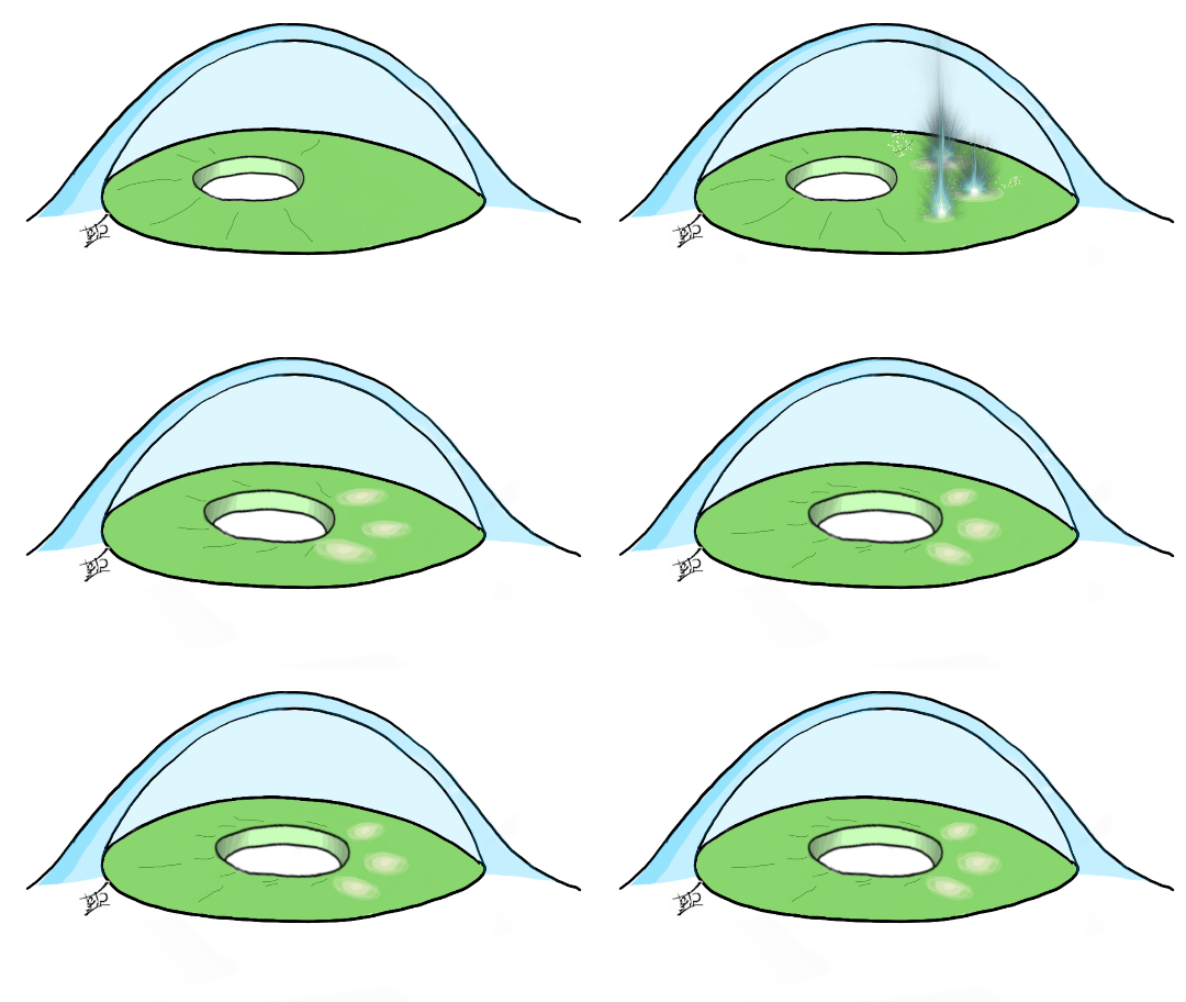
Pupilloplasty
All content on Eyewiki is protected by copyright law and the Terms of Service. This content may not be reproduced, copied, or put into any artificial intelligence program, including large language and generative AI models, without permission from the Academy.
Surgical Therapy
Many old, new and emerging novel techniques for iris repair are utilized.
Because of the better understanding of the importance of pupil size and regularity of pupillary aperture, pupilloplasty is increasingly used by ophthalmic surgeons to optimize patient visual outcomes.
Background
The first published pupilloplasty technique was made by McCannel in 1976 who tried to repair an iris defect after ICCE to manage a subluxated, iris- sutured IOL inside the anterior chamber with full air bubble by using sutures retrieved through three small incisions.[1] In 1985, Alpar modified the McCannel technique by using Healon ( 1% sodium hyaluronate) in cat eyes with improved corneal protection, less change in pachymetry, and less decrease in endothelial cell counts.[2] Siepser sliding knot technique was introduced in 1994.
Osher described a modification of the Siepser slip-knot that allowed the knot to become locked, decreasing the chance of suture failure in 2005. [3]
Patient Selection
Indications
Indications for surgery can be divided into these five main categories:
1. Functional or optical indications:
A. Symptomatic iris defects can be considered for surgical intervention, symptoms can range from glare, shadow images and diplopia. Symptomatic iris defects can be a consequence of either:
- Congenital (iris coloboma)
- Acquired
- Iatrogenic (iris lesion removal)
- Traumatic (traumatic mydriasis or direct injury resulting in pupil irregularity or tissue loss)
- Complicated intraocular surgery (can cause atonic dilated pupil or iris tissue loss and injury)
B. Decentered pupil can be re-centered with laser pupilloplasty, this is usually used in cases of decentered pupil with implanted multifocal intraocular lens.[4] [5]
2. To provide structural support for lens implantation:
Pupilloplasty can be performed to provide the necessary support required for iris claw lens (Artisan) implantation as an option if there was no adequate capsular support to implant in-bag or sulcus IOL.[6]
3. To prevent post-operative complications as in PKP (penetrating keratoplasty)
In some cases of floppy iris that is expected to adhere to the peripheral edge of a corneal graft causing peripheral anterior synechiae, pupilloplasty is performed to tighten the iris preventing it from causing synechial adhesions that would increase the risk of angle closure and graft failure.[7]
4. To relieve appostional angle closure or PAS (peripheral anterior synechiae)
Single pass four through pupilllplasty can be used to break PAS and angle apposition angle closure glaucoma whether primary, post trauma, plateau iris syndrome, Urrets-Zavalia syndrome, and in cases associated with long standing silicone oil in the anterior chamber.[8]
5. To provide more cosmetically acceptable pupil
Although pupilloplasty is rarely performed solely for cosmesis, it can be considered, especially in large colobomas in a colored iris and it can provide functional and cosmetic repair.[9]
Contraindications
Relative contraindications include:
- Phakic eye with a clear lens due to the possibility of lens touch and cataract formation.
- An atrophic iris due to the possibility of an iridodialysis and increased iris damage.[10]
TECHNIQUES
These can be categorized into surgical and non-surgical methods:
INTRAOCULAR SURGICAL METHODS
Various techniques had been advocated for pupillary reconstruction, all shared a common goal; restoration of shape regularity and/or centration of the pupillary aperture aiming for a better quality of vision. Techniques will be described in order of popularity.
Siepser sliding knot technique
This knot, and its recent modifications, utilizes a sliding knot, which is created outside the eye and slid into place atop the iris defect.
With the advent of more sophisticated small incision surgical techniques, the repair of a torn or irregular iris and malformed pupil has changed radically. The collective use of the Siepser Double Sliding Knot and Pupilloplasty technique offers an excellent recovery of the physiologic function and cosmesis post-operatively in patient with pre-existing traumatic iris dysfunctions. The Siepser Double Sliding Knot method offers a "closed chamber" slipping suture technique for iris repairs. Unlike preceding methods, the Siepser Double Sliding Knot technique allows sutures to be tied within the eye without impairing delicate uveal tissue thus keeping a well-maintained anterior chamber during the process.[11] [12]
Many modifications in the original technique had been described, the main goal was to reduce corneal damage by decreasing the number of incisions and to reduce the manipulation exerted on the delicate iris tissue.
Pupil cerclage technique
In this technique a running suture is passed around the pupillary margin to create a purse-string suture aiming for a smaller pupillary aperture size in selected cases with symptomatic abnormally dilated pupil (such as traumatic mydriasis or atonic pupil). A long, curved transchamber needle on 10-0 polypropylene is passed in and out of the anterior chamber through limbal paracentesis openings while weaving the needle through the iris near the pupillary margin to form the cerclage. The technique minimizes stretching and distortion of the iris and creates a pupil that is precisely sized and rather round.[13] Pupil cerclage technique re-establishes the pupil with a precise regulation of the pupil size without distortion of its natural round shape. New sliding knot allows surgeon to reduce the risk of iatrogenic iris damage and to make a security permanent knot.[14]
Single-pass four-throw (SFT)
This technique is regarded as the simplest form of surgical pupillplasty, with short learning curve. In this surgical technique for pupilloplasty, after a single pass of needle through the edges of iris defect along the pupillary margin, the suture end is passed through the loop with 4 throws, creating a helical configuration in modified Siepser slipknot technique that is self-retaining and self-locking.
The pupillary margin is grasped with an end-opening forceps and pulled toward the center of the pupil. The pupillary stretch is performed every 2-clock hours around the entire pupillary margin. The 10-0 suture attached to the long arm of the needle is passed through the proximal iris tissue while the end-opening forceps grasp it. The 10-0 needle is then passed through the distal iris tissue, and the tip of the 10-0 needle is then docked into the barrel of the 26-gauge needle introduced from the paracentesis incision from the opposite side. The 26-gauge needle is withdrawn from the anterior chamber, and this draws along with it the 10-0 needle out of the eye. A Sinskey hook is passed through the paracentesis incision, and the loop of the suture is grasped with an end-opening forceps and is withdrawn from the eye. The suture end is passed through the loop four times and both the suture ends are then pulled from either side of the eye. The approximating loop slides inside the eye and brings both the iris leaflets together. The suture ends are then cut with the microscissors. The procedure of SFT is repeated in the opposite quadrant, and a minimum of 4-point traction is achieved.
Surgical pupilloplasty with single-pass four-throw technique helps to alleviate the appositional closure along with the breakage of peripheral anterior synechia, thereby increasing the aqueous outflow and decreasing intraocular pressure.
when used in ACG , iris traction would be proportional to the degree of angle closure. For example, if the PAS are more than 270 ‘, 6 traction points are recommended to make 3 SFT while when there is less than 270 synchiae, 4 points traction is sufficient.[15] [16]
Pinhole pupilloplasty
In this procedure the same technique for SFT (single pass four through) is utilized with a modification aimed to achieve a smaller (pinhole) pupillaery aperture (of approximately 1.3 mm in diameter) Based on the fact that obtaining a pinhole-sized pupil will filter the stray light from the periphery of the cornea in cases with higher-order corneal aberrations. The pinhole effect blocks distorted and unfocused light rays and isolates more focused central and paracentral rays through the central aperture, thereby reducing aberrations of the optical system as a whole and it inceases the depth of focus enhancing visual acuity and its image quality both for near and far.
There are technical challenges to be met while performing this procedure as the pinhole should be sized properly and it should be centered on the Purkinje image of the microscope light.
McCannel technique
The McCannel technique involves passing a 10-0 polypropylene suture through two paracenteses, which had been created perpendicular to the edge of an iris defect. A full thickness stab incision is then created between the paracenteses through the peripheral cornea. A hook introduced through the stab incision is used to bring both ends of the suture out of the eye where it is then tied pulling the pupillary edges together to close the defect.[19]
LASER SURGICAL METHODS
Argon laser pupilloplasty
Argon laser pupilloplasty can be used to adjust the position of a decentered pupil, or to change the size of miosed pupil. In cases of decentered pupil the procedure is performed by applying laser spots to the midperipheral iris in sectoral fashion to pull the pupil in a particular direction aiming for re-centration. This technique was especially useful in cases of multifocal intraocular lenses. Another implication of this procedure is to change the size of pupil by applying circumferential laser spots in multiple rows that would cause contraction of the iris tissue pulling the pupilllary margin toward the periphery causing a mydriatic effect, again this procedure was performed in cases of miosed pupil with implanted multifocal IOL (because miosis will obstruct light from reaching the multifocal rings of multifocal IOL resulting in functional disruption of multifocality and deteriorating th visual quality)[20] [21]
Argon laser pupilloplasty may be used in combination with other procedures such as Argon Laser Peripheral Iridoplasty to relieve the pupillary block when the pupillary sphincter cramps Linear cuts can be made across the iris fibers argon laser burns delivered through an Abraham iridectomy lens. Intrinsic iris tension will then cause the linear cuts to spread apart. This allows enlargement, reshaping, or repositioning of the pupil and large laser iridotomies with minimal burn energies and a very high percentage of success. [22]
Complications
- Iritis
- Hyphema
- Iris dialysis
- Cataract (mechanical injury in phakic cases)
- Retinal detachment
- Impaired night vision (in pinhole pupilloplasty)
References
- ↑ McIntyre DJ. The McCannel suture: a bimanual technique. American IntraOcular Implant Society Journal. 1979 Apr 1;5(2):150-3.
- ↑ Alpar JJ. The use of Healon in McCannel suturing procedures. Transactions of the ophthalmological societies of the United Kingdom. 1985;104:558-62.
- ↑ Osher RH, Snyder ME, Cionni RJ. Modification of the Siepser slip-knot technique. J Cataract Refract Surg 2005;31:1098-100. [Crossref] [PubMed]
- ↑ Yao Y, Jhanji V. Simplified pupilloplasty technique through a corneal paracentesis to manage small iris coloboma or traumatic iris defect. Annals of Eye Science. 2018;2:31-31.
- ↑ Frisina R, Parrozzani R, Tozzi L, Pilotto E, Midena E. Pupil cerclage technique for treatment of traumatic mydriasis. European Journal of Ophthalmology. 2019;:112067211983930.
- ↑ Ye P, He F, Shi J, Liu J, Liang G, Wu J et al. Artisan iris-fixated intraocular lens implantation in combination with pupilloplasty—correction of aphakia with pathologically large pupil and insufficient capsular support. Canadian Journal of Ophthalmology. 2017;52(2):155-160.
- ↑ Arundhati A, Hui Jun B, Janardhan P, Tan D. Iris Reconstruction in Penetrating Keratoplasty—Surgical Techniques and a Case-control Study to Evaluate Effect on Graft Survival. American Journal of Ophthalmology. 2008;145(2):203-209.e2.
- ↑ Narang P, Agarwal A, Kumar DA. Single-pass four-throw pupilloplasty for angle-closure glaucoma. Indian journal of ophthalmology. 2018 Jan;66(1):120.
- ↑ Cionni R, Karatza E, Osher R, Shah M. Surgical technique for congenital iris coloboma repair. Journal of Cataract & Refractive Surgery. 2006;32(11):1913-1916.
- ↑ Narang P, Agarwal A. Single-Pass Four-Throw Technique for Pupilloplasty. European Journal of Ophthalmology. 2016;27(4):506-508.
- ↑ Lian R, Siepser S, Afshari N. Iris reconstruction suturing techniques. Current Opinion in Ophthalmology. 2020;31(1):43-49.
- ↑ Jordan E. TRAUMATIC IRIS RECONSTRUCTION USING PUPILLOPLASTY FOLLOWING THE SIEPSER DOUBLE SLIDING KNOT TECHNIQUE [Internet]. Aaopt.org. 2020 [cited 20 March 2020]. Available from: https://www.aaopt.org/detail/knowledge-base-article/traumatic-iris-reconstruction-using-pupilloplasty-following-siepser-double-sliding-knot-technique
- ↑ Ogawa GS. The iris cerclage suture for permanent mydriasis: a running suture technique. Ophthalmic Surg Lasers. 1998 Dec;29(12):1001-9. Erratum in: Ophthalmic Surg Lasers 1999 May;30(5):412. PubMed PMID: 9854714.
- ↑ Frisina R, Parrozzani R, Tozzi L, Pilotto E, Midena E. Pupil cerclage technique for treatment of traumatic mydriasis. European Journal of Ophthalmology. 2019;:112067211983930.
- ↑ Narang P, Agarwal A. Single-Pass Four-Throw Technique for Pupilloplasty. European Journal of Ophthalmology. 2016;27(4):506-508.
- ↑ Narang P, Agarwal A, Kumar D. Single-pass four-throw pupilloplasty for angle-closure glaucoma. Indian Journal of Ophthalmology. 2018;66(1):120.
- ↑ Narang P, Agarwal A, Ashok Kumar D, Agarwal A. Pinhole pupilloplasty: Small-aperture optics for higher-order corneal aberrations. J Cataract Refract Surg. 2019;45(5):539–543. doi:10.1016/j.jcrs.2018.12.007
- ↑ Narang P, Holladay J, Agarwal A, Jaganathasamy N, Kumar D, Sivagnanam S. Application of Purkinje images for pinhole pupilloplasty and relevance to chord length mu. Journal of Cataract & Refractive Surgery. 2019;45(6):745-751.
- ↑ Lian R, Siepser S, Afshari N. Iris reconstruction suturing techniques. Current Opinion in Ophthalmology. 2020;31(1):43-49.
- ↑ Laser Pupilloplasty: a Useful Technique? [Internet]. CRSToday. 2020 [cited 19 March 2020]. Available from: https://crstoday.com/articles/2009-jun/crst0609_02-php/
- ↑ Zhou W, Zhao F, Shi D, Qadri M, Jiang L, Ma L. Argon Laser Peripheral Iridoplasty and Argon Laser Pupilloplasty: Alternative Management for Medically Unresponsive Acute Primary Angle Closure. Journal of Ophthalmology. 2019;2019:1-7.
- ↑ Wise J. Iris Sphincterotomy Iridotomy, and Synechiotomy by Linear Incision with the Argon Laser. Ophthalmology. 1985;92(5):641-645.


