Esotropia
All content on Eyewiki is protected by copyright law and the Terms of Service. This content may not be reproduced, copied, or put into any artificial intelligence program, including large language and generative AI models, without permission from the Academy.
Disease Entity
Strabismus/Ocular misalignment
Disease
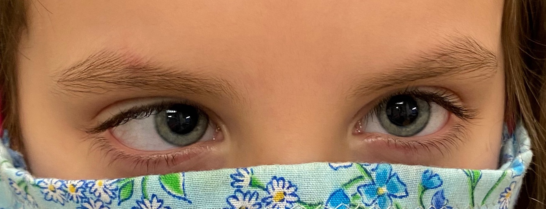
An esotropia is an eye misalignment in which one eye is deviated inward toward the nose. The deviation may be constant or intermittent. The deviating eye may always be the same eye or may alternate between the two eyes. A comitant esotropia has nearly the same degree of deviation in every position of gaze while an incomitant esotropia with have a deviation that varies between gazes. Evaluating the comitance can help determine the cause of the esotropia.
Comitant
Infantile esotropia
Infantile esotropia may also be referred to as "congenital esotropia". This is an esodeviation, often constant, that presents in the first 6 months of life. It is associated with a large angle deviation, fusion maldevelopment nystagmus (latent nystagmus), a crossfixation pattern of fixation, overelevation in adduction, dissociated vertical deviation, a normal accommodative convergence to accommodation ratio, and age-appropriate refractive errors. The cause is unknown.
Ocular instabiltiy of infancy
Transient misalignment of the eyes (esodeviation or exodeviation) that is common up to the age of 3 months and possibly longer in premature infants; this should not be confused with infantile esotropia.
Accommodative esotropia
Accommodative esotropia is the most common subtype of esotropia. Accommodative esotropia is an inward turning of one or both eyes that occurs with activation of the accommodative reflex. Accommodation is a dynamic process in which the curvature of the eye’s natural lens is temporarily adjusted to improve focus at near or in eyes that are hyperopic (far-sighted). Accommodative convergence describes the normal convergence of the eyes in response to accommodation of the lens. The amount of accommodative convergence relative to the degree of accommodation is called the AC/A ratio. There are two main types of accommodative esotropia: 1) Refractive (normal AC/A ratio) accommodative esotropia caused by uncorrected or under-corrected hyperopia and 2) Non-refractive (high AC/A ratio) accommodative esotropia caused by excessive convergence of the eyes in response to accommodation for near focus, regardless of refractive error. A patient may have a high AC/A ratio in addition to having refractive accommodative esotropia. Onset most commonly occurs between 2 and 4 years of age, but may occur between 4 months and 7 years of age. Most patients with refractive accommodative esotropia have moderate hyperopia (typically ≥2.00 D, average of +4.00 D).
Partially accommodative esotropia
Alignment is greatly improved with refractive correction, but a deviation of ≥ 10 PD persists. These patients may require surgical correction of their residual deviation.
Acquired non-accommodative esotropia
There are multiple subtypes of acquired non-accommodative esotropia.
Basic
Develops after 6 months of age and is not accommodative. Children may initially have diplopia if onset is acute. Acute onset of esotropia may indicate and underlying neurologic disorder, especially in the setting of divergence insufficiency, headache, nausea or vomiting, small V pattern incomitance, abnormal head position. The prognosis for recovery of binocular vision with prism or surgery is good, as patients previously had the ability to fuse. The cause is unknown.
Cyclic
A comitant, intermittent esotropia occurs at regular intervals, classically every other day. This form of strabismus is rare and the cause unknown. Surgical treatment it typically effective.
Divergence insufficiency type esotropia
Primary divergence insufficiency is most often associated with adult patients 50 years and older, is characterized by an esodeviation greater at distance than near. Fusional divergence amplitudes are reduced at both distance and near fixation, and this esotropia is comitant in primary and lateral gazes. Imaging may show thinning, elongation, and rupture of the connective tissue between the lateral and superior rectus muscles and sagging and elongation of the lateral rectus muscle[1]. Secondary divergence insufficiency is associated with pontine tumors, elevated intracranial pressure and head trauma and be secondary to mild CN VI palsies.
Microtropia/Monofixation Syndrome
This is characterized by patients with a central scotoma in one eye together with peripheral fusion. These patients typically lack fine stereopsis and maintain a mild to moderate degree of fusional amplitudes.
Sensory esotropia
Unilateral reduced visual acuity, due to various organic causes, presents a barrier to fusion. In children under 4 years of age, the blind or poorer-seeing eye will generally become esotropic. Older children or adults with sensory visual deprivation will generally develop a sensory exotropia.
Spasm of the near synkinectic reflex
Variable esotropia in conjunction with increased accommodation and miosis are suggestive of spasm of the near reflex. It is thought to be related to stress and anxiety. Typically, the patient will show limited abduction on version testing, but have normal abduction when tested with the other eye covered. Cycloplegia and bifocal glasses may be tried. Botox can be considered if the spasm persists.[2]
Consecutive esotropia
This occurs when a person who was formerly exotropic becomes esotropic. Sometimes this is a result of surgical overcorrection for exotropia.
Incomitant
Cranial nerve VI Palsy
Damage to CN VI anywhere along its course can lead to a CN VI palsy. There are numerous potential causes, including congenital, microvascular ischemia, trauma, intracranial tumor, elevated intracranial pressure, stroke, infectious, and inflammatory causes.
Heavy Eye Syndrome
Heavy eye syndrome is classically characterized by esotropia and hypotropia in patients with high myopic. It is hypothesized that HES results from inferior shift of the lateral rectus and nasal shift of the superior rectus.
Congenital Cranial Dysinnervation Disorders
Type 1 and type 3 Duane syndrome are due to absent or dysplastic abducens motor neurons with resulting synkinesis on the lower division of the oculomotor nerve that innervates the medial rectus. In type 1, the patient cannot abduct the affected eye. In type 3, the patient cannot abduct or adduct the affected eye. Duane syndrome is associated with narrowing of the interpalpebral fissure with adduction of the eye and upshoots and downshoots on adduction.
Möbius syndrome most commonly affects the abducens nerve and the facial nerve leading to esotropia and facial droop, but may also affect CN III, IV, V, IX, X, and XII.
Thyroid Eye Disease
Thyroid eye disease leads to enlargement of the extraocular muscles and restrictive strabismus. The medial rectus muscle is the second most common extraocular muscle affect. Forced duction testing will be positive for restriction of the medial rectus and forced generation and saccadic velocity will be normal. Findings associated with thyroid eye disease include: intraocular pressure may increase when looking away from the restriction, proptosis, lid retraction, compressive optic nerve dysfunction, conjunctival hyperemia, chemosis, and corneal affections due to exposure
Post-surgical
Due to a slipped lateral rectus muscle or medial rectus restriction following large resection.
Orbital causes of restriction
Muscle entrapment following medial orbital wall fracture, orbital mass, orbital scarring, orbital or periocular implant
Myasthenia Gravis
Myasthenia gravis is an autoimmune disease in which antibodies destroy neuromuscular connections resulting in muscle weakness and fatigability. The type and degree of strabismus may vary significantly over time and patients may have associated ptosis and weakness of other muscles.
Nystagmus and Esotropia
Nystagmus Blockage Syndrome
Nystagmus blockage syndrome describes a an esotropia resulting from overconvergence that dampens nystagmus seen in infantile nystagmus syndrome. Infantile nystagmus syndrome (INS) is a conjugate, horizontal, uniplanar nystagmus that worsens with attempted fixation and improves with convergence. INS may be idiopathic or result from sensory defects.
Fusion Maldevelopment Nystagmus
Infantile onset strabismus or decreased vision in one eye can lead to fusion maldevelopment with associated nystagmus. The nystagmus is conjugate, horizontal jerk nystagmus that beats in the direction of the fixing eye. It may only manifest under monocular conditions or can be seen when only one eye is fixing, such as with suppression. The nystagmus may dampen with accommodation .
Risk Factors
Hyperopia, anisometropia, neurodevelopmental disorders, hydrocephalus, prematurity, craniofacial abnormalities, chromosomal abnormalities, and a positive family history of strabismus increase the risk of having esotropia.
Epidemiology
There is no predilection for esotropia in terms of gender. About half of children with an esotropia will develop amblyopia.
Diagnosis
Physical examination
All patients with esotropia need a complete ophthalmologic examination, including visual acuity, binocular function and stereopsis, motility evaluation, strabismus measurements at near, distance, and cardinal positions of gaze, measurement of fusional amplitudes, cycloplegic refraction and examination of the anterior and posterior segments. Some cases may require a 4 prism diopter base out test for microtropia, strabismus measurements after Marlowe’s prolonged occlusion test, or strabismus measurements with +3.00 lenses at near fixation. Consider sustained upgaze and repeated blinking to fatigue the muscles and bring out strabismus and ptosis in myasthenia. A rest test or ice test may improve strabismus and ptosis in myasthenia gravis.
Symptoms
Esotropia can lead to diplopia in adults and with acute onset in children. Children will typically learn to suppress the deviated eye to avoid diplopia; the suppression can lead to amblyopia.
A complete exam of the eyes is necessary to determine the likely cause of esotropia. Careful attention should be paid to the deviation in different positions of gaze and focal distances to determine if the deviation is comitant or incomitant and if it is greater at distance or near.
For comitant deviations: Hyperopia or a high AC/A ratio in patients with a comitant deviation is suggestive of an accommodative cause and refractive correction can be tried to help clinch the diagnosis. A greater deviation at distance is consistent with divergence insufficiency and could be related to underlying neurologic causes, particularly in children. Onset before 6 months and associated DVD, fusional maldevelopment nystagmus, inferior oblique overaction are consistent with a diagnosis of infantile esotropia. Associated myopia on manifest refraction and with lower myopia or hyperopia on cycloplegic refraction along with miosis and abduction deficit only seen binocularly suggest spasm of the near reflex. A cause of decreased vision other than amblyopia on exam is suggestive of a sensory strabismus.
For incomitant deviations: Limited abduction with associated decreased force generation and decreased saccadic velocity are consistent with CN VI palsy. Findings of a congenital CN VI palsy with an associated CN VII palsy or other cranial nerve palsy suggests Möbius syndrome. Limited abduction with narrowing of the interpalpebral fissure on adduction and possibly upshoots and downshoots on adduction is consistent with Duane syndrome. Esotropia with hypotropia in a patient with high myopia may be secondary to heavy eye syndrome and MRI imaging can be used to evaluate if the angle between the superior rectus and lateral rectus is enlarged. Restriction can be be evaluated for using forced duction testing. Other signs or history, such as lid retraction and proptosis in thyroid eye disease, or a history of orbital fracture, may point to the cause; however, imaging of the orbits may be helpful for confirming the diagnosis and pre-operative planning.
Myasthenia gravis should be considered when the deviation is variable or there is associated ptosis. Ice testing to improve ptosis, rest test to improve alignment or ptosis, serum titers for anti-ACh Receptor antibodies and anti-MUSK antibodies, edrophonium testing, and single fiber EMG can aid in this diagnosis.
Differential diagnosis
Management
Nonsurgical
Nonsurgical treatments include patching, correction of full hyperopic refractive error, and divergence orthoptic exercises for divergence insufficiency. Fresnel prisms or prism glasses can be used to relieve diplopia or asthenopia in certain patients. Sometimes sensory esotropia can be helped by treating the underlying cause (amblyopia, cataract, media opacity).
Associated amblyopia therapy
The timing of amblyopia treatment in relation to eye muscle realignment surgery is debatable. Some surgeons treat amblyopia before performing surgery to create a stronger visual drive to maintain alignment. However, it has been found that those with mild to moderate amblyopia are as likely to maintain alignment as those who had amblyopia treated prior to surgery. Surgical realignment may improve amblyopia in some cases. [3]
Chemodenervation
Chemodenervation with botulinum toxin has been shown to be effective in the treatment of infantile,[4][5][6][7] partially accommodative esotropia with[8] and without[5] high AC:A ratio, and acute esotropia[9][10]. Longevity of results may not be as high in patients receiving chemodenervation compared to incisional surgery. Botulinum may be more effective in infantile esotropia when the angle of deviation is <30-35 PD[11] and also in smaller angle deviation in partially accommodative esotropia.[12] Regardless of treatment with chemodenervation or surgery, acute esotropia patients with prompt treatment and no amblyopia have better outcomes.[10]
Surgery
Surgery is performed on the extraocular muscles in an attempt to give binocular single vision, to relieve diplopia, or to restore the eyes to their regular state of alignment. The prognosis for surgical success is best if the patient has an intermittent, as opposed to constant, esotropia, alternating fixation, and treatment of amblyopia. In certain cases, amblyopia may not be fully corrected due to the strabismus, and surgery may be needed prior to full correction of the amblyopia. The optimal time for surgical intervention is as early as possible, within a few months of the onset, prior to any degenerations of the lateral geniculate nucleus. In case of associated amblyopia it is recommended to treat it prior to surgery to improve the long-term success rate of eye alignment.
The surgical approach for comitant esotropia is typically a bilateral medial rectus recession or unilateral recess-resect procedure. The surgical approach in patients with an incomitant cause of strabismus will vary with the type, and involve an alternative approach, such as vertical rectus muscle transposition for CN VI palsy or loop myopexy for treatment of heavy eye syndrome.
References
- ↑ Chaudhuri Z, Demer JL. Sagging eye syndrome: connective tissue involution as a cause of horizontal and vertical strabismus in older patients. JAMA Ophthalmol. 2013 May;131(5):619-25. doi: 10.1001/jamaophthalmol.2013.783. PMID: 23471194; PMCID: PMC3999699.
- ↑ Kaczmarek BB, Dawson E, Lee JP. Convergence spasm treated with botulinum toxin. Strabismus. 2009 Jan-Mar;17(1):49-51. doi: 10.1080/09273970802678511. PMID: 19301195.
- ↑ Lam GC, Repka MX, Guyton DL. Timing of amblyopia therapy relative to strabismus surgery. Ophthalmology. 1993 Dec;100(12):1751-6. doi: 10.1016/s0161-6420(13)31403-1. PMID: 8259271.
- ↑ Issaho DC, Carvalho FRS, Tabuse MKU, Carrijo-Carvalho LC, de Freitas D. The Use of Botulinum Toxin to Treat Infantile Esotropia: A Systematic Review With Meta-Analysis. Invest Ophthalmol Vis Sci. 2017 Oct 1;58(12):5468-5476. doi: 10.1167/iovs.17-22576. PMID: 29059315.
- ↑ 5.0 5.1 AlShamlan FT, Alghazal F. Comparison of Dose Increments of Botulinum Toxin A with Surgery as Primary Treatment for Infantile Esotropia and Partially Accommodative Esotropia. Clin Ophthalmol. 2022 Aug 27;16:2843-2849. doi: 10.2147/OPTH.S382499. PMID: 36061630; PMCID: PMC9432566.
- ↑ Song D, Qian J, Chen Z. Efficacy of botulinum toxin injection versus bilateral medial rectus recession for comitant esotropia: a meta-analysis. Graefes Arch Clin Exp Ophthalmol. 2023 May;261(5):1247-1256. doi: 10.1007/s00417-022-05882-5. Epub 2022 Nov 2. PMID: 36322214.
- ↑ Niyaz L, Yeter V, Beldagli C. Success rates of botulinum toxin in different types of strabismus and dose effect. Can J Ophthalmol. 2023 Jun;58(3):239-244. doi: 10.1016/j.jcjo.2021.12.002. Epub 2022 Jan 14. PMID: 35038409.
- ↑ Tejedor J, Gutiérrez-Carmona FJ. Botulinum toxin in the treatment of partially accommodative esotropia with high AC/A ratio. PLoS One. 2020 Feb 28;15(2):e0229267. doi: 10.1371/journal.pone.0229267. PMID: 32109950; PMCID: PMC7048305.
- ↑ Lang LJ, Zhu Y, Li ZG, Zheng GY, Peng HY, Rong JB, Xu LM. Comparison of botulinum toxin with surgery for the treatment of acute acquired comitant esotropia and its clinical characteristics. Sci Rep. 2019 Sep 25;9(1):13869. doi: 10.1038/s41598-019-50383-x. PMID: 31554874; PMCID: PMC6761114.
- ↑ 10.0 10.1 Cheung CSY, Wan MJ, Zurakowski D, Kodsi S, Ekdawi NS, Russell HC, Shetty S, Dumitrescu AV, Dagi LR, Shah AS, Hunter DG; AACE Outcomes Consortium. A Comparison of Chemodnervation to Incisional Surgery for Acute, Acquired, Comitant Esotropia: An International Study. Am J Ophthalmol. 2024 Mar 4;263:160-167. doi: 10.1016/j.ajo.2024.02.036. Epub ahead of print. PMID: 38447598.
- ↑ de Alba Campomanes AG, Binenbaum G, Campomanes Eguiarte G. Comparison of botulinum toxin with surgery as primary treatment for infantile esotropia. J AAPOS. 2010 Apr;14(2):111-6. doi: 10.1016/j.jaapos.2009.12.162. PMID: 20451851.
- ↑ Alarfaj, Motazz A.1,2; Alsarhani, Waleed K.3,4; Alrashed, Saleh H.5; Alarfaj, Faris A.1; Ahmad, Khabir6; Awad, Abdulaziz1; Sesma, Gorka1,. Factors Affecting the Efficacy of Botulinum Toxin Injection in the Treatment of Infantile and Partially Accommodative Esotropia. Middle East African Journal of Ophthalmology 29(3):p 122-126, Jul–Sep 2022. | DOI: 10.4103/meajo.meajo_39_23
General references
- Baker JD, Parks MM. Early onset accommodative esotropia. Am J Ophthalmol 1980: 90:11.
- Cassin B, Beecham B, Friedberg K. Stereoacuity, fusional amplitudes and AC/A ratio in accommodative esotropia. Am Orthopt J 1976; 26:60-64.
- Costenbader F. Infantile esotropia clinical characteristics and diagnosis. Am Orthopt J 1968; 18:5-10.
- Foster RS, Paul O, Jampolsky A. Management of infantile esotropia. Amer J Ophthalmol 1974; 82:291-299.
- Helveston E. Origins of congenital esotropia. Am Orthopt J. 1986; 36:40-48.
- Ing MR. Early surgical alignment for congenital esotropia. J Ped Ophthalmol Strab 1984; 20:11-18.


