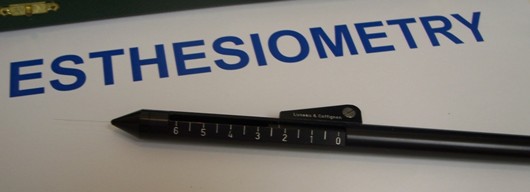
Corneal Esthesiometry
All content on Eyewiki is protected by copyright law and the Terms of Service. This content may not be reproduced, copied, or put into any artificial intelligence program, including large language and generative AI models, without permission from the Academy.
Esthesiometry (es-the-si-om-e-try) is the measurement of sensation, specifically tactile[1]. The measurement of corneal sensation evaluates the ophthalmic branch of the fifth cranial nerve (trigeminal)[2]. An esthesiometer or aesthesiometer is a device used to measure sensation. To test for corneal sensation there are qualitative and quantitative methods. The most commonly used method in clinical practice which is qualitative in nature, is the use of a cotton-tipped applicator. Topical anesthetics should not be used prior to testing corneal sensation.
History
The first esthesiometer was described in 1894 by von Frey and was built using horse hairs of different lengths[3]. In 1932 Francheschetti, improved on von Frey’s version and then in 1956 Boberg-Ans described a device using a single nylon thread with a constant diameter but variable length.[3] Cochet-Bonnet improved on the Boberg-Ans version and developed two different models.[3] One model uses a diameter of 0.08 mm which allows pressure of 2 to 90 mg/0.005 mm2 and the second model uses a diameter of 0.12 mm with pressure ranging from 11 to 200mg/0.0113 mm2[3].
Uses
Corneal esthesiometry is typically used clinically to evaluate for neurotrophic keratitis. In research, esthesiometry has been used for various purposes; including, measuring the duration of an analgesic on the cornea or indicating the corneal health in long-term contact wearers[2][4][5].
Qualitative Method
The qualitative method is most commonly used in clinic and often achieved with a cotton-tipped applicator because it is easily accessible. Alternatively, a small piece of dental floss may be used. [6]No topical anesthetics should be used prior to performing the test. A wisp of the cotton-tipped applicator is used to compare sensation in each eye. It is recommended to approach the patient from the side and test all four quadrants. Record the sensation in each location as normal, reduced, or absent[7].
Quantitative Method
There are various quantitative methods that are typically reserved for research or complicated cases. The most common quantitative method is the handheld esthesiometer (Cochet-Bonnet). Other methods reported include[2]:
- Non-contact air puff technique
- Chemical stimulation using capsaicin
- Thermal stimulation with a carbon dioxide laser
Handheld esthesiometer (Cochet-Bonnet)
The handheld esthesiometer (Cochet-Bonnet) is a device that contains a thin, retractable, nylon monofilament that extends up to 6 cm in length. Variable pressure can be applied by the device by adjusting the length. The monofilament ranges from 60 mm to 5 mm and as the length is decreased the pressure increases from 11 mm/gm to 200 mm/gm[8]. The Luneau Cochet-Bonnet Aesthesiometer tends to be one of the easier instruments to find and purchase.
Steps for using the handheld esthesiometer:
- Extend the filament to full length of 6 cm
- Retract the filament incrementally in 0.5 cm steps until the patient can feel its contact
- Record the length (NOTE: The shorter the length indicates decreased sensation)
- Compare the fellow cornea
- Repeat steps 1-4 in each quadrant: superior, temporal, inferior, nasal
- Sterilize the filament and retract back into the device to protect it from damage
Corneal Sensation
The nasociliary nerve from the ophthalmic branch (V1) of the fifth cranial nerve (CN V), the trigeminal nerve, provides most of the sensory innervation for the cornea. Key points to note about ocular sensation include:
- Greatest in the central cornea except in elderly patients where it is more sensitive in the periphery[2]
- Drops rapidly as distance increases from the limbus[2]
- Falls with increasing age[3]
- Is not affected by iris color[2]
- More sensitive in the temporal limbus than the inferior limbus[2]
- Reduction has been reported in Diabetes Type 1 and Type 2[7]
- Decreases during pregnancy[9]
Corneal Hypoesthesia Differential Diagnosis
Corneal hypoesthesia can occur from any etiology that causes damage directly to the nerves of the cornea or CN V. Important etiologies to consider include[7]:
- Herpes simplex keratitis
- Herpes zoster ophthalmicus
- Corneal surgery (PK, LASIK, PRK, RK, large limbal incisions)
- PRP[10]
- Neurosurgery or other surgical trauma (ablation of the trigeminal ganglion, irradiation near or on the eye, other intracranial surgery causing unintended CN V damage)
- Topical medications (anesthetics, NSAIDs, atropine, ß-blockers, and carbonic anhydrase inhibitors)[3][11]
- Exposure keratopathy secondary to lagophthalmos (exophthalmos, facial nerve palsy, eyelid coloboma, and ectropion)
- Cocaine abuse
- Cerebrovascular events
- Aneurysms
- Tumors (acoustic neuroma, neurofibroma, or angioma)
- Multiple sclerosis, Parkinson's disease, and other neurological conditions[12]
- Hansen disease (leprosy)
- Genetic causes such as familial dysautonomia (Riley-Day syndrome) (dermatome in remaining area of CN V is not involved) or congenital trigeminal anesthesia (dermatome of CN V is involved)
References
- ↑ An Encyclopedia Britannica Company. Merriam-Webster. http://www.merriam-webster.com/medical/esthesiometry. Accessed 01 DEC 2011.
- ↑ 2.0 2.1 2.2 2.3 2.4 2.5 2.6 Faulkner WJ, Varley GA. Corneal diagnostic techniques. In: Krachmer JH, Mannis MJ, Holland EJ, eds. Cornea. 2nd ed. Vol. 1 Philadelphia: Elsevier/Mosby; 2005:229-235.
- ↑ 3.0 3.1 3.2 3.3 3.4 3.5 Martin XY. Safran AB. Corneal hypoesthesia. Survey of Ophthalmology. 1988:33(1):28-40.
- ↑ Brennan NA, Bruce AS. Esthesiometry as an indicator of corneal health. Optom Vis Sci. 1991 Sep;68(9):699-702.
- ↑ Trevithick JR, Dzialoszynski T, Hirst M, Cullen AP. Esthesiometric evaluations of corneal anesthesia and prolonged analgesia in rabbits. Lens Eye Toxic Res. 1989;6(1-2):387-93.
- ↑ Salinger, C, "Neurotrophic Keratitis: What's in the Toolbox," Ophthalmology Management, June 1, 2020.
- ↑ 7.0 7.1 7.2 External Disease and Cornea, Section 8. Basic and Clinical Science Course, AAO, 2010.
- ↑ Western Ophthalmics Corporation. Luneau Cochet-Bonnet Aesthesiometer. http://west-op.com/aesthesiometer.html. Accessed 01 DEC 2011.
- ↑ Millodot MI. The influence of pregnancy on the sensitivity of the cornea. Br J Ophthalmol. 1977 Oct 1;61(10):646-9. http://doi.org/10.1136/bjo.61.10.646
- ↑ Rogell GD. Corneal hypesthesia and retinopathy in diabetes mellitus. J Ophthalmol. 1980;87(3):229-233. doi:10.1016/s0161-6420(80)35257-3.
- ↑ Ocular Surgery News, July 25, 2023, "Noncontact esthesiometer may offer easier method of measuring corneal sensitivity." This article reported on a poster at ARVO that demonstrated that patients on glaucoma drops had reduced corneal sensation compared to controls.
- ↑ Reddy VC, Patel SV, Hodge DO, & Leavitt JA. Corneal sensitivity, blink rate, and corneal nerve density in progressive supranuclear palsy and Parkinson disease. Cornea. 2013;32(5):631-635. doi:10.1097/ico.0b013e3182574ade.


