Conjunctival Telangiectasia
From EyeWiki
All content on Eyewiki is protected by copyright law and the Terms of Service. This content may not be reproduced, copied, or put into any artificial intelligence program, including large language and generative AI models, without permission from the Academy.
All contributors:
Assigned editor:
Review:
Assigned status Up to Date

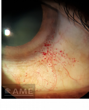
Conjunctival Telangiectasia (Image courtesy of Dr Amanda Mohanan Earatt)
Disease Entity
The presence of small dilated blood vessels near the surface of the mucous membranes of the conjunctiva.
Etiology
Primary Telangiectatic Disorders
- Ataxia Telangiectasia / Louis Bar Syndrome
- Hereditary Hemorrhagic Telangiectasia / Osler - Weber - Rendu Syndrome
- Bloom Syndrome
- Generalised Essential Telangiectasia
Ocular Manifestation of a Systemic Disease
- Rosacea
- Xeroderma Pigmentosum
- Fabry Disease
- Hyperviscosity Syndrome
- Alport syndrome
- Diabetes mellitus
Trauma
Post Radiotherapy
Idiopathic
Clinical diagnosis
Primary Telangiectatic Disorder
Ataxia Telangiectasia / Louis Bar Syndrome[1][2]
- Autosomal recessive inheritance
- Conjunctival telangiectasia is seen in 91% of patients and develops between the ages of 3 and 5 years. Involvement is initially interpalpebral but away from the limbus; it eventually becomes generalized.
- Other features to look for:
- Telangiectasia on eyelid skin, external ear, nares and subsequently in other sun exposed areas
- Ocular motor apraxia, strabismus, nystagmus
- Progressive cerebellar ataxia
- Immunoglobulin deficiency can cause recurrent respiratory tract infection
Hereditary Hemorrhagic Telangiectasia / Osler - Weber - Rendu Syndrome[3][4][5]
- Autosomal dominant inheritance
- Usually, onset of symptoms occurs in the 4th decade of life
- Spider-like angiomatous malformations can be seen mainly on the palpebral conjunctiva leading to recurrent subconjunctival hemorrhage or recurrent hemolacria
- Other features to look for:
- Iris vascular malformation, retinal telangiectasia and AVM; can cause BRAO and intraoperative choroidal hemorrhage[6]
- Multiple telangiectasia at the level of skin, lips, oral cavity, fingers, nasopharynx
- ArterioVenous Malformations (AVM) involving lung, brain, GIT, liver, and spinal cord may cause recurrent epistaxis, GI bleeding, hematuria and consequent anemia; stroke
Bloom Syndrome[7]
- Conjunctival telangiectasia seen predominantly on the bulbar conjunctiva
- Other features to look for:
- Erythematous, telangiectatic and scaly rash over malar region and other sun-exposed areas including the backs of the hands and neck.
- Short stature, high-pitched voice;
- Distinct facial features, including a long, narrow face, micrognathism, prominent nose and ears
- Hypo-pigmented and hyperpigmented areas on skin, cafe-au-lait spots, and telangiectasia
- Immunoglobulin deficiency causing recurrent respiratory tract infection
Generalised Essential Telangiectasia[8]
- Conjunctival telangiectasia seen predominantly on the bulbar conjunctiva
- More common in women, with age of onset usually in the late 30s
- Generalized development of dilated venules, which start at the lower extremities and progressively spread to the rest of the body
Ocular Manifestation of a Systemic Disease
Rosacea[9]
- Ocular Features
- Blepharitis
- Lid margin and conjunctival telangiectasia
- Chalazia and hordeolum,
- Punctate Epithelial Erosions, keratitis
- Other Clinical Features
- Flushing, telangiectasia, erythema, papules and pustules, and rhinophyma
Xeroderma Pigmentosum[10][11]
- Ocular Features
- Eyelid atrophy and tumors
- Corneal sicca and opacification
- Exposure keratitis
- Pterygium
- Chronic conjunctival injection,
- Conjunctival telangiectasia
- Symblepharon
- Other Clinical Features
- Sensitivity to UV radiation resulting in inflammation and neoplasia in sun exposed areas of the skin, mucous membranes, and ocular surfaces
Fabry Disease[12]
- Ocular Features
- Telangiectasia and sludging of blood usually seen in inferior bulbar conjunctiva
- Cornea verticillata
- Posterior lens cataract
- Retinal vascular tortuosity
- Other Clinical Features
- Pale, waxy complexion, thick eyelids and thickening of facial features
- Angiokeratomas, small bluish-black non-blanching telangiectasia, in the bathing trunk area, oral cavity and hands
- Episodic ‘‘Fabry crises’’ of agonizing, neuropathic pain
- GI symptoms like vomiting and diarrhea
Hyperviscosity Syndrome[13]
- Sickle cell anemia
- Multiple myeloma
- Polycythemia rubra vera
Alport syndrome[14]
- Ocular Features
- Perilimbal conjunctival telangiectasia, usually at 3 and 9 o’ clock
- PPMD
- Anterior lenticonus
- Dot fleck retinopathy
- Hematuria, hearing loss
- Diabetes mellitus
Treatment
Usually no treatment required; cautery has been used in case of recurrent bleed in Hereditary Hemorrhagic Telangeictasia[5]
Additional Resources
https://www.ncbi.nlm.nih.gov/medgen/66780
References
- ↑ Ferreira MG, Nascimento FA, Teive HAG. Cerebellar Ataxia and Ocular Conjunctival Telangiectasia: Look Again. Neurology: Clinical Practice. 2021;11(4):e587-e588. doi:10.1212/CPJ.0000000000000977
- ↑ Ataxia-Telangiectasia in Ophthalmology Clinical Presentation: History, Physical, Causes. Accessed November 9, 2021. https://emedicine.medscape.com/article/1219140-clinical
- ↑ Knox FA, Frazer DG. Ophthalmic presentation of hereditary haemorrhagic telangiectasia. Eye. 2004;18(9):947-949. doi:10.1038/sj.eye.6701360
- ↑ Gómez-Acebo I, Prado SR, De La Mora Á, et al. Ocular lesions in hereditary hemorrhagic telangiectasia: genetics and clinical characteristics. Orphanet Journal of Rare Diseases. 2020;15(1):168. doi:10.1186/s13023-020-01433-5
- ↑ Jump up to: 5.0 5.1 Goldberg SH, Bullock JD. Hereditary Hemorrhagic Telangiectasia. Ophthalmic Plastic & Reconstructive Surgery. 1990;6(2):136-138.
- ↑ Abdolrahimzadeh S, Formisano M, Marani C, Rahimi S. An update on the ophthalmic features in hereditary haemorrhagic telangiectasia (Rendu-Osler-Weber syndrome). Int Ophthalmol. 2022 Jun;42(6):1987-1995. doi: 10.1007/s10792-021-02197-y. Epub 2022 Jan 16. PMID: 35034241; PMCID: PMC9156511.
- ↑ Sahn EE, Hussey III RH, Christmann LM. A Case of Bloom Syndrome with Conjunctival Telangiectasia. Pediatric Dermatology. 1997;14(2):120-124. doi:10.1111/j.1525-1470.1997.tb00218.x
- ↑ Extensive Acquired Telangiectasias: Comparison of Generalized Essential Telangiectasia and Cutaneous Collagenous Vasculopathy | Actas Dermo-Sifiliográficas. Accessed November 9, 2021. https://www.actasdermo.org/en-extensive-acquired-telangiectasias-comparison-generalized-articulo-S1578219017300227
- ↑ Case Report: Ocular rosacea: an underdiagnosed cause of relapsing conjunctivitis-blepharitis in the elderly. Accessed November 9, 2021. https://www.ncbi.nlm.nih.gov/pmc/articles/PMC4170302/
- ↑ Goyal JL, Rao VA, Srinivasan R, Agrawal K. Oculocutaneous manifestations in xeroderma pigmentosa. British Journal of Ophthalmology. 1994;78(4):295-297. doi:10.1136/bjo.78.4.295
- ↑ Schelini MC, Chaves LFOB, Toledo MC, et al. Xeroderma Pigmentosum: Ocular Findings in an Isolated Brazilian Group with an Identified Genetic Cluster. Journal of Ophthalmology. 2019;2019:e4818162. doi:10.1155/2019/4818162
- ↑ Rothstein K, Gálvez JM, Gutiérrez ÁM, Rico L, Criollo E, De-la-Torre A. Ocular findings in Fabry disease in Colombian patients. biomedica. 2019;39(3):434-439. doi:10.7705/biomedica.3841
- ↑ Hekimsoy HK, Şekeroğlu MA. A Rare Coexistence of Isolated Unilateral Conjunctival Telangiectasia and Retinal Vascular Tortuosity. Case Reports in Ophthalmological Medicine. 2020;2020:e8814961. doi:10.1155/2020/8814961
- ↑ Decock C, Laey J, Leroy B, Kestelyn P. Alport syndrome and conjunctival telangiectasia. Bulletin de la Société belge d’ophtalmologie. 2003;290:29-31.

