Kawasaki Disease
All content on Eyewiki is protected by copyright law and the Terms of Service. This content may not be reproduced, copied, or put into any artificial intelligence program, including large language and generative AI models, without permission from the Academy.
Kawasaki disease is the most common cause of acquired heart disease in children in developed countries, and ocular inflammation is an important early clinical sign. Though long-term ocular morbidity is rare, the systemic manifestations of Kawasaki disease can be fatal.
Disease Entity
Kawasaki disease (KD) is predominantly a vasculitis of childhood, though it rarely can occur in adults. Signs include ≥5 days of fever plus the presence of conjunctivitis, mucositis, skin rash, extremity changes, and cervical lymphadenopathy.
The disease was first described in Japan by a Japanese pediatrician named Tomisaku Kawasaki in 1967. It was approximately three years later when physicians began describing the various cardiac sequelae of the disease which are a leading cause of pediatric acquired heart disease worldwide.
Epidemiology
The incidence of KD varies geographically, with the highest incidence in Japan of approximately 265 cases per 100,000 children under 5 years of age, followed by 134 and 83 cases per 100,000 in Korea and Taiwan respectively. In the United States, the annual incidence of KD is about 18 per 100,000.[2] Boys are more commonly affected than girls, and the vast majority of cases occur between the ages of 6 months and 5 years. Incidence tends to be higher during winter months.
Etiology
While the etiology of KD remains unknown, the leading theory is that exposure to a pathogen (most likely viral) triggers an inflammatory cascade resulting in immune system dysregulation in a genetically susceptible child. Evidence that some individuals have a genetic predisposition include varying incidence among different ethnicities, higher incidence in children with a family history of KD, and single-nucleotide polymorphisms in a variety of genes that have been implicated through genome-wide association studies.[3][4] There is currently no evidence to suggest that the disease is associated with administration of routine vaccines.
Pathophysiology
Kawasaki Disease is a medium vessel vasculitis of childhood which typically affects muscular arteries, often with a predilection for the coronary arteries. It is thought that respiratory or gastrointestinal exposure to an unknown pathogen results in activation of B-cells within the associated lymphoid tissue, which then differentiate into IgA-producing plasma cells. These plasma cells enter circulation and along with T-cells, neutrophils, macrophages, and eosinophils, cause infiltrative damage of arterial walls. Aberrant activation of these infiltrating cells potentiates the inflammatory response within arterial walls leading to compromised structural integrity, vessel dilation, and aneurysms.[5]
The systemic inflammatory response that occurs in KD can affect various ocular tissues including the conjunctiva, cornea, sclera, uvea, vitreous, retina, optic nerve, extraocular muscles, and any vasculature in or around the eye.[6] In one study, acute bilateral conjunctivitis was directly correlated with presence of a skin rash, and this was thought to be due to overlap between inflammatory pathways present in the conjunctiva and skin.[7] Histology of conjunctival swabs taken from patients in the acute phase of the disease sometimes shows abundant neutrophils surrounding conjunctival epithelial cells referred to as “neutrophilic rosetting.”[8]
Diagnosis
Diagnosis is based on a specific set of clinical findings that often follow an expected timeline. The Kawasaki Disease Research Committee and the American Heart Association have both developed guidelines for diagnosing KD. While the AHA guidelines specify that a fever must last at least 5 days, the Japanese guidelines suggest that KD can be diagnosed in the absence of a fever.[9]
Clinical presentation and systemic manifestations:
KD is a clinical diagnosis classically characterized by ≥5 days of fevers (which cannot be explained by any other disease), plus four or more of the following physical exam findings:
- Conjunctivitis, typically bilateral, bulbar, and nonexudative
- Mucositis causing cracked red lips and/or “strawberry tongue”
- Polymorphous widespread rash
- Extremity changes including erythema, desquamation, and swelling of the hands and feet
- Cervical lymphadenopathy, which is often absent and is the least common feature
There are no confirmatory lab tests that can be used to diagnose Kawasaki Disease, though there are several characteristic lab findings often present:
- Elevated positive acute phase reactants (ESR >40mm/h, CRP >3g/dL, Ferritin above normal range, thrombocytosis which typically peaks in the third week)
- Decreased albumin (< 3g/dL)
- Leukocytosis >15,000 per mm3 with neutrophilia and left shift
- Low hemoglobin (often normocytic normochromic anemia)
- Hyponatremia
- Elevated ALT and/or GGT
- CSF showing mononuclear pleocytosis with normal glucose and protein
Clinical features omitted from the diagnostic criteria include uveitis, perineal and periungual desquamation, irritability, sterile pyuria, peripheral arthritis, hydropic gallbladder, myocarditis, interstitial pneumonitis, hepatitis, and pancreatitis.[9][10] Prodromal symptoms of diarrhea, vomiting, abdominal pain, cough, rhinorrhea, and decreased oral intake are common 7-10 days prior to the onset of fever and mucocutaneous features.[10]
KD can cause a variety of acquired pediatric cardiovascular diseases including coronary artery dilation and aneurysms, decreased coronary artery compliance, ischemic heart disease/ myocardial infarction, myocarditis, pericarditis, pericardial effusions, valvular disease, aortic root dilation, arrythmias, and sudden cardiac death.[11] These complications affecting the coronary arteries, myocardium, cardiac contractility, and overall cardiac function can occur acutely at the time of presentation or many years after resolution of the acute disease.[12]
KD can also present with an atypical or incomplete presentation, in which some but not all diagnostic criteria are met, often making it harder for clinicians to diagnose at an initial encounter. Patients that present with atypical KD are at increased risk of coronary artery aneurysms and long term cardiac complications due to a higher rate of missed diagnosis and delays in treatment.[13] It is therefore essential that the diagnosis of Kawasaki disease be considered on the differential in any patient with prolonged fever and 1 or more of the common clinical features.[3]
Ocular manifestations
Over 90% of patients with KD develop ocular abnormalities. The following list outlines the various ocular manifestations and relevant considerations for each finding:
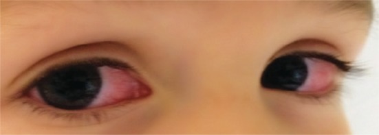
- Conjunctivitis is the most common (90%) and is bilateral, bulbar, nonexudative, limbic sparing, and typically present within a few days of fever onset.[10] It is directly correlated with the presence of skin rash.[7]
- Anterior uveitis is present in up to 70% of patients and usually arises within a week of fever onset. It is characteristically Grade 1+ or 2+ and bilateral, and sometimes associated with keratic precipitates. Presence of uveitis early in the acute phase can help establish the diagnosis particularly in cases of incomplete KD. Uveitis was shown in at least one study to be significantly correlated with coronary artery dilation.[7] Intermediate uveitis has also less commonly been reported.
- Posterior segment changes can include papilledema, papillitis, vitreous opacities, vitritis, retinitis, and retinal ischemia from inflammatory damage or thrombosis of retinal vasculature.[14][15][16]

- Ptosis can rarely be observed 1 to 4 weeks after the onset of symptoms. In reported cases, ptosis has been self-limited, resolving anywhere from 5 days to 4 weeks after onset.[18][17][19][20][21] Elevated levels of acetylcholine receptor antibodies can be seen, suggesting a myastheniform condition.[21] Proposed pathophysiology of ptosis in KD includes destruction of arteries supplying the levator muscles, myositis, or immune-mediated nerve involvement.[20][21]
- Palsies of the oculomotor nerve, abducens nerve, and facial nerve have been reported.[22][23][24][25]
- Myopia is more common in patients with a history of KD. A population-based cohort study conducted in Taiwan in 2017 found a statistically significant increase in myopia in a cohort of 532 KD patients compared to a cohort of 2128 non-KD patients.[6] It is thought that myopic shift is induced by ocular inflammation.
Other reported ocular manifestations include:
- Superficial punctate keratitis
- Disciform keratitis[26]
- Periorbital vasculitis associated with eyelid edema[27]
- Subconjunctival hemorrhage
- Dacryocystitis[28]
Differential diagnosis
The broad differential diagnosis of KD includes viral infections (adenovirus, echovirus, EBV, and measles), bacterial infections (scarlet fever, acute rheumatic fever, Rocky Mountain spotted fever, and leptospirosis), toxin-mediated illnesses (staphylococcal scalded skin syndrome and toxic shock syndrome), hypersensitivity reactions (e.g. Stevens-Johnson syndrome), rheumatic disease (juvenile idiopathic arthritis, polyarteritis nodosa, and reactive arthritis), and multisystem inflammatory syndrome in children (MIS-C) that is associated with the SARS-CoV-2 virus.2 Children with KD may have a concurrent viral or bacterial infection.
MIS-C is a severe presentation of COVID-19 and can feature many of the same ocular manifestations as KD including conjunctivitis, eyelid edema, anterior uveitis, retinitis, cotton wool spots, and optic neuritis.
Adenovirus infection and Stevens-Johnson syndrome are more likely to cause an exudative (membranous or pseudomembranous) conjunctivitis. This finding in conjunction with the absence of prolonged fever and other clinical manifestations of KD should steer clinicians toward an alternative diagnosis. Conjunctivitis is typically absent in streptococcal scarlet fever and juvenile idiopathic arthritis, though uveitis is often present in the latter.
Management
Echocardiography to assess for coronary artery dilation is mandatory, and is usually performed at the time of diagnosis, prior to discharge, and at 2 week and 6 week follow up appointments.
Ophthalmologists are often consulted during the patient’s hospital admission to assess for surface pathology, anterior chamber inflammation, and posterior segment changes.
Systemic treatment
Initial therapy includes intravenous immunoglobulin (IVIG), whichhas been shown to reduce the risk of developing coronary artery lesions if administered within 10 days of symptom onset.[10]
Aspirin is often used for its anti-inflammatory and antiplatelet activity, though it has not been shown to reduce the risk of developing coronary artery lesions. Aspirin should be avoided in KD patients with potential concurrent influenza or varicella infection due to the possibility of precipitating Reye syndrome.
Additional agents including glucocorticoids and tumor necrosis factor inhibitors (Infliximab) can be added in patients whose fever is refractory to IVIG 36 hours after administration.
Ocular treatment
Ocular inflammation in Kawasaki disease is almost always self-limited, resolving over a course of a few days to 8 weeks. Symptomatic ocular inflammation can be treated with topical corticosteroids. Cycloplegic agents can be added to reduce pain and photophobia and to prevent the formation of posterior synechiae.
Complications
Usually, ocular manifestations of KD resolve after treatment and do not cause long term problems. However, patients who have recovered from KD do commonly develop a myopic shift.
Retinal damage from vasculitis or thrombus formation within the branches of the ophthalmic artery can cause permanent visual impairment.
Dacrycystitis and conjunctival scarring are rarely reported long term complications of KD.
The reported cases of neuromuscular abnormalities (e.g. ptosis, cranial nerve palsies) during the acute and subacute phase of KD were self-limited and did not require long term follow-up.
Prognosis
Mortality from Kawasaki Disease is very low, estimated to be about 0.1 to 0.3% of patients. Fatal outcomes are usually the result of myocardial infarction, arrhythmia, or ruptured coronary aneurysm. Persistent ocular pathology is rare, and most patients do not require consistent follow up with an ophthalmologist outside of recommended annual exams.
References
- ↑ Rossi Fde S, Silva MF, Kozu KT, et al. Extensive cervical lymphadenitis mimicking bacterial adenitis as the first presentation of Kawasaki disease. Einstein (Sao Paulo). 2015;13(3):426-429. doi:10.1590/S1679-45082015RC2987
- ↑ Elakabawi K, Lin J, Jiao F, Guo N, Yuan Z. Kawasaki Disease: Global Burden and Genetic Background. Cardiol Res. Feb 2020;11(1):9-14. doi:10.14740/cr993
- ↑ 3.0 3.1 Rife E, Gedalia A. Kawasaki Disease: an Update. Curr Rheumatol Rep. Sep 13 2020;22(10):75. doi:10.1007/s11926-020-00941-4
- ↑ Onouchi Y. The genetics of Kawasaki disease. Int J Rheum Dis. Jan 2018;21(1):26-30. doi:10.1111/1756-185x.13218
- ↑ Takahashi K, Oharaseki T, Yokouchi Y. Pathogenesis of Kawasaki disease. Clin Exp Immunol. May 2011;164 Suppl 1(Suppl 1):20-2. doi:10.1111/j.1365-2249.2011.04361.x
- ↑ 6.0 6.1 Kung YJ, Wei CC, Chen LA, et al. Kawasaki Disease Increases the Incidence of Myopia. Biomed Res Int. 2017;2017:2657913. doi:10.1155/2017/2657913
- ↑ 7.0 7.1 7.2 Shiari R, Jari M, Karimi S, et al. Relationship between ocular involvement and clinical manifestations, laboratory findings, and coronary artery dilatation in Kawasaki disease. Eye (Lond). Oct 2020;34(10):1883-1887. doi:10.1038/s41433-019-0762-y
- ↑ Al-Abbadi MA, Abuhammour W, Harahsheh A, Abdel-Haq NM, Hasan RA, Saleh HA. Conjunctival changes in children with Kawasaki disease: cytopathologic characterization. Acta Cytol. May-Jun 2007;51(3):370-4. doi:10.1159/000325749
- ↑ 9.0 9.1 Singh S, Jindal AK, Pilania RK. Diagnosis of Kawasaki disease. Int J Rheum Dis. Jan 2018;21(1):36-44. doi:10.1111/1756-185x.13224
- ↑ 10.0 10.1 10.2 10.3 McCrindle BW, Rowley AH, Newburger JW, et al. Diagnosis, Treatment, and Long-Term Management of Kawasaki Disease: A Scientific Statement for Health Professionals From the American Heart Association. Circulation. Apr 25 2017;135(17):e927-e999. doi:10.1161/cir.0000000000000484
- ↑ Bayers S, Shulman ST, Paller AS. Kawasaki disease: part II. Complications and treatment. J Am Acad Dermatol. Oct 2013;69(4):513.e1-8; quiz 521-2. doi:10.1016/j.jaad.2013.06.040
- ↑ Conti G, Giannitto N, De Luca FL, et al. Kawasaki disease and cardiac involvement: an update on the state of the art. J Biol Regul Homeost Agents. Jul-Aug 2020;34(4 Suppl. 2):47-53. special issue: focus on pediatric cardiology.
- ↑ Ha KS, Jang G, Lee J, et al. Incomplete clinical manifestation as a risk factor for coronary artery abnormalities in Kawasaki disease: a meta-analysis. Eur J Pediatr. Mar 2013;172(3):343-9. doi:10.1007/s00431-012-1891-5
- ↑ Anand S, Yang YC. Optic disc changes in Kawasaki disease. J Pediatr Ophthalmol Strabismus. May-Jun 2004;41(3):177-9. doi:10.3928/0191-3913-20040501-12
- ↑ Font RL, Mehta RS, Streusand SB, O'Boyle TE, Kretzer FL. Bilateral retinal ischemia in Kawasaki disease. Postmortem findings and electron microscopic observations. Ophthalmology. May 1983;90(5):569-77. doi:10.1016/s0161-6420(83)34522-x
- ↑ Klig JE. Ophthalmologic complications of systemic disease. Emerg Med Clin North Am. Feb 2008;26(1):217-31, viii. doi:10.1016/j.emc.2007.10.003
- ↑ 17.0 17.1 Lin Y, Wang L, Li A, Zhang H, Shi L. Eyelid ptosis and muscle weakness in a child with Kawasaki disease: a case report. BMC Pediatr. Nov 27 2021;21(1):526. doi:10.1186/s12887-021-02979-4
- ↑ Zhao SH, Wang R, Ma LQ, Yang Y. Ptosis of both palpebra superiors caused by Kawasaki disease in a child. Zhongguo Dang Dai Er Ke Za Zhi. Feb 2007;9(1):83.
- ↑ Falcini F, La Torre F, Conti G, Vitale A, Messina MF, Calcagno G. Intermittent bilateral superior palpebra ptosis in a 20-month-old infant. Clin Exp Rheumatol. 2011:360. vol. 2.
- ↑ 20.0 20.1 Hameed A, Alshara H, Schleussinger T. Ptosis as a complication of Kawasaki disease. BMJ Case Rep. Jun 9 2017;2017doi:10.1136/bcr-2017-219687
- ↑ 21.0 21.1 21.2 Sánchez Marcos E, Flores Perez P, Jimenez García R. Refusal to Walk and Ptosis as an Atypical Presentation of Kawasaki Disease. Pediatr Infect Dis J. Apr 11 2022;doi:10.1097/inf.0000000000003551
- ↑ Emiroglu M, Alkan G, Kartal A, Cimen D. Abducens nerve palsy in a girl with incomplete Kawasaki disease. Rheumatol Int. 2016:1181-3. vol. 8.
- ↑ Bushara K, Wilson A, Rust RS. Facial palsy in Kawasaki syndrome. Pediatr Neurol. Nov 1997;17(4):362-4. doi:10.1016/s0887-8994(97)00103-3
- ↑ Wurzburger BJ, Avner JR. Lateral rectus palsy in Kawasaki disease. Pediatr Infect Dis J. Nov 1999;18(11):1029-31. doi:10.1097/00006454-199911000-00025
- ↑ Thapa R, Mallick D, Biswas B, Chakrabartty S. Transient unilateral oculomotor palsy and severe headache in childhood Kawasaki disease. Rheumatol Int. Jan 2011;31(1):97-9. doi:10.1007/s00296-009-1154-6
- ↑ Kadyan A, Choi J, Headon MP. Disciform keratitis and optic disc swelling in Kawasaki disease: an unusual presentation. Eye (Lond). 2006:976-7. vol. 8.
- ↑ Felz MW, Patni A, Brooks SE, Tesser RA. Periorbital vasculitis complicating Kawasaki syndrome in an infant. Pediatrics. Jun 1998;101(6):E9. doi:10.1542/peds.101.6.e9
- ↑ Mauriello JA, Jr., Stabile C, Wagner RS. Dacryocystitis following Kawasaki's disease. Ophthalmic Plast Reconstr Surg. 1986;2(4):209-11. doi:10.1097/00002341-198601070-00007


