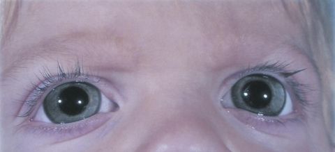
Megalocornea
All content on Eyewiki is protected by copyright law and the Terms of Service. This content may not be reproduced, copied, or put into any artificial intelligence program, including large language and generative AI models, without permission from the Academy.
Disease Entity
OMIM Numbers
- 249300 – Megalocornea
- 309300 – MGC1
- 300350 – CHRDL1
Disease/Description
Megalocornea (MGC1) is a rare developmental defect characterized by nonprogressive, usually symmetric, bilateral enlargement of the diameter of the cornea (≥13 mm).[2] The cornea is clear and of normal or slightly below normal thickness. Keratometry reveals normal to above normal steepness. The condition is also known as “anterior megalophthalmos” since the entirety of the anterior segment is enlarged.[3]In addition to an enlarged cornea, patients present with a very deep anterior chamber and a normal to thin corneal thickness.[4]
Epidemiology
Due to the X-linked recessive inheritance of congenital megalocornea, males are more often affected (90%) than females. The incidence of megalocornea is unknown.[5]
Inheritance and Molecular Genetics
MGC1 is most often transmitted in a X-linked recessive inheritance but can rarely be inherited in both autosomal dominant and autosomal recessive patterns.
MGC1 is caused by a mutation in Chordin-like 1 (CHRDL1) on Xq23.[4] CHRDL1 codes for the protein ventroptin, an antagonist to bone morphogenetic protein 4 (BMP-4). BMP-4 is mainly expressed in the anterior retina, and the interaction between ventroptin and BMP-4 is important for the development of corneal stroma and endothelium. A mutation in the CHRDL1 gene may lead to unregulated growth causing megalocornea.[4][6]
CHRDL1 is a gene that is 12 exons in length, and cytogenetic mapping of the gene places its location at Xq23.[4][6] However, other mutations have been observed between Xq21.3-q22.[7]
Embryogenesis
The definitive mechanism behind the formation of megalocornea is currently unknown, but a common theory is that failure of anterior cup fusion allows for more than average corneal growth.[8] Posterior placement of the iris-lens diaphragm and normal endothelial density from a primary overgrowth (as opposed to low density from a distended cornea as seen in glaucoma) seem to support this theory.[9] Other theories include arrested development of congenital glaucoma and abnormal collagen synthesis. [10]
Clinical Presentation
Megalocornea often occurs as an isolated condition, or can present with multiple ocular and systemic associations. Clinical outcomes for primary megalocornea are generally good.[11] Endothelial cell density is normal. The steeper cornea usually results in with the rule astigmatism and myopia. Also known as megalophthalmos, these eyes may have a larger than average ciliary ring and anterior segment. This can result in phacodonesis, iridodonesis, and ectopia lentis. Iris stretching can lead to transillumination defects and increased ™ pigmentation and iris processes, thus predisposing these eyes to glaucoma. [10]
Megalocornea can also be a presenting symptom of a larger developmental disease. Frank-Ter Haar Syndrome is a rare autosomal recessive disease of skeletal dysplasia that presents with megalocornea and developmental delay.[12] Megalocornea is rarely associated with Marfan Syndrome and can be a helpful diagnostic sign in infants and young children.[13] Neuhauser Syndrome, also known as Megalocornea-Mental Retardation Syndrome, is a rare inherited defect that is described by a classic triad of primary megalocornea, intellectual disability, and hypotonia.[14] Megalocornea can also be associated with craniosynostosis and rare cases of albinism and Down Syndrome.[15][16][17]
Ocular Findings
Primary megalocornea often does not present with ocular symptoms other than blurred vision secondary to a refractive error. In some cases, however, patients can manifest premature cataract formation, retinal detachment, glaucoma, lens subluxation, and primary congenital glaucoma.[5][11]
Diagnosis
Diagnosis of megalocornea is generally made in young patients and requires thorough examination of the eye, often under general anesthesia. Megalocornea is defined as nonprogressive bilateral enlargement (13 mm or greater) of the cornea. Deep anterior chambers can also be helpful in making the diagnosis.[2]
Differential Diagnosis[9][11][12][13][14][15][16][17]
Congenital glaucoma is usually the primary differential diagnosis given the concern for buphthalmos. Patients with megalocornea will not exhibit ocular hypertension, Haabs striae, optic disc changes seen in congenital glaucoma. A sharply demarcated limbal region can also be seen in megalocornea (and not in congenital glaucoma) and can be used as a distinguishing feature. [10]
Primary megalocornea
- Frank-Ter Haar Syndrome
- Marfan Syndrome
- Neuhauser Syndrome (Megalocornea-mental retardation syndrome)
- Primary Congenital Glaucoma
- Crouzon Syndrome (craniosynostosis)
- Albinism
- Down Syndrome
- Keratoglobus
Management/Therapeutic Considerations
Symptomatic primary megalocornea typically requires the correction of refractive errors. Many patients with myopia or astigmatism can develop unimpaired vision with regular follow-up and corrective lenses.
Detection of, monitoring, and treating associated ocular abnormalities such as cataracts, glaucoma, and retinal detachments, are imperative to optimizing ocular health in these patients.
Cataract surgery in megalocornea patients is complex due to the large size of the anterior segment and the weakened zonules. These patients may be at higher risk for poor dilation, lens subluxation, and posterior capsular rupture. The weakened zonules result in difficulty supporting an artificial intraocular lens (IOL).[18] Instead of a standard IOL, patients with megalocornea may benefit from a iris-claw IOL or the Artisan lens that clips into the iris to maintain the position of the lens post-operatively (video can be accessed from references).[9][19]
Additional Resources
Anterior Segment Developmental Anomalies (ASDA)
References
- ↑ American Academy of Ophthalmology. Megalocornea. https://www.aao.org/image/megalocornea-5 Accessed July 05, 2019.
- ↑ Jump up to: 2.0 2.1 Ito YA, Walter MA. Genomics and anterior segment dysgenesis: a review. Clin Experiment Ophthalmol. 2014;42(1):13-24. doi:10.1111/ceo.12152
- ↑ Skuta GL, Sugar J, Ericson ES. Corneal Endothelial Cell Measurements in Megalocornea. Arch Ophthalmol. 1983;101(1):51-53. doi:10.1001/archopht.1983.01040010053007
- ↑ Jump up to: 4.0 4.1 4.2 4.3 Webb TR, Matarin M, Gardner JC, et al. X-Linked Megalocornea Caused by Mutations in CHRDL1 Identifies an Essential Role for Ventroptin in Anterior Segment Development. Am J Hum Genet. 2012;90(2):247-259. doi:10.1016/j.ajhg.2011.12.019
- ↑ Jump up to: 5.0 5.1 Roche O, Dureau P, Uteza Y, Dufier JL. [Congenital megalocornea]. J Fr Ophtalmol. 2002;25(3):312-318. http://www.ncbi.nlm.nih.gov/pubmed/11941259. Accessed June 10, 2019.
- ↑ Jump up to: 6.0 6.1 Sakuta H, Suzuki R, Takahashi H, et al. Ventroptin: A BMP-4 Antagonist Expressed in a Double-Gradient Pattern in the Retina. Science (80- ). 2001;293(5527):111-115. doi:10.1126/science.1058379
- ↑ Chen JD, Mackey D, Fuller H, Serravalle S, Olsson J, Denton MJ. X-linked megalocornea: close linkage to DXS87 and DXS94. Hum Genet. 1989;83(3):292-294. http://www.ncbi.nlm.nih.gov/pubmed/2571565. Accessed June 11, 2019.
- ↑ Mann I. Developmental Abnormalities of the Eye. 2nd ed. Philadelphia: J.B.Lippincott Co.; 1957.
- ↑ Jump up to: 9.0 9.1 9.2 Welder J, Oetting TA. Megalocornea. EyeRounds.org - Ophthalmology - The University of Iowa. https://webeye.ophth.uiowa.edu/eyeforum/cases/121-megalocornea.htm. Published 2010. Accessed June 13, 2019.
- ↑ Jump up to: 10.0 10.1 10.2 Mark J. Mannis and Edward J. Holland. Cornea. St. Louis, Mo.: Mosby/Elsevier, 2011
- ↑ Jump up to: 11.0 11.1 11.2 Chlasta-Twardzik E, Nowińska A, Wąs P, Jakubowska A, Wylęgała E. Traumatic cataract in patient with anterior megalophthalmos. Medicine (Baltimore). 2017;96(30):e7160. doi:10.1097/MD.0000000000007160
- ↑ Jump up to: 12.0 12.1 Femitha P, Joy R, Gane BD, Adhisivam B, Bhat BV. Frank -Ter Haar Syndrome in a Newborn. Indian J Pediatr. 2012;79(8):1091-1093. doi:10.1007/s12098-011-0599-2
- ↑ Jump up to: 13.0 13.1 Morse RP, Rockenmacher S, Pyeritz RE, et al. Diagnosis and management of infantile marfan syndrome. Pediatrics. 1990;86(6):888-895. http://www.ncbi.nlm.nih.gov/pubmed/2251026. Accessed June 10, 2019.
- ↑ Jump up to: 14.0 14.1 Gutiérrez-Amavizca BE, Juárez-Vázquez CI, Orozco-Castellanos R, Arnaud L, Macías-Gómez NM, Barros-Nuñez P. Neuhauser syndrome: a rare association of megalocornea and mental retardation. Review of the literature and further phenotype delineation. Genet Couns. 2013;24(2):185-191. http://www.ncbi.nlm.nih.gov/pubmed/24032289. Accessed June 11, 2019.
- ↑ Jump up to: 15.0 15.1 Alshamrani AA, Al-Shahwan S. Glaucoma With Crouzon Syndrome. J Glaucoma. 2018;27(6):e110-e112. doi:10.1097/IJG.0000000000000946
- ↑ Jump up to: 16.0 16.1 Awaya S, Tsunekawa F, Koizumi E, Miyake Y, Yokoyama K. [Studies of X-linked recessive ocular albinism of the Nettleship-Falls type--with special reference to the association of megalocornea]. Nihon Ganka Gakkai Zasshi. 1988;92(1):146-150. http://www.ncbi.nlm.nih.gov/pubmed/3389256. Accessed June 12, 2019.
- ↑ Jump up to: 17.0 17.1 Rogers GL, Polomeno RC. Autosomal-dominant inheritance of megalocornea associated with down’s syndrome. Am J Ophthalmol. 1974;78(3):526-529. http://www.ncbi.nlm.nih.gov/pubmed/4278003. Accessed June 12, 2019.
- ↑ Wang Q-W, Xu W, Zhu Y-N, Li J-Y, Zhang L, Yao K. Misdiagnosis induced intraocular lens dislocation in anterior megalophthalmos. Chin Med J (Engl). 2012;125(17):3180-3182. http://www.ncbi.nlm.nih.gov/pubmed/22932204. Accessed June 12, 2019.
- ↑ Oetting TA, Newsom TH. Bilateral Artisan lens for aphakia and megalocornea: Long-term follow-up. J Cataract Refract Surg. 2006;32(3):526-528. doi:10.1016/j.jcrs.2005.12.060


