Telecanthus
All content on Eyewiki is protected by copyright law and the Terms of Service. This content may not be reproduced, copied, or put into any artificial intelligence program, including large language and generative AI models, without permission from the Academy.
Disease Entity
ICD-10: Q10.3: Telecanthus
Disease
Telecanthus is an uncommon palpebral anomaly condition defined as an increased distance between the medial canthi.[1]
Etiology

Telecanthus is produced by an abnormal insertion or abnormally length of the medial canthal tendons. Telecanthus may occur in isolation or in association with blepharophimosis. [1] Telecanthus is often associated with many congenital disorders such as Down syndrome, fetal alcohol syndrome, Cri du Chat syndrome, Klinefelter syndrome, Turner syndrome, Ehlers-Danlos syndrome and Waardenburg syndrome.
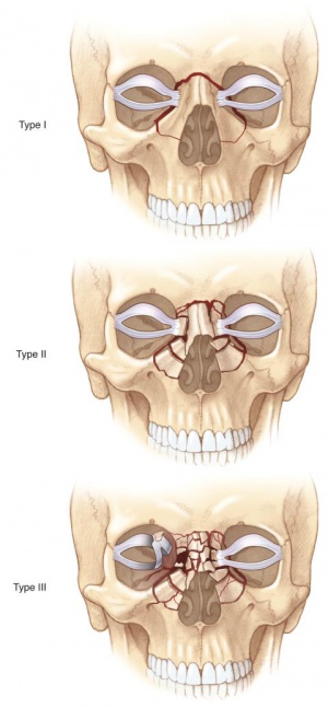
Traumatic telecanthus is the result of facial injuries due to either the medial canthal tendon being lacerated or avulsed from its bony attachments around the lacrimal crest or displacement of the nasoorbital component following a nasoorbitoethmoidal fracture.
Diagnosis
Physical examination
The average interpupillary distance is 60–62 millimeters (mm), which corresponds to an intercanthal distance of approximately 30–31 mm. In telecanthus, there is an increased distance between the medial canthi of the eyes, while the inter-pupillary distance is normal. Tessier distinguished among varying severities of hypertelorism as first degree (30–34 mm interorbital distance [IOD]), second degree (35–40 mm IOD), and third degree (>40 mm IOD).[3]
Differential diagnosis
Telecanthus should be differentiated from hypertelorism (hypertelorbitism), which is an increased distance between the bony orbits taken at the region of the dacryon (lacrimal crest) and an associated increase in interpupillary distance.[3]
Management
Surgery
The treatment of telecanthus involves shortening and refixation of the medial canthal tendons to the anterior lacrimal crest, or insertion of a trans-nasal suture. It is well known that the treatment is difficult due to recurrence by loosening of tendon. A transnasal wiring is an effective and most frequently used method in achieving the highest fixation force.[4]
References
- ↑ 1.0 1.1 Brad Bowling. Kanski's Clinical Ophthalmology. Eight edition. Chapter 1: 57-59
- ↑ American Academy of Ophthalmology. Naso-orbital-ethmoidal (NOE) fractures. https://www.aao.org/image/naso-orbital-ethmoidal-noe-fractures Accessed December 06, 2019.
- ↑ 3.0 3.1 Resident Manual of Trauma to the Face, Head, and Neck. First edition. Retrieved 12 January 2015.
- ↑ Tae-Gon Kim, et al. Medial Canthopexy Using Y-V Epicanthoplasty Incision in the Correction of Telecanthus. Annals of Plastic Surgery & Volume 72, Number 2, Feb 2014

