All content on Eyewiki is protected by copyright law and the Terms of Service. This content may not be reproduced, copied, or put into any artificial intelligence program, including large language and generative AI models, without permission from the Academy.

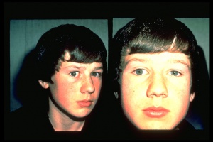
Ocular torticollis is defined by abnormal head posture as a compensatory mechanism for an ocular abnormality.
Overview

Ocular torticollis is an abnormal head posture adopted in order to optimize vision and/or maintain binocularity. The incidence is approximately 3% in pediatric ophthalmology practice.[1] It may consist of any combination of head tilt, face turn, or chin elevation or depression.[2] While ocular torticollis may occur at any age, it typically manifests early in life and may be more pronounced over time as the visual system matures.[3]
Etiology
In general, the etiology of torticollis may be ocular, neurologic or orthopedic. When due to an ocular cause, the head position occurs to compensate for an ocular condition. This may include a condition such as infantile nystagmus syndrome to improve the vision or incomitant strabismus to improve binocularity. The chart below details possible ocular and non-ocular etiologies.
A prospective, consecutive case series examining children with an abnormal head posture on routine pediatric examinations found an ocular etiology for the torticollis in 25 of 63 (39.7%) children.[4] Studies have confirmed that the most common causes of ocular torticollis are incomitant strabismus and nystagmus.[5][6] When due to ocular causes, a prospective study of 188 patients found that incomitance accounted for 62.7% of the head postures, while nystagmus was the etiology in 20.2%.[7] This was similar to a later study which found that 330 of 630 (52.4%) cases of ocular torticollis were due to incomitant strabismus and 120 (19%) were due to nystagmus.[8] Of the patients in this study with incomitance, most were due to were due to A/V pattern (116, 35.2%) or superior oblique palsy (59, 17.9%).
Diagnosis
Ocular torticollis is diagnosed primarily by history and a detailed neuro-ophthalmic clinical exam. Diagnostic evaluation may include measuring the degree of head tilt and face turn. The extraocular motility and alignment should be examined in all gaze positions, with particular focus on positions opposite to those favoured. Of note, if occlusion of one eye eliminates the abnormal head position, this suggests the torticollis provides binocular fusion. An external examination can identify nystagmus and ptosis, although subtle nystagmus may be visible only during slit-lamp or fundus examination. A dilated fundus examination may also be helpful for evaluating torsion. Retinoscopy can be performed to assess for a refractive error with particular attention to oblique axis astigmatism. Additional maneuvers that may be considered include: Hess-Lancaster or Lee screen to assess for antagonist muscle contracture; stereoscopic tests and prisms to test for motor fusion; and the Bielschowsky three step test to diagnose a vertical muscle palsy.
Differential Diagnosis[2][9]
Ocular Torticollis
- Attempt to improve visual resolution
- Nystagmus
- Infantile nystagmus syndrome (congenital motor or sensory nystagmus; null point)
- Fusion maldevelopment nystagmus syndrome (manifest latent nystagmus; less in adduction)
- Periodic alternative nystagmus (alternating null point)
- Spasmus nutans
- Acquired adult jerk nystagmus
- Ptosis
- Monocular blindness (with fusion maldevelopment nystagmus syndrome, or for centration of remaining field)
- Refractive error
- Homonymous hemianopia
- Nystagmus
- Motility Disorders
- A- or V- pattern esotropia or exotropia
- Paretic strabismus
- Superior oblique palsy
- Duane retraction syndrome
- Sixth nerve palsy
- Third nerve palsy
- Inferior oblique palsy
- Restrictive strabismus
- Brown syndrome
- Thyroid eye disease
- Orbital fracture
- Congenital fibrosis of extraocular muscles
- Supranuclear disorders
- Monocular elevation deficiency
- Dorsal midbrain syndrome
- Gaze palsy
- Dissociated vertical deviation
- Ocular tilt reaction
Non-ocular Torticollis
- Orthopedic
- Congenital muscular torticollis
- Trauma
- Inflammatory myositis
- Skeletal abnormalities (such as Klippel-Feil syndrome, plagiocephaly, cervical spine subluxation, occipito-cervical stenosis)
- Fibrotic shortening of the sternocleidomastoid muscle
- Neurologic
- Syringomyelia
- Dystonia
- Deafness in one ear
- Psychiatric disturbance
Management
Management is directed towards treating the etiology of ocular torticollis. Early diagnosis and correction of underlying etiology is important to prevent musculoskeletal changes such as neck and facial asymmetry. The strategy is to increase the field of binocular fusion and decrease the tilt or turn. For example, in the setting of nystagmus, it is important to identify the fixating eye and null point as these two components determine the direction and magnitude of the face turn. Surgery then involves moving the null point of the fixating eye to primary position and correcting any residual or induced strabismus by repositioning the non-fixating eye.
References
- ↑ Boricean ID, Bărar A. Understanding ocular torticollis in children. Oftalmologia. 2011;55(1):10-26.
- ↑ Jump up to: 2.0 2.1 Mitchell PR. Ocular torticollis. Trans Am Ophthalmol Soc. 97, 697–769 (1999).
- ↑ Yoon JA, Choi H, Shin YB, Jeon H. Development of a questionnaire to identify ocular torticollis. Eur J Pediatr. 2021 Feb;180(2):561-567.
- ↑ Nucci P, Kushner BJ, Serafino M, Orzalesi N. A multi-disciplinary study of the ocular, orthopedic, and neurologic causes of abnormal head postures in children. Am J Ophthalmol. 2005 Jul;140(1):65-8.
- ↑ Kushner BJ. Ocular causes of abnormal head postures. Ophthalmology. 1979 Dec;86(12):2115-25.
- ↑ Mitchell PR. Ocular torticollis. Trans Am Ophthalmol Soc. 1999;97:697-769.
- ↑ Kushner BJ. Ocular causes of abnormal head postures. Ophthalmology. 1979 Dec;86(12):2115-25.
- ↑ Mitchell PR. Ocular torticollis. Trans Am Ophthalmol Soc. 1999;97:697-769.
- ↑ American Academy of Ophthalmology. “Torticollis: Differential Diagnosis and Evaluation.” Basic and Clinical Science Course: 2020-2021, 83 (2020).

