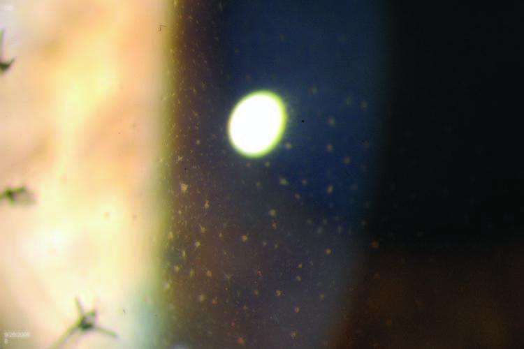
Herpes Simplex Uveitis
All content on Eyewiki is protected by copyright law and the Terms of Service. This content may not be reproduced, copied, or put into any artificial intelligence program, including large language and generative AI models, without permission from the Academy.
Disease Entity
Herpes simplex virus (HSV) associated uveitis is a common cause of unilateral hypertensive anterior uveitis. Herpetic anterior uveitis causes approximately 5-10% of uveitis cases.[2]
Etiology
Herpes simplex iritis is due to the Herpes simplex virus, a double-stranded DNA virus. The most common subtype associated with ocular infection is HSV-1. It can lay dormant in the trigeminal ganglion and becomes reactivated that manifests as skin lesions, keratitis, or anterior uveitis.[3] Anterior uveitis is more common during reactivation vs primary disease.
Epidemiology
HSV associated anterior uveitis is more common in patients in their 40-50's affecting both genders equally.[4]
Risk Factors
Patients may have a medical history of cold sores or fever blisters. [4] A history of immunosuppression is associated with a higher risk of HSV reactivation.
General Pathology
HSV is a double-stranded DNA virus that can be separated into two types: HSV-1 and HSV-2. HSV-1 is more commonly the cause of cold sores and ocular infections, while HSV-2 is a more common cause of genital herpes. While it is possible for ocular HSV infections to occur during primary infection, it is more often that symptomatic ocular HSV infections occur at times of recurrence; the virus reactivates from the trigeminal ganglion and travels along sensory nerve branches to the eye. Reactivation may theoretically occur in response to systemic triggers such as stress, fever, or immunocompromised conditions.
Primary HSV-1 infection is associated with an immune response mediated by mononuclear inflammatory cells, primarily composed of T cells. Plasma cells, macrophages, and natural killer cells have also been detected in infected tissues. Despite antibody and cell-mediated immunity, reactivation of HSV-1 can lead to ocular infection. Upon reactivation, HSV-1 has the ability to downregulate MHC-1 and become resistant to Fas-mediated apoptosis. Viral particles can also secrete TGF-β1, which can downregulate IFN-γ-induced MHC-2 expression. This consequently decreases CD4+ T cell activation and thus allows HSV-1 to further evade immune detection upon reactivation in the eye.
Primary prevention
There is no evidence of primary prevention strategies for HSV iritis or ocular HSV.
Diagnosis
Clinical Findings / Signs
Keratouveitis and iritis are uncommon manifestations of primary HSV ocular infection. In a study of a large population in the Northern California region of Kaiser Permanente, iritis was the presenting sign in 1% of patients with an initial episode of HSV eye disease. [5]
Most commonly, in patients with iritis due to HSV, a keratouveitis is present. Findings in those cases may include a combination of these findings: (from anterior to posterior) corneal epithelial and/or stromal edema, stromal keratitis, keratic precipitates, endotheliitis, and anterior chamber cells and flare. Iritis can also occur alone, without evidence of keratitis, and may present as an unilateral hypertensive anterior uveitis.
Keratic precipitates (KP) may take several forms. They can be granulomatous, nongranulomatous, or stellate. Often they may be present in a patch on the endothelial surface, underlying a localized patch of corneal edema. They can be regional, in the inferior one-third of the cornea or diffuse. When stellate KP are present, they are typically diffuse. [6]
It is also important to note that fine, stellate KPs along with mild anterior chamber cell are also associated with Fuchs heterochromic iridocyclitis (FHI), and there has been evidence to suggest that HSV (and other infectious etiologies) may potentially cause FHI. In FHI, there may be prominent iris and angle vessels visible due to iris atrophy.
High intraocular pressure is a common complication of HSV iritis and can serve as a diagnostic hallmark. The IOP can be very high, in the range of 50-60 mm Hg during an episode of acute iritis. High IOP is due to trabeculitis, as well as inflammatory cells clogging the trabecular meshwork. Although antiglaucoma therapy may be required in the acute setting, once the inflammation is controlled, typically the intraocular pressure will normalize and the patient will not require ongoing antiglaucoma treatment. By contrast, prolonged or recurrent inflammation may cause peripheral anterior synechiae (PAS), which can lead to secondary glaucoma. Other signs include posterior synechiae and iridoplegia. [3]
Spontaneous hyphema can occur in HSV iritis, as well as layers of hyphema mixed with hypopyon, known as a “candy-cane hypopyon”.
Although patchy iris atrophy may be present following an episode of HSV keratouveitis or iritis, inflammatory iris lesions are not typically seen. Disease is more common a unilateral process, but can be bilateral as well.
Symptoms
Typical symptoms of iritis with photophobia and pain are seen in HSV iritis. If there is an accompanying high IOP, headache, deeper pain, and decreased vision due to corneal edema may be present. In patients with keratouveitis, pain and decreased vision may be present because of the keratitis and edema. If there is significant damage to cranial nerve 5, from prior episodes of HSV, the patient may not have significant pain.
Clinical diagnosis
The diagnosis of HSV iritis is suggested by a unilateral anterior uveitis, accompanied by high intraocular pressure. The presence of patchy iris transillumination defects further suggests HSV iritis, but the absence of transillumination defects does not rule it out. A prior history of HSV ocular disease is also strongly suggestive of the role of HSV in iritis. Other suggestive features are cornea hypoesthesia and the appearance of the keratic precipitates. As stated previously, the presence of stellate KPs with mild anterior chamber cell, prominent iris and angle vessels, and iris heterochromia may be indicative of FHI. In addition, the potential diagnosis of Posner-Schlossman syndrome needs to be carefully considered, especially when clinical exam findings demonstrate episodes of anterior chamber cell with extremely elevated IOP, with quiescence and normal IOP in between episodes. Some studies have suggested HSV as a possible infectious etiology for Posner-Schlossman syndrome, although cytomegalovirus is also implicated.
Diagnostic procedures / Laboratory Testing
Because of the high prevalence of positive HSV antibodies in most populations, serology is only helpful in the diagnosis in that a negative HSV antibody titer will rule out the possibility that iritis is due to HSV. Polymerase chain reaction for HSV DNA from an anterior chamber tap may be helpful in diagnosis with a high sensitivity (91.3%) and specificity (98.8).[7]
Management
Medical therapy
Topical corticosteroids are the mainstay of treatment for HSV iritis. Cycloplegic agents may be helpful to decrease symptoms of photophobia and decrease or lyse posterior synechiae.
Topical antiviral agents may help to prevent dendritic keratitis during treatment with corticosteroids, in patients with keratouveitis. In general topical antivirals are of little use in the treatment of HSV iritis, since these agents do not penetrate well into the anterior chamber. In fact, topical antiviral agents may be detrimental due to their topical toxicity. Systemic antiviral agents, such as acyclovir, famciclovir, or valacyclovir, attain excellent anterior chamber drug levels and may be beneficial in cases of iritis. The HEDS study trial of acyclovir in the treatment of HSV iritis was stopped prior to meeting the number of subjects needed according to the sample size estimates.[8] This was done because of problems with recruitment. In the findings that were published, there was a statistically suggestive trend for more rapid resolution of iritis with the use of systemic acyclovir, 400 mg five times per day.
Appropriate doses of systemic antiviral agents for treating active ocular disease are: acyclovir, 400 mg five times per day; valacyclovir, 1000 mg three times per day; famciclovir 250 mg three times per day. Dosing should be adjusted for patients with a history of renal dysfunction. Dosing should also be adjusted for pediatric patients. A dosing table by Liu et. al shows recommended treatment and prophylactic acyclovir doses for pediatric patients with ocular HSV.
Local injections or systemic corticosteroids are not typically recommended for control of HSV anterior uveitis, partly due to the risk of worsening HSV activity due to immunosuppression.
Some patients may require very slow tapering of topical corticosteroids and may require long term, low dose topical corticosteroid therapy to control inflammation.
Long term, low dose, systemic antiviral therapy may be beneficial for some patients, in order to decrease the frequency of recurrences of iritis. At present, controlled studies to show this are lacking. One might expect the HEDS study findings on prophylaxis may be generalizable to iritis, but this cannot be certain given the low number of strictly iritis patients.[9] Oral doses for prophylaxis for ocular herpes simplex disease are acyclovir, 400 mg twice per day or valacyclovir, 500-1000 mg daily.
Surgery
There are no surgical treatments for HSV iritis. If adequate medical management of high IOP is not possible or secondary glaucoma develops due to the development of PAS, glaucoma surgery may be indicated. One should keep in mind, however, that in most patients, the intraocular pressure will significantly improve once inflammation is controlled.
References
- ↑ American Academy of Ophthalmology. Herpes simplex virus keratouveitis. https://www.aao.org/education/image/herpes-simplex-virus-keratouveitis-2 Accessed May 23, 2024.
- ↑ Rathinam SR. Global variation and pattern changes in epidemiology of uveitis cases. Indian J Ophthalmol. 2007;55:173–183
- ↑ Jump up to: 3.0 3.1 Chan NS, Chee SP. Demystifying viral anterior uveitis: A review. Clin Exp Ophthalmol. 2019 Apr;47(3):320-333. doi: 10.1111/ceo.13417. Epub 2018 Nov 13. Review. PubMed PMID: 30345620.
- ↑ Jump up to: 4.0 4.1 Chan NS, Chee SP. Demystifying viral anterior uveitis: A review. Clin Exp Ophthalmol. 2019 Apr;47(3):320-333. doi: 10.1111/ceo.13417. Epub 2018 Nov 13.Review. PubMed PMID: 30345620.
- ↑ Gritz DC, Wong IG. Incidence and prevalence of uveitis in Northern California; the Northern California Epidemiology of Uveitis Study. Ophthalmology. 2004;111(3):491-500. doi:10.1016/j.ophtha.2003.06.014
- ↑ American Academy of Ophthalmology. Viral Uveitis. Basic and Clinical Science Course, Section 9. Uveitis and Ocular Inflammation. San Francisco: American Academy of Ophthalmology; 2021-2022:247-250.
- ↑ Sugita S, Ogawa M, Shimizu N, et al. Ophthalmology. 2013;120:1761–1768.
- ↑ U.S. National Library of Medicine. Herpetic Eye Disease Study (HEDS) I. clinicaltrials.gov, NCT00000138 Accessed March 07, 2023
- ↑ Acyclovir for the prevention of recurrent herpes simplex virus eye disease. Herpetic Eye Disease Study Group. N Engl J Med. 1998 Jul 30;339(5):300-6. PubMed PMID: 9696640.


