Strabismus Fixus
All content on Eyewiki is protected by copyright law and the Terms of Service. This content may not be reproduced, copied, or put into any artificial intelligence program, including large language and generative AI models, without permission from the Academy.
Disease Entity
Strabismus fixus convergens was first described as a type of retraction syndrome.[1][2] It was originally thought to be a congenital structural anomaly (fibrosis of the medial rectus), but was later determined to differ from retraction syndromes.[3]
Disease
Strabismus fixus can be associated with a number of conditions including congenital fibrosis, amyloidosis and high myopia.
Etiology
The underlying etiology of strabismus fixus still remains unclear. Yokoyama et al[4] provided the most recent explanation that the enlarged globe in high myopia herniates superotemporally and retroequatorially through the muscle cone. Other etiologies include congenital, fibrosis of extraocular muscle secondary to myositis or myopathies and infiltration of extraocular muscle by amyloid.
Pathophysiology
- Congenital: Congenital strabismus fixus is believed to be caused by fibrosis which explains loss of elasticity of medial rectus
- Lateral rectus palsy: Lateral rectus palsy causes the secondary contracture and fibrosis of medial rectus
- High myopia: Patients with high myopia have a weak intermuscular septa with an elongated globe which causes the superior rectus to slip medially towards medial rectus and lateral rectus to slip inferiorly, thus creating an esotropia and hypotropia. This has also been called heavy eye syndrome
- Amyloidosis: Infiltration of extraocular muscle by amyloid causes the loss of elasticity of the extraocular muscle
- Mechanical: Orbital factors that limit or prevent movement
- Other: Unspecific, progressive fibrosis, myopathy or myositis
General Pathology
Histologic studies have demonstrated the disappearance of myofibrils in the recti of highly myopic eyes, with added tissue changes of homogenization, acidophilia pyknosis and karyolysis. These findings suggest involvement of the rectus muscles in some myopathic process in pathological myopia. However it is still controversial as to which muscle is primarily affected in pathological myopia resulting in such ocular imbalances.
Diagnosis
Patient Characteristics
Patients are usually older and/or with high myopia, although cases of strabismus fixus in the absence of high myopia have been described.
Signs
Extraocular movements
As a rule the eyes are anchored in a position of extreme adduction and is fixed in this position with limitation of extraocular movements laterally.
Visual acuity
Patients with bilateral strabismus fixus tend to prefer one eye for vision. The inability to move the eye can result in an anomalous head posture.
Diagnostic procedures
Forced duction test: Done under topical anesthesia with forceps, forced ductions will confirm the immobility of eye and associated fibrosis and loss of elasticity.
Differential diagnosis
Acquired bilateral sixth nerve palsy.
Congenital bilateral sixth nerve palsy resembles cross fixation and sometimes it may become impossible to decide whether the origin is myogenic or neurogenic.
Acquired bilateral sixth nerve palsy is commonly seen in middle-aged individuals with history of diabetes, head injury, or increased intracranial pressure symptoms. At a later stage, when secondary changes have set in, it may be difficult to differentiate it from strabismus fixus.
Investigations
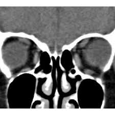
Computed tomography (CT) of the orbit can demonstrate relative localization of extra ocular muscle with respect to the globe and presence of any slippage of extra ocular muscle in patient of myopic strabismus fixus.
MRI Scan : In cases of congenital strabismus fixus, adherences among the extraocular muscles, posterior Tenon's capsule, and the globe within the muscle cone on MRI will be seen.
Management
Treatment is mainly surgical.
For convergent strabismus fixus, complete disinsertion of medial rectus is done. In addition to this, the surgeon may consider lateral rectus resection and/or recession of conjunctiva. Although this will limit abduction beyond midline, it will help in cosmetic and functional improvement.
For myopic strabismus fixus (heavy eye syndrome), loop myopexy, the procedure proposed by Yamaguchi et al[5] unites the lateral and superior rectus muscles with non-absorbable suture 15 mm behind the limbus. This procedure normalizes the vector forces of these muscles and eliminates the mechanical disturbance in eye movement. It can be combined with medial rectus muscle recession in cases of contracture as demonstrated by a positive forced duction test. The disadvantage of suture myopexy includes muscle strangulation, which may affect anterior ciliary circulation and may rarely cause cheese-wiring of the muscle.
References
- ↑ Aebli R : (1933) Retraction syndrome, Arch Ophthalmol 10:602=610.
- ↑ Bielschowsky.A : (1939) Lectures on motor anomalies, Am J. Ophthalmol 22:357-367.
- ↑ Villaseca A : (1959) Strabismus fixus, Am J Ophthalmol 48:761-762
- ↑ Yokoyama T, Tabuchi H, Ataka S, Shiraki K, Miki T, Mochizuki K. The mechanism of development in progressive esotropia with high myopia. In: de Faber JT, editor. Transactions of the 26 th Meeting European Strabismological Association Barcelona, Spain, September 2000. Lisse, Netherland: Swets and Zeitlinger Publishers; 2000. p. 218-21.
- ↑ Yamaguchi M, Yokoyama T, Shiraki K. Surgical procedure for correcting globe dislocation in highly myopic strabismus. Am J Ophthalmol 2010;149:341-6.e2.


