Nerve Supply of the Eyelids
All content on Eyewiki is protected by copyright law and the Terms of Service. This content may not be reproduced, copied, or put into any artificial intelligence program, including large language and generative AI models, without permission from the Academy.
The eyelid is supplied by three cranial nerves (III, V, VII) and sympathetic nerve fibers.
Motor
Eyelid muscle innervation is achieved by cranial nerve VII (the facial nerve), cranial nerve III (the oculomotor nerve), and sympathetic nerve fibers.
The facial nerve (CNVII) innervates the orbicularis oculi, frontalis, procerus, and corrugator supercilii muscles, and supports eyelid protraction. The temporal and zygomatic branches of the facial nerve supply the orbicularis oculi, the main eyelid protractor. The facial nerve also supplies the corrugator supercilii and the procerus, both of which contribute to brow depression and secondarily contribute to upper eyelid protraction.
The oculomotor nerve (CNIII) innervates the main upper eyelid retractor, the levator palpebrae superiorus, via its superior branch. The inferior division of CNIII also innervates the inferior rectus muscle, which by extension via the capsulopalpebral fascia causes lower eyelid retraction in downgaze. Sympathetic fibers contribute to upper eyelid retraction by innervation of the superior tarsal muscle, also known as Müller's muscle. Sympathetic fibers also innervate the inferior tarsal muscle, contributing to lower lid retraction.
Sensory
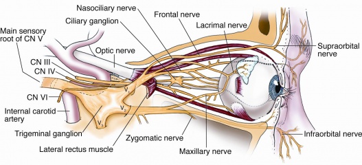
Cranial nerve V (the trigeminal nerve) supplies somatosensory innervation to the eyelid via its ophthalmic (V1) and maxillary (V2) divisions. Terminal branches of the ophthalmic division supply the upper eyelid as the lacrimal, supraorbital, and supratrochlear nerves (lateral to medial), and the medial aspect of both upper and lower lids as the infratrochlear nerve. Terminal branches of the maxillary division supply the lower eyelid as the zygomaticofacial and infraorbital nerves. The zygomaticofacial nerve supplies the lateral lower lid and the infraorbital nerve supplies the lower eyelid proper.
Anatomical Course of the Nerves
CN III
In addition to innervating the levator palpebrae supriorus, the somatomotor fibers of the oculomotor nerve also supply the medial, superior, and inferior rectus muscles, and the inferior oblique muscle. The oculomotor nerve also carries parasympathetic fibers that supply the intrinsic muscles of the eye and transmits propriosensation from the extraocular muscles.
Somatomotor innervation of the levator palpebrae superiorus originates in a subnucleus of the somatic portion of the oculomotor nucleus in the midbrain near the dorsal midline. Its fascicles combine with other somatomotor and parasympathetic divisions to form the oculomotor nerve, which exits the brainstem between the posterior cerebral and superior cerebellar arteries. After exiting the brainstem, the oculomotor nerve runs medial and inferior to the tentorium edge and then enters the roof of the cavernous sinus. In the cavernous sinus, the oculomotor nerve travels on the lateral wall and receives sympathetic fibers from the internal carotid artery plexus. The nerve then exits the skull via the superior orbital fissure through the oculomotor foramen and divides into superior and inferior divisions. The superior division carries the somatomotor supply to the levator palpebrae superiorus. The superior division passes into the orbit medial to the optic nerve and inferior to the superior rectus muscle. It then divides into several fiber bundles, which pass through or travel medial to the superior rectus, finally inserting onto the inferior surface of the levator palpebrae superious. In this manner, the superior division innervates both the levator palpebrae superiorus and the superior rectus muscles. The inferior division of the oculomotor nerve travels inferiorly to supply the medial rectus, inferior rectus and the inferior oblique muscle. The inferior division also carries autonomic fibers destined for the ciliary ganglion and intrinsic muscles of the eye.
CN V
Sensation to the eyelids is supplied by terminal branches of the ophthalmic and maxillary divisions of the trigeminal nerve. The cell bodies of the terminal branches originate in the trigeminal ganglion.
The ophthalmic division (V1) arises from the anterior aspect of the trigeminal ganglion and its sensory supply to the eyelid terminates as the lacrimal, supraorbital and supratrochlear nerves supplying the upper eyelid, and the infratrochlear nerve, supplying the medial upper and lower eyelids. After leaving the trigeminal ganglion, the V1 division passes through the cavernous sinus on its lateral wall. As it passes through the cavernous sinus, it receives sympathetic fibers from the internal carotid artery plexus and divides into the lacrimal, frontal, and nasociliary nerves.
- The lacrimal nerve enters the orbit through the superior orbital fissure (via V1), courses along the superior border of the lateral rectus muscle, and enters the lacrimal gland. A portion of the lacrimal nerve terminates in the lacrimal gland, but some fibers pass through or around the gland to innervate the conjunctiva and lateral upper eyelid skin.
- The frontal nerve enters the orbit through the superior orbital fissure (via V1), courses between the levator palpebrae superiorus and the orbital wall, and then divides into the supratrochlear and supraorbital nerves. The supratrochlear nerve courses medially, exits the orbit between the trochlea and supraorbital notch, and innervates the conjunctiva and skin of the medial third of the upper eyelid. The supraorbital nerve travels along the orbital midline and exits superomedially with the supraorbital artery to supply the conjunctiva and skin of the central two-thirds of the upper eyelid. The supraorbital nerve supplies additional sensation to the forehead and scalp. Lateral branches of the supraorbital nerve travel in the muscular fascia deep to the facial muscles in the forehead, but rise to become superficial at the level of the scalp. Medial branches of the supraorbital nerve pass through facial muscles more proximally, and travel superficially in the forehead and scalp.
- The nasociliary nerve enters the orbit through the superior orbital fissure (via V1) and travels lateral to the optic nerve. It gives off sensory branches to the ciliary ganglion, crosses the optic nerve medially, and gives off the long ciliary branches. The nasociliary nerve then gives off sensory branches to the ethmoid sinus, and proceeds to run along the superior border of the medial recuts muscle as the infratrochlear nerve. It exits the orbit above the medial canthal tendon and supplies the medial structures of the external eye, including the medial conjunctiva, caruncle, lacrimal sac, and medial upper and lower eyelids.
The maxillary division (V2) arises from the posterolateral aspect of the trigeminal ganglion and its sensory supply to the eyelid terminates as the zygomaticofacial nerve supplying the lateral lower eyelid and the infraorbital nerve supplying the lower eyelid. After leaving the trigeminal ganglion, the V2 division enters the cavernous sinus and travels along its lateral wall before exiting the skull via the foramen rotundum. Then, it enters the pterygopalatine fossa and gives off palatine branches, nasal branches, and the zygomatic nerve. The terminal branch enters the orbit via the inferior orbital fissure as the infraorbital nerve. The infraorbital nerve runs along the orbital floor with the zygomatic nerve in the infraorbital groove. The infraorbital nerve then enters the infraorbital canal and exits the orbit via infraorbital foramen with the infraorbital artery. After exiting, the infraorbital nerve forms the external nasal, internal nasal, labial, and palpebral branches. The palpebral branch innervates the lower eyelid and conjunctiva via medial and lateral branches.
Prior to exiting the orbit, zygomatic nerve divides. It forms the zygomaticotemporal and zygomaticofacial nerves. The zygomaticotemporal nerve exits the orbit via the zyogmaticotemporal foramen and supplies the skin of the lateral temple. The zygomaticofacial nerve exits the orbit via the zygomaticofacial canal and supplies the lateral cheek and lateral lower eyelid.
CN VII
Somatomotor innervation of the orbicularis oculi, frontalis, procerus, and corrugator supercili is supplied by the facial nerve (CNVII). The motor neurons originate in the pons. Their fibers hook medially around the abducens nucleus in the medial pons before exiting at the cerebellopontine angle near the anterior inferior cerebellar artery. The motor division then enters the internal auditory canal, passes through the geniculate ganglion, travels next to the mastoid air cells, and exits the skull via the stylomastoid foramen. After exiting the stylomastoid foramen, the facial nerve ascends enters the parotid gland, crosses the external carotid artery, and divides into the upper temporofacial and lower cervicofacial divisions.
The upper temporofacial division divides into the temporal, zygomatic, and buccal branches deep in the orbicularis muscle. The temporal and zygomatic branches supply the frontalis and orbicularis muscles on their deep surfaces. The temporal branch becomes superficial as it travels towards the lateral orbit and innervates additional muscles of facial expression. Additional innervation of the orbicularis comes from terminal branches of the facial nerve running in the fascial plane posterior to the orbicularis. The zygomatic and buccal branches may also co-innervate orbicularis and upper facial muscles. The buccal branch may supply further innervation to the eyelid muscles; a superficial branch of the buccal passes medially and superiorly to supply the superomedial orbicularis, procerus, and corrugators.
Sympathetic
Sympathetic supply to the eyelid muscles originates in the hypohthalamus. Hypothalamic neurons travel ipsilaterally in the brainstem and synapse in the spinal cord. Second-order neurons exit the spinal cord, travel in the sympathetic chain, and synapse in the superior cervical ganglion. Third-order neurons then travel along the carotid artery, follow the internal carotid artery and enter the cavernous sinus. From here, the fibers most likely travel from the internal carotid artery to the ophthalmic division of the trigeminal and enter the orbit via the superior orbital fissure to supply the superior and inferior tarsal muscles. However, the exact pathway of the sympathetic fibers distal to the cavernous sinus remains controversial.
References
1)"Orbital Nerves." Atlas of Clinical and Surgical Orbital Anatomy. By Jonathan J. Dutton. Elsevier Inc., 2011. 2ed. p51-82.
2)Dutton, Jonathan J. "Clinical Anatomy of the Eyelids." Ophthalmology. By Myron Yanoff and Jay S. Duker . Elsevier Inc, 2013. 4th ed. 1255-1257.e1
3)Harris, Paul and Bryan Mendelson. "Eyelid and Midcheek Anatomy." Putterman's Cosmetic Oculoplastic Surgery. Elsevier Inc., 2008. p45-63.
4)Lin, Lily Koo. "Eyelid Anatomy and Function." Ocular Surface Disease: Cornea, Conjunctiva and Tear Film. By Edward Holland, Mark Mannis, and W. Barry Lee. Elsevier Inc., 2013. p11-15.


