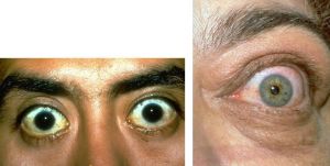
Upper Eyelid Retraction
All content on Eyewiki is protected by copyright law and the Terms of Service. This content may not be reproduced, copied, or put into any artificial intelligence program, including large language and generative AI models, without permission from the Academy.
Disease Entity
International Classification of Disease (ICD)
ICD-9-CM 374.41 Lid retraction or lag
ICD-10-CM H02.531 Eyelid retraction right upper eyelid
ICD-10-CM H02.534 Eyelid retraction left upper eyelid
Background
Upper eyelid retraction is defined by abnormally high resting position of the upper lid. This produces visible sclera between the eyelid margin and corneal limbus, which produces the appearance of a stare with an accompanying illusion of exophthalmos. Eyelid retraction can lead to lagophthalmos and exposure keratitis, which can cause mild ocular surface irritation to vision-threatening corneal decompensation. The most common causes of upper eyelid retraction include thyroid eye disease, recession of superior rectus muscle, and contralateral ptosis.
Anatomy
The eyelid retractors in the upper eyelid are the levator palpebrae superioris muscle and Müller’s muscle. The upper eyelid normally rests 1 to 2 mm below the corneoscleral limbus. Upper eyelid retraction is present when the upper eyelid is displaced superiorly with exposed sclera between the eyelid margin and corneoscleral limbus.
Clinical Exam
Upon examination of the patient, observe for upper scleral show. Clinical measurements can be used to assess for eyelid asymmetry and retraction. The distance from upper eyelid margin to corneal light reflex (margin reflex distance, MRD1) can be used to assess for elevated upper eyelid position. MRD1 is normally 4-5mm and may be increased in patients with upper eyelid retraction. Additionally, the midpupil to upper lid margin distance (MPLD) can be used to assess for lid retraction. An MPLD greater than 5.3 mm is considered as eyelid retraction. [1][2]Attention to MLPD asymmetry is also important. If asymmetry greater than 1.0 mm and if both eyelids are within normal range (3.5 – 5.5 mm), the differential diagnosis is between retraction of the higher eyelid and ptosis of the lower eyelid. [1]
Etiologies
The most common cause of unilateral or bilateral upper eyelid retraction is Graves’ ophthalmopathy, or thyroid eye disease. Early in Graves’ disease, eyelid malposition may result from increased sympathetic activity. With time, the levator palpebrae superioris and Müller’s muscle become hypertrophic, fibrotic, and adherent to orbital tissues. Patients with thyroid eye disease often have associated globe proptosis and lid lag along with eyelid retraction. Lower lid retraction is usually not seen in patients with Graves’ ophthalmopathy without concomitant retraction of the upper lids. Upper eyelid retraction in thyroid eye disease often has temporal flare, when retraction is more pronounced at the lateral aspect of the eyelid. [3]
If thyroid eye disease has been ruled out, Bartley proposed three categories of upper eyelid retraction: neurogenic, myogenic, and mechanistic causes.[4]
Congenital
- Congenital eyelid retraction is a rare condition characterized by abnormally elevated upper or lower eyelids present at birth, often leading to misdiagnosis or unnecessary investigations It affects one side in most cases and is usually recognized by cosmetic asymmetry, with symptoms like redness or soreness being less common and related to corneal exposure.[5]
- The condition is congenital and remains stable throughout life without progressive changes, as confirmed by patient history, photographs, and clinical follow-ups.
- The underlying cause is unclear but may involve congenital abnormalities in the levator muscle, its innervation, or associated structures, such as a thicker or shorter aponeurosis or orbital septum.
- There is no history of thyroid issues, trauma, or systemic diseases in most cases, and the affected eyelids often exhibit normal levator function, with some showing lagophthalmos on downgaze or associated mild squints.
Neurogenic
- The eye-popping reflex, which is sudden widening of the palpebral fissures in response to sudden reduction of ambient lighting, is present in normal infants during 14-18 weeks of age.
- Unilateral acquired ptosis (associated with levator aponeurosis dehiscence or disinsertion) can cause contralateral eyelid retraction. [6] This is due to Hering’s law of equal innervation. As the patient strains to elevate the ptotic lid, increased firing of neurons results in the overelevation of the contralateral lid. [7] To diagnose, cover or elevate the ptotic eyelid and observe for improvement of the eyelid retraction.
- Collier’s sign describes lid retraction that occurs from dorsal midbrain syndrome caused by neurological diseases such as pinealoma, hydrocephalus, subthalamic or midbrain arteriovenous malformations, disseminated sclerosis and encephalitis.[4]
- Parinaud syndrome is the combination of lid retraction, paralysis of vertical gaze, convergence retraction nystagmus on attempted upgaze and pupillary light near dissociation.[8]
- Paradoxical lid retraction may occur in myasthenia gravis.[9]
Myogenic
- Thyroid Eye Disease – see above
- Congenital upper eyelid retraction
- Postsurgical – superior rectus recession, blepharoptosis repair, enucleation
Mechanistic
- Mechanical causes of upper eyelid retraction often resolve with correction of the underlying abnormality. Prominence of the globe can occur with high myopia, buphthalmos, proptosis (orbital mass or idiopathic), craniosynostosis, orbital floor fractures (upper lid retracted from traction on connective tissue sheath), and as a postoperative finding from scleral buckle surgery, blepharoplasty, orbicularis myectomy, and glaucoma filtering operation with prominent bleb.
- Cutaneous scarring from eyelid neoplasms ,herpes zoster ophthalmicus, atopic dermatitis, scleroderma or burns can “distract” one or both eyelids from normal position.
- Blowout fractures of the orbital floor may cause upper eyelid retraction on either neurogenic or mechanistic basis – hypotropia of the globe can stimulate increased innervation of the superior rectus and levator palpebrae superioris muscles or traction of the connective sheath of the levator can elevate the upper lid mechanistically.
- Contact lens use may be associated with upper lid retraction from possible mechanical irritation of the palpebral conjunctiva.[4]
Clinical Significance and Management
Eyelid retraction can cause lagophthalmos and subsequent corneal and ocular surface disease, from dry eye symptoms to exposure keratopathy. Exposure keratopathy may progress to corneal ulceration and perforation. Thus, management of eyelid disease is vital to preserve vision. Treatment of upper eyelid retraction is aimed at correcting the underlying cause. If thyroid disease is suspected, serological tests should be ordered for thyroid hormone levels, thyrotropin receptor antibodies, and orbital imaging studies.[1] Ocular lubrication using artificial tears, ointments or punctal plugs to relieve irritation from corneal exposure can be used in mild cases of upper eyelid retraction. Mild eyelid retraction in thyroid eye disease can resolve spontaneously with time.
There are a variety of surgical techniques to correct eyelid retraction if the condition persists or the eyelid retraction causes an immediate threat to the cornea or vision. These techniques include release or recession of eyelid retractors, with or without use of spacers or grafts. Surgical intervention ranges from temporary suture tarsorrhaphy for ocular surface protection to eyelid-lengthening procedures to correct retraction and decrease scleral show.
If temporary eyelid closure is required to reduce keratopathy or to treat corneal ulceration, suture tarsorrhaphies with foam bolsters may be used until permanent surgery can be performed.[10] Temporary suture tarsorrhaphies may be placed using one or more double-armed 5-0 nylon sutures placed in mattress fashion with foam bolsters to protect the eyelid skin.
Recession of the upper eyelid can also be performed. Measurement of eyelid retraction should be stable for at least 6 months prior to surgery in thyroid eye disease. Upper eyelid retraction can be corrected by excision or recession of Müller’s muscle, recession of the levator aponeurosis with or without hang-back sutures or other spacer, measured myotomy of the levator muscle, or full-thickness transverse blepharotomy. Upper eyelid spacers include fascia lata, donor sclera, ear cartilage, or alloplastic materials. [11][12][13][14][15]
To address the possibility of overcorrection or undercorrection, patients are usually examined at one week after surgery. At the one week visit swelling is reduced, and wounds may be teased open with a new incision. Dissected tissue can be located with a minimum of bleeding under local anesthesia, and corrections to lid height and contour may be made between one to two weeks. Undercorrections rarely improve with decreased swelling and should be addressed at the earliest convenient time. Overcorrections of the eyelid height may improve as postoperative swelling subsides, and observation for an additional week may be considered prior to revision. [10]
When the prerequisite for surgical correction for eyelid retraction is not met (stable measurement of upper eyelid retraction < 6 months, euthyroid status) or if the patient does not desire surgical intervention, there are therapeutic alternatives. Transconjunctival Botulinum toxin A injections have been used for the medical management of upper eyelid retraction due to thyroid eye disease.[16] Additionally, triamcinolone acetonide deep fornix and subconjunctival injections and hyaluronic acid filler subconjunctival injection have been described for the treatment for upper eyelid retraction in thyroid eye disease. [17][18]
Summary
Upper eyelid retraction presents with an elevated resting position of the upper lid with subsequent scleral show. Eyelid retraction can lead to lagophthalmos and exposure keratitis, which can cause mild ocular surface irritation to vision-threatening corneal decompensation. There are various etiologies for upper eyelid retraction. Management should be aimed at correcting the underlying cause and workup for thyroid disease. Ocular lubrication with eyedrops and ointments is important to protect the ocular surface and cornea. Various therapeutic injections are available for upper eyelid retraction management prior to surgical intervention. Surgical planning requires consideration of functional and cosmetic concerns.
References
- ↑ 1.0 1.1 1.2 Bartley GB, Gorman CA. Diagnostic Criteria for Graves’ Ophthalmopathy. Am J Ophthalmol. 1995;119(6):792-795.
- ↑ Nerad JA, Carter KD, Alford M. Section 5 - Disorders of the Eyelid: Blepharoptosis and Eyelid Retraction. In: Nerad JA, Carter KD, Alford M, eds. Oculoplastic and Reconstructive Surgery. Mosby; 2008:101-129. Accessed December 1, 2021.
- ↑ Nerad JA, Carter KD, Alford M. Section 5 - Disorders of the Eyelid: Blepharoptosis and Eyelid Retraction. In: Nerad JA, Carter KD, Alford M, eds. Oculoplastic and Reconstructive Surgery. Mosby; 2008:101-129. Accessed December 1, 2021.
- ↑ 4.0 4.1 4.2 Bartley GB. The Differential Diagnosis and Classification of Eyelid Retraction. Ophthalmology. 1996;103(1):168-176.
- ↑ Collin JR, Allen L, Castronuovo S. Congenital eyelid retraction. Br J Ophthalmol. 1990 Sep;74(9):542-4. doi: 10.1136/bjo.74.9.542. PMID: 2393644; PMCID: PMC1042204.
- ↑ Meyer DR, Wobig JL. Detection of Contralateral Eyelid Retraction Associated with Blepharoptosis. Ophthalmology. 1992;99(3):366-375.
- ↑ Gay AJ, Salmon ML, Windsor CE. Hering’s law, the levators, and their relationship in disease states. Arch Ophthalmol Chic Ill 1960. 1967;77(2):157-160.
- ↑ Rismondo V, Borchert M. Position-dependent Parinaud’s Syndrome. Am J Ophthalmol. 1992;114(1):107-108.
- ↑ Kansu T, Subutay N. Lid retraction in myasthenia gravis. J Clin Neuroophthalmol. 1987;7(3):145-150.
- ↑ 10.0 10.1 Chang EL, Rubin PAD. Upper and lower eyelid retraction. Int Ophthalmol Clin. 2002;42(2):45-59.
- ↑ Ben Simon GJ, Mansury AM, Schwarcz RM, Modjtahedi S, McCann JD, Goldberg RA. Transconjunctival Müller muscle recession with levator disinsertion for correction of eyelid retraction associated with thyroid-related orbitopathy. Am J Ophthalmol. 2005;140(1):94-99.
- ↑ Demirci H, Hassan AS, Reck SD, Frueh BR, Elner VM. Graded full-thickness anterior blepharotomy for correction of upper eyelid retraction not associated with thyroid eye disease. Ophthal Plast Reconstr Surg. 2007;23(1):39-45.
- ↑ Ceisler EJ, Bilyk JR, Rubin PAD, Burks WR, Shore JW. Results of Müllerotomy and Levator Aponeurosis Transposition for the Correction of Upper Eyelid Retraction in Graves Disease. Ophthalmology. 1995;102(3):483-492.
- ↑ Collin JR, O’Donnell BA. Adjustable Sutures in Eyelid Surgery for Ptosis and Lid Retraction. Br J Ophthalmol. 1994;78(3):167-174.
- ↑ Grove AS. Eyelid Retraction Treated by Levator Marginal Myotomy. Ophthalmology. 1980;87(10):1013-1018.
- ↑ Ozturk Karabulut G, Fazil K, Saracoglu Yilmaz B, et al. An algorithm for Botulinum toxin A Injection for Upper eyelid retraction associated with thyroid eye disease: long-term results. Orbit. 2021;40(5):381-388.
- ↑ Luo LH, Gao LX, Wang W, Miao H, Ma XM, Li DM. [Triamcinolone acetonide deep fornix injection for the treatment of upper eyelid retraction in patients with thyroid-associated ophthalmopathy]. Zhonghua Yan Ke Za Zhi Chin J Ophthalmol. 2020;56(7):524-529.
- ↑ Hassan Hussien M, Abd El-Wahed Hassan E, El-Haddad NSEDM. Comparison between hyaluronic acid filler and botulinum toxin type A in the treatment of thyroid upper eyelid retraction. Ther Adv Ophthalmol. 2020;12.


