Congenital and Acquired Epiblepharon
All content on Eyewiki is protected by copyright law and the Terms of Service. This content may not be reproduced, copied, or put into any artificial intelligence program, including large language and generative AI models, without permission from the Academy.
Disease Entity
Congenital and acquired Epiblepharon
Disease
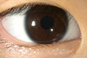
Epiblepharon is commonly a developmental eyelid/eyelid margin condition characterised by overriding of the anterior lamella at the lid margin. Typically it presents as an abnormal horizontal skin fold at the lower eyelid margin, resulting in misdirected lashes towards the ocular surface - against both the conjunctiva and cornea. More frequently seen in children and young adults of East Asian descent, it is much more common in the lower eyelids compared to the upper eyelids (Fig 1)
Primarily a congenital or developmental condition[1] [2], an Acquired Epiblepharon may also be seen in east Asian children and adults following orbital conditions like orbital tumors (primary or secondary), Thyroid Eye Disease (TED), acute orbital hematoma, and other causes of unilateral or bilateral proptosis. Other ethnic groups with some east Asian characteristics, like Hispanics, Latin americans and Inuits etc may also manifest this. Spectrum of clinical consequences include wet eyes with tearing, light sensitivity, punctate to diffuse keratopathy - conjunctival and/or corneal epithelial defects, infections and ulceration, thinning and scarring. It is often missed and misdiagnosed in non-east Asian populations and frequently mismanaged as well.
Etiology
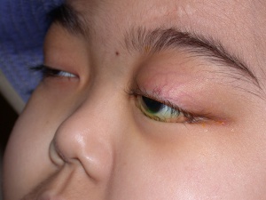
While in East Asian children it is frequently developmental and an exaggeration of normal anatomy, in other ethnic groups it is a true abnormality. Developmental epiblepharon results from absent or poor attachment of the lower and/or upper eyelid retractors to the overlying skin and subcutaneous tissues. This, along with the circumferential sphincter-like orbicularis oculi fibres, results in overriding of the preseptal over the tethered pretarsal orbicularis causing a skin fold, which in turn results in an inward or posterior rotation of the eyelashes against the medial bulbar conjunctival and infero-medial and central cornea. A low attachment of the orbital septum to the upper (elevator aponeurosis) and lower eyelid retractors also allows the orbital fat to prolapse towards the lid margin causing imbalance between the anterior and posterior lamellae with consequences at the anterior lid margin (Fig 2). Significantly long and thick eyelashes with persistent conjunctival and corneal touch result in varying degrees of symptomatic kerato-conjunctivopathy. Long standing keratopathy may even result in irreversible corneal scarring. Other factors such as orbicularis hypertrophy, relatively poor attachment of the pretarsal orbicularis, and pre-existing tight orbits play a role as well..
Secondary or Acquired Epiblepharon often arises from secondary increases in intraorbital pressure eg. Congenital Glaucoma[3], Thyroid Eye Disease[4] [5], orbital tumors (Fig 2), acute orbital hemorrhage, obesity etc, in patients at risk with tight eyelids and orbits typical of East Asians.
Risk Factors
The well known and most common risk factor are children and young adults of East Asian descent, typically with parents who have either a weak or absent upper eyelid creases ('double eyelids'). Thus children with absent or poor eyelid creases and those with higher Body Mass Index (BMI) - overweight and obesity, are at greater risk[6] [7] . While patients with soft finer lashes may be minimally symptomatic, those with thick stubby lashes are more symptomatic from resultant keratopathy. The tighter eyelids in East Asian orbits is also a risk factor.
Types of epiblepharon
Primary, congenital or developmental epiblepharon: This is the most common type typically seen in children and young adults of East Asian descent. Occasionally patients may present at a much later age having had symptoms for years but misdiagnosed as other conditions. In other ethnic groups, this is a transient developmental abnormality which usually resolves spontaneously.
Secondary or acquired epiblepharon often arises from secondary increases in intraorbital pressure eg. Thyroid Eye Disease(TED), orbital tumors (Fig 2), acute orbital hemorrhage, obesity etc, in patients at risk with tight orbits and eyelids typical of East Asians, but may be seen at any age and some other ethnicities as well. It may be transient if and when the underlying orbital condition resolves eg. haemorrhage, acute oedema, etc or long lasting.
Upper and Lower Epiblepharon: While lower epiblepharon is more common and symptomatic, upper epiblepharon may be present in 15-20% of children and young adults who have lower epiblepharon. Thus examining children in primary gaze and in adduction, down gaze (lower epibelpharon) and upgaze (upper epiblepharon) is crucial for complete assessment along with assessment of corneal staining.
Severity and Grading
Khwarg et al (1997)have proposed a practical classification based on the degree and extent of eyelid skin fold, lash-corneal touch and keratopathy.[8]
Primary Prevention
Developmental epiblepharon - none. However clinical signs may be partially ameliorated in children with BMI reduction. Acquired epiblepharon- prevention and better management of underlying orbital condition with tight orbits in East Asians- thyroid eye disease (medical or surgical decompression), orbital haemorrhage (medical or surgical evacuation), Orbital tumors (orbitotomy with tumor removal).
Diagnosis
The diagnosis of epiblepharon is primary clinical, based on history, clinical suspicion and dynamic slit lamp exam of the eye with and without corneal staining. It should however be differentiated from entropion, trichiasis and/or distichiasis. Lower epiblepharon may be manifest in primary gaze, but more typically in down gaze and in adduction. Likewise upper epiblepharon is more obvious in upgaze. A milder form of upper epiblepharon is lash ptosis with which may predispose to an epiblepharon on forced blink or eye closure. (Fig 4a, 4b)
History
The classic history and presentation is a young child of East Asian descent, who presents with rubbing, frequent blinking, tearing and/or discharge from one or both eyes, where other conditions like nasolacrimal duct obstruction and allergic conjunctivitis have been ruled out. Most children may have received a course of topical antibiotics or antiallergics without much response. Occasionally, younger and older adults may have had similar symptoms since childhood or even present with having had repeated epilations or trimmed eyelashes.
Patients with an acquired epiblepharon often present with typical clinical features of an underlying orbital disease i.e., thyroid eye disease, orbital tumor or inflammatory disease, orbital haemorrhage.
Physical Examination
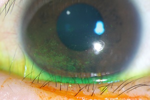
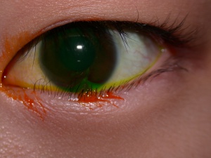
A high degree of suspicion is often necessary to make a diagnosis. External examination may reveal a narrow palpebral fissure, weak or absent upper eyelid crease, any of the variants of medial epicanthal fold and occasionally wet sticky eyes. The eyelid margin and the posterior lamella are in their normal position. Slit lamp exam often shows a variable degree of the eyelid skin overriding the lower eyelid margin. The lashes are directed against the medial bulbar conjunctiva and infero-medial cornea, especially in down gaze and adduction. Fluorescein staining of the cornea and conjunctiva is usually positive varying from fine superficial punctate keratopathy to confluent coarse keratopathy. Rarely, in older children and adults with long standing duration, corneal scarring may be present as well
Upper epiblepharon manifests as misdirected medial eyelashes with intermittent corneal-conjunctival touch especially in upward and inward gazes. The typical eyelid skin fold is not seen with upper epiblepharon unless in severe cases.
Varying forms of medial epicanthal folds may also be present, which may or may not be addressed at the time of surgical management.
Symptoms
In early stages and very young children, this is often asymptomatic. Common symptoms include Irritation, foreign body sensation, frequent blinking or rubbing of eyesitching, Tearing with/without discharge
Signs
The most common signs of Epiblepharon include:
- Variable lower eyelid skin fold, more prominent medially
- Eyelid margin and posterior lamella are in normal position
- Lash corneal-conjunctival touch, especially in downgaze and adduction in lower epiblepharon and up gaze in upper epiblepharon.
- Varying spectrum and severity of medial epicanthal fold may also be present.
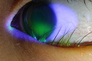
- Fine, intermediate to coarse and confluent keratopathy that stains with fluorescein. (Fig 3a, 3b) . Spectrum of corneal changes include fine to coarse epitheliopathy with staining, recurrent infections, thinning and scarring.
Associated orbital signs such as proptosis and increased orbital pressure may be present in acquired epiblepharon.
Abnormal eyelashes may be bent or broken. Occasional stumps from clipped lashes may be seen (Fig 4)
Cornea scarring in severe and long standing keratopathy.
Upper epiblepharon typically manifests with a weak or absent eyelid crease with downward rotation of the eyelashes with lash-corneal touch in primary or up gaze.
Thus these symptoms are mistaken for chronic and allergic conjunctivitis, congenital nasolacrimal duct obstruction in very young children.
Clinical diagnosis
The diagnosis of Epiblepharon is clinical. It may be easily missed in early and mild disease in very young children especially when not looked for. In a symptomatic child, the presence of eyelashes against the cornea and conjunctival in any gaze is diagnostic of this disease. Associated keratopathy determines the threshold for medical and when indicated surgical intervention.
Diagnostic procedures
Epiblepharon is primarily a clinical diagnosis. The main diagnostic procedure is fluorescein staining with documentation by slit lamp photography regarding the nature of severity of keratopathy.
Laboratory test
None
Differential diagnosis
The differential diagnosis for Epiblepharon and their differentiating features are as follows:
- Trichiasis is an acquired condition resulting from a malpositioned eyelash misdirected against the globe.
- Distichiasis is a developmental condition with an aberrant additional row of eyelashes arising from Meibomian gland orifices.
- Entropion of lower eyelid: True inward rotation of posterior lamella and lid margin.
- Nasolacrimal duct obstruction: symptoms since birth, more commonly unilateral, tearing and discharge.
- Allergic conjunctivitis: stringy mucus discharge, conjunctival injection, papillary conjunctival changes.
Natural history
Most children are asymptomatic as the eyelashes are fine and may not induce keratopathy even with lash-corneal touch. As the lashes get thicker and longer and with increasing skin fold, especially in those with higher body mass index. Keratopathy may evolve from asymptomatic fine punctate erosions to confluent keratopathy, and even corneal scarring. A higher astigmatism rate has also been reported[9]Interestingly, with the development of targeted therapy for TED, regression of Acquired Epiblepharon has been reported following treatment.[9]
Management
Developmental epiblepharon: Children may be initially managed conservatively with lubricants and regular follow up until resolution. Indications for surgical intervention include persistent symptoms, significant keratopathy despite conservative management or inability to frequently apply lubricants especially in young children..
Transient epiblepharon may be symptomatically managed with copious lubrication or temporising procedures until underlying orbital disease is addressed.
Minimally invasive procedures include tissue filler injection into the valley of the skin fold or everting sutures result in transient improvement albeit with higher recurrence rates.
Surgical management is often indicated when symptoms and signs are persistent and severe. Although both transconjunctival and transcutaneous procedures have been described, the latter is most commonly performed and reportedly most successful.
Lower epiblepharon: The most commonly recommended procedures is the modified Hotz procedure, which involves excision of an ellipse of redundant skin mostly medially, with sutures placed to ensure outward rotation of the eyelashes with wound closure.[10] [11] other modifications include lower eyelid retractor release with lash margin rotation sutures have also been described.
Acquired epiblepharon: While most acquired epiblepharon may resolve with resolution or treatment of the underlying primary orbital pathology eg. Orbital tumors, hematoma, etc, Apart from lash repositioning procedures, addressing tight lower eyelids may be considered.[12] In more severe cases, orbital decompression may be indicated as it addresses the primary underlying condition.[13]
Outcomes and complications
Success rates are over 95% with high satisfaction rates. Potential complications include undercorrections, especially medially, overcorrections resulting in transient ectropion and visible lid scar, all of which can be prevented with a good technique.
Prognosis
The prognosis is generally excellent in patients who have minimal intermittent lash-corneal touch with fine lashes and those who receive adequate and timely surgical correction. Revision and repeat surgeries are rarely indicated.
References
- ↑ Sundar G, Young SM, Tara S, et al. Epiblepharon in East Asian patients: the Singapore experience. Ophthalmology 2010; 117(1): 184-9.
- ↑ Revere KE, Foster JA, Katowitz WR, Katowitz JA. Developmental Eyellid Abnormalities in Pediatric Oculoplastic Surgery 2nd Ed. Ed Katowitz JA, Katowitz WR. Springer 2018. ISBN 978-3-319-60812-9.
- ↑ Kim N, Yoo YJ, Choung HK, Khwarg SI. Epiblepharon in congenital glaucoma: case-control study. Br J Ophthalmol. 2017 Dec;101(12):1654-1657.
- ↑ Chang EL, Hayes J, Hatton M, et al. Acquired lower epiblepharon in patients with thyroid eye disease. Ophthal Plast Reconstr Surg 2005; 21: 192-6.
- ↑ Park SW, Khwarg SI, Kim N, Lee MJ, Choung HK. Acquired lower eyelid epiblepharon in thyroid-associated ophthalmopathy of Koreans. Ophthalmology. 2012 Feb;119(2):390-5.
- ↑ Takahashi Y, Kono S, Vaidya A, Yokoyama T, Kakizaki H. Severe corneal involvement secondary to congenital lower eyelid epiblepharon. Graefes Arch Clin Exp Ophthalmol. 2022 Dec 23.
- ↑ Medsinge A, Duncan K, Yu JY. The association between epiblepharon and obesity: an experience at tertiary care center in Western Pennsylvania/North America. Int Ophthalmol. 2021 Mar;41(3):991-994
- ↑ Khwarg SI, Lee YJ. Epiblepharon of the lower eyelid: classification and association with astigmatism. Korean J Ophthalmol 1997; 1:111-7.
- ↑ Jump up to: 9.0 9.1 Chen KW, Phelps PO. Acquired Epiblepharon Alleviated With Teprotumumab Treatment in Active Thyroid Eye Disease Patient. Ophthalmic Plast Reconstr Surg. 2021 Nov-Dec 01;37(6):e195-e196.
- ↑ Kim JS, Jin SW, Hur MC, et al. The clinical charateristics and surgical outcomes of epiblepharon in Korean children: a 9-year experience. J Ophthalmol 2014; 156-601.
- ↑ Woo KI, Kim YD. Management of epiblepharon: state of the art. Curr Opin Ophthalmol. 2016 Sep;27(5):433-8.
- ↑ Zhao J, Hodgson NM, Chang JR, Campbell AA, McCulley TJ. Thyroid Eye Disease-Related Epiblepharon: A Comparative Case Study. Asia Pac J Ophthalmol (Phila). 2020 Jan-Feb;9(1):44-47.
- ↑ Almousa R, Sundar G. Acquired Epiblepharon Treated by Lateral Orbital and fat Decompression. Middle East Afr J Ophthalmol. 2011 Jan;18(1):80-1.


