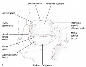
Suspensory Ligament of the Eye (Lockwood’s Ligament)
All content on Eyewiki is protected by copyright law and the Terms of Service. This content may not be reproduced, copied, or put into any artificial intelligence program, including large language and generative AI models, without permission from the Academy.
First described by surgeon Charles Barrett Lockwood, the suspensory ligament of the eye forms a support hammock below the globe extending from the lateral orbital tubercle to the medial canthal tendon.[1] [2]It is formed by the fusion of the capsulopalpebral fascia just anterior to the inferior oblique. The caspulopalpebral fascia is a lower eyelid retractor which extends anteriorly from the inferior rectus, splitting around the inferior oblique to fuse with the orbital septum and insert on the inferior border of the inferior tarsal plate.[3][4]
Lockwood’s ligament supports the position of the globe in its position within the orbit, preventing any displacement. The globe is also supported by the medial and lateral check ligaments, septum, and adipose tissue.[5]The course of the ligament determines the shape of the lower conjunctival fornix.[6]
Weakening and descent of Lockwood ligament may cause the globe to descend, reducing the space between the globe and the floor of the orbit.[5]
Components
Kakizaki et al in 2004 described 3 components of Lockwood’s ligament: the main Lockwood ligament, the arcuate expansion, and the inferior ligament.[7]
The main Lockwood ligament inserts onto Whitnall tubercle on the lateral orbital wall.[2] The analog of the main Lockwood ligament in the upper eyelid is Whitnall's ligament. The arcuate expansion of Lockwood ligament typically defines the boundaries of the middle and lateral fat pads of the lower lid and inserts on the inferolateral orbital rim.[8]
References
- ↑ Camirand A, Doucet J, Harris J. Anatomy, pathophysiology, and prevention of senile enophthalmia and associated herniated lower eyelid fat pads. Plast Reconstr Surg. 1997;100:1535-1546.
- ↑ 2.0 2.1 Lockwood CB. The anatomy of the muscles, ligaments, and fasciae of the orbit, including an account of the capsule of Tenon, the check ligaments of the recti, and the suspensory ligament of the eye. J Anat Physiol. 1885;20:1-25.
- ↑ Fink WH. Ligament of lockwood in relation to surgery of the inferior oblique and inferior rectus muscles. Arch of Ophthalmol. 1948;39(3):371-82.
- ↑ Hawes MJ, Dortzbach RK. THe microscopic anatomy of the lower eyelid retractors. Arch Ophthalmol. 1982;100:1313.
- ↑ 5.0 5.1 Kakizaki H, Malhotra R, Madge SN, Selva D. Lower eyelid anatomy - an update. Ann Plast Surg 2009;63:344-351.
- ↑ Kakizaki H, Iwaki M. A new trapezoid lower eyelid clamp to shape the lower conjunctival fornix. Eur J Plast Surg. 2008;31:161-164.
- ↑ Kakizaki H, Zako M, Nakano T, Asamoto K, Iwaki M. Three ligaments reinforce the lower eyelid. Okajimas Folia Anat Jpn. 2004;81:97-100.
- ↑ Yousif NJ, Sonderman P, Dzwierzynski WW, et el. Anatomic considerations in transconjunctival blepharoplasty. Plast Reconstr Surg. 1995;96:1271-1276.


