Superior Transverse Ligament (Whitnall’s Ligament)
All content on Eyewiki is protected by copyright law and the Terms of Service. This content may not be reproduced, copied, or put into any artificial intelligence program, including large language and generative AI models, without permission from the Academy.
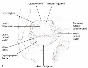
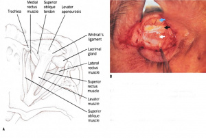
Whitnall’s ligament was first described in 1910 by Dr. Samuel Ernest Whitnall as a superior transverse ligament above the musculotendinous junction of the levator palpebrae superioris.[1][2] It is formed by a collection of muscle sheaths from the levator palpebrae superioris muscle that assemble into a ligament in the area where the levator palpebrae muscle becomes an aponeurosis.
It attaches to the superior rectus muscle tendon sheath, the medial side of the trochlea, and to the lateral orbital margin.[3] Functionally, the ligament is composed of two distinct parts: the transverse superior fascial expansion inferior to the levator palpebrae superioris and the superior transverse ligament superior to the levator palpebrae superioris.[4][5]
Whitnall’s Ligament acts as a suspensory ligament to support the upper eyelid, the lacrimal gland, and the superior orbit.[3][6] It also acts as a fulcrum, changing the direction of the levator aponeurosis from anterior-posterior to superior-inferior.[3][4] When Whitnall’s ligament functionally fails, the levator palpebrae muscle becomes prolonged and sunken, causing decreased elevation of the eyelid.[3]
References
- ↑ Lukas JR, Priglinger S, Denk M, Mayr R. Two fibromuscular transverse ligaments related to the levator palpebrae superioris: Whitnall's ligament and an intermuscular transverse ligament. Anat Rec. 1996 Nov;246(3):415-22. doi: 10.1002/(SICI)1097-0185(199611)246:3<415::AID-AR13>3.0.CO;2-R. PMID: 8915464.
- ↑ Whitnall SE. A ligament acting as a check to the action of the levator palpebrae superioris muscle. J Anat Physiol. 1910;45:131–139.
- ↑ Jump up to: 3.0 3.1 3.2 3.3 Lim HW, Paik DJ, Lee YJ. A cadaveric anatomical study of the levator aponeurosis and Whitnall's ligament. Korean J Ophthalmol. 2009;23(3):183-187.
- ↑ Jump up to: 4.0 4.1 Codère F, Tucker NA, Renaldi B. The anatomy of Whitnall ligament. Ophthalmology. 1995 Dec;102(12):2016-9. doi: 10.1016/s0161-6420(95)30761-0.
- ↑ Ettl A, Priglinger S, Kramer J, Koornneef L. Functional anatomy of the levator palpebrae superioris muscle and its connective tissue system. Br J Ophthalmol. 1996 Aug;80(8):702.
- ↑ Anderson RL, Dixon RS. The role of Whitnall's ligament in ptosis surgery. Arch Ophthalmol. 1979;97:705–707.


