Pulfrich Phenomenon
All content on Eyewiki is protected by copyright law and the Terms of Service. This content may not be reproduced, copied, or put into any artificial intelligence program, including large language and generative AI models, without permission from the Academy.
Phenomenon Entity
The Pulfrich phenomenon (also known as the Pulfrich effect) is a neuro-ophthalmic observation in which two-dimensional (2-D) objects are perceived to be three-dimensional (3-D) due to slight discrepancies in signal transmission time between a set of eyes and the visual cortex [1]. The relative delay in conduction in one of the optic pathways causes a discrepancy in visual perception between the two eye and may cause difficulties in daily activities, such as sports and driving [1][2].
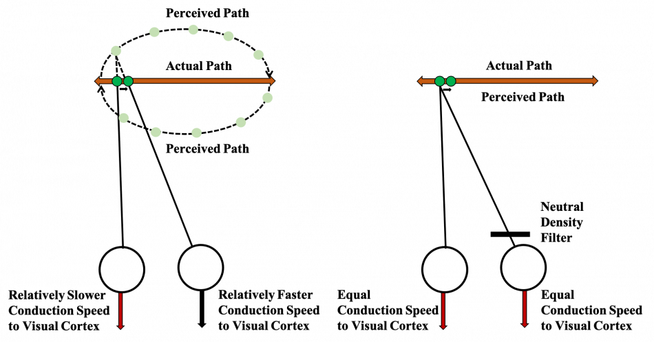
The Pulfrich phenomenon is classically described in demyelinating optic neuritis but has also been observed in other ocular conditions (e.g., cataracts) [1][3]. Individuals who experience this effect often notice it with moving objects that travel within a single plane. A well-known illustration to elucidate this phenomenon involves a patient viewing a pendulum swinging side-to-side [4]. As the pendulum swings side-to-side, an unaffected binocular individual will track the motion of the pendulum with the same rate of retinal signal transmission to the visual cortex, thus interpreting the motion as uniplanar. An individual who experiences the Pulfrich Phenomenon has a relative conduction delay in one of the optic pathways when viewing the pendulum. The perceived distance of the pendulum differs when processed by the visual cortex, presenting with a misjudgment in object depth. Therefore, the swinging pendulum appears to be moving in a circular direction due to this disparity, causing a 3D image from a single planar movement. This delay can also be elucidated with a uniocular dark lens, also known as a neutral density filter, which can cause a delay in signal latency to the visual cortex. Studies have shown that a factor of ten reduction in unilateral retinal illumination produces a 15-millisecond delay (Figure 1.) [5].

History of the Pulfrich Phenomenon
The Pulfrich phenomenon was first described in 1922 by Carl Pulfrich, an expert in stereoscopy [6]. Pulfrich had named the phenomenon “stereo effect” but over time the phenomenon has become better known as the “Pulfrich effect”. Since the documentation of this effect and the ability to reproduce it with filters, the Pulfrich phenomenon has played a role in major media in the induction of 3-D visual effects for 2-D images (e.g., 3-D glasses).
Etiology and Treatment
The Pulfrich phenomenon is a result of a delay in afferent signal conduction to the visual cortex [7]. Thus, there are various etiologies behind this effect. A common pathology that displays the Pulfrich effect is demyelinating optic neuritis (e.g., multiple sclerosis). In multiple sclerosis, there is an autoimmune demyelination of the central nervous system due to inflammatory attacks against oligodendrocytes, including the oligodendrocytes that myelinate the optic nerves. The primary role of myelin is to increase the speed of electrical conduction across the axon. If one of the optic nerves is affected in multiple sclerosis, this will cause a discrepancy in the electrical conduction of the optic pathway to the visual cortex, resulting in the Pulfrich phenomenon. Interestingly another reported cause of the Pulfrich phenomenon is unilateral cataract [8]. A study consisting of 29 patients with unilateral cataract and contralateral pseudophakia was conducted to analyze the Pulfrich effect with a computer image of a pendulum. Rotation of the uniplanar pendulum was seen in these subjects but was also neutralized with density filters in the pseudophakic eye.
In most cases, treatment of the underlying condition causing the unilateral conduction delay resolves the symptoms (e.g., cataract surgery for unilateral cataract). However, in some cases where the underlying etiology is irreversible, there have been treatments developed to reduce and/or eliminate this phenomenon. As previously mentioned, a neutral density filter over one eye may produce the Pulfrich effect in non-affected individuals by increasing the latency in retinal processing. In patients with Pulfrich phenomenon, a similar method with a neutral density filter can be applied to the unaffected or less affected eye [9]. A longitudinal study with uniocular contact lens or tinted spectacle in the lesser affected eye of Pulfrich phenomenon patients reported elimination of the effect for years [10]. This attenuation through a light-filtering lens allows for the signal transmission of the lesser affected eye to match the slower conduction of the more affected eye.
Confirmation Testing
While the Pulfrich phenomenon may have different etiologies, visual evoked potential (VEPs) may be used to confirm the visual latency that causes this effect [10][11]. VEPs measure evoked electrical potentials in the visual cortex after stimuli (e.g., flash) are presented to each eye [12]. These tests can measure the conduction speed after the flash and provide useful information in quantifying the current function of the optic nerve pathway.
Prognosis
The Pulfrich phenomenon can be treated by addressing the reversible causes of vision loss (e.g., cataracts) or by using a neutral density filter for irreversible causes (e.g., optic neuritis) [8][10]. In some cases, the underlying etiology for the Pulfrich phenomenon may progressively worsen further slowing the transmission in the affected eye and widening the disparity between the two optic nerve pathways [4]. Such cases may require changes in the neutral density filter for treating the Pulfrich Phenomenon [1][4].
Summary
Clinicians should be aware of the Pulfrich phenomenon and that it may present in patients with demyelinating optic neuritis or unilateral cataract. Treatment of the underlying etiology is generally sufficient, but some patients require neutral density filters to balance out the transmission asymmetry to relieve symptoms.
References
- ↑ Jump up to: 1.0 1.1 1.2 1.3 Farr, J., et al., The Pulfrich Phenomenon: Practical Implications of the Assessment of Cases and Effectiveness of Treatment. Neuroophthalmology, 2018. 42(6): p. 349-355 DOI: 10.1080/01658107.2018.1446537.
- ↑ Heron, G., M. McQuaid, and E. Morrice, The Pulfrich effect in optometric practice. Ophthalmic Physiol Opt, 1995. 15(5): p. 425-9.
- ↑ O'Doherty, M. and I. Flitcroft, An unusual presentation of optic neuritis and the Pulfrich phenomenon. BMJ Case Rep, 2009. 2009 DOI: 10.1136/bcr.08.2008.0647.
- ↑ Jump up to: 4.0 4.1 4.2 Sokol, S., The Pulfrich stereo-illusion as an index of optic nerve dysfunction. Surv Ophthalmol, 1976. 20(6): p. 432-4 DOI: 10.1016/0039-6257(76)90068-0.
- ↑ Lit, A., The magnitude of the Pulfrich stereophenomenon as a function of binocular differences of intensity at various levels of illumination. Am J Psychol, 1949. 62(2): p. 159-81.
- ↑ Pitz, A.P.E., The Historical Origin of the Pulfrich Effect: A Serendipitous Astronomic Observation at the Border of the Milky Way. Neuro-Ophthalmology, 2009 DOI: https://doi.org/10.1080/01658100802590829.
- ↑ Reynaud, A. and R.F. Hess, Interocular contrast difference drives illusory 3D percept. Sci Rep, 2017. 7(1): p. 5587 DOI: 10.1038/s41598-017-06151-w.
- ↑ Jump up to: 8.0 8.1 Scotcher, S.M., et al., Pulfrich's phenomenon in unilateral cataract. Br J Ophthalmol, 1997. 81(12): p. 1050-5 DOI: 10.1136/bjo.81.12.1050.
- ↑ Heron, G. and G.N. Dutton, The Pulfrich phenomenon and its alleviation with a neutral density filter. Br J Ophthalmol, 1989. 73(12): p. 1004-8 DOI: 10.1136/bjo.73.12.1004.
- ↑ Jump up to: 10.0 10.1 10.2 Heron, G., K.J. Thompson, and G.N. Dutton, The symptomatic Pulfrich phenomenon can be successfully managed with a colored lens in front of the good eye--a long-term follow-up study. Eye (Lond), 2007. 21(12): p. 1469-72 DOI: 10.1038/sj.eye.6702459.
- ↑ Heron, G., L. McCulloch, and N. Dutton, Visual latency in the spontaneous Pulfrich effect. Graefes Arch Clin Exp Ophthalmol, 2002. 240(8): p. 644-9 DOI: 10.1007/s00417-002-0501-z.
- ↑ Creel, D.J., Visually evoked potentials. Handb Clin Neurol, 2019. 160: p. 501-522 DOI: 10.1016/B978-0-444-64032-1.00034-5.

