Kestenbaum Index
All content on Eyewiki is protected by copyright law and the Terms of Service. This content may not be reproduced, copied, or put into any artificial intelligence program, including large language and generative AI models, without permission from the Academy.
Introduction
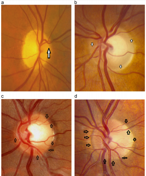
The Kestenbaum capillary number index is defined as the number of capillaries observed on the optic disc. The normal count is approximately 10. In optic atrophy, the number of these capillaries reduces to less than 6, while more than 12 suggests a hyperemic disc. [1] Kestenbaum's sign consists of a quantifiable measure of the vascular profile of the optic disc, which can aid to quantify the degree of optic nerve atrophy, as well as the diagnosis of borderline optic atrophy.[2]
Background
Kestenbaum's index was initially described in 1947 by Alfred Kestenbaum as a method to quantify the degree of optic atrophy. [2]The original reference describes the technique to perform the test: “In order to set a numerical value on the degree of atrophy, the vessels which pass over the margin of the disk may be counted. One starts at the twelve o'clock point and counts all vessels crossing the margin, counting separately the arterioles, the veins and the small vessels. “Small vessels” means the vessels which cannot be recognized as arteries or as veins. The number of vessels passing over the margin in normal eyes is fairly constant. Without dilatation of the pupil, usually 9 large vessels (4-5 veins and 4-5 arteries) and about 10 small vessels can be seen.”
Clinical application
Optic nerve atrophy develops as result from dysfunction and subsequent axonal degeneration at the optic nerve head, most commonly due to glaucomatous optic neuropathy and optic neuritis. Presence of optic nerve pallor has traditionally been considered a hallmark of optic nerve damage resulting from atrophy. The etiology of optic atrophy can often be difficult to establish. One study claimed that retrospective review of history and examination findings typically explain the etiology, when initially the diagnosis was unexplained, whereas neuroimaging is only diagnostic in approximately 20% of cases. This further demonstrates the relevance of clinical examination and the need for basic observation. [2]
In chronic open-angle glaucoma, disease progression is associated with vascular dysregulation in the form of local vasospasm and systemic hypertension. The surface of a healthy optic disc contains a large number of capillaries which are derived from branches of the retinal arterioles and are continuous with the peripapillary retinal capillaries.[2] These retinal capillaries are supplied by the central retinal artery, unless a cilioretinal artery is present, in 5–40% of patients. Kestenbaum's sign provides a quantifiable measure of the vascular appearance of the optic disc and would further support the contention that end stage disease has a underlying vascular etiology.[2]

Whilst the use of high-yield imaging technology to aid diagnosis and quantification of optic atrophy has an ever increasing role in the diagnostic criteria of these disorders, measuring capillary number count could be implemented as well to raise or lower suspicion of disc abnormality in a borderline case.[2] Although Kestenbaum's capillary number index has not been correlated with quantitative imaging techniques of optic nerve viability, such as OCT and fluorescein angiography, it can be used as a rapid and useful preliminary assessment of the optic nerve. This index may be integrated as a simple measure into the clinical evaluation of the optic nerve head for glaucoma.
References
- ↑ Devendra V. Venkatramani, Gangaprasad Muthaiah Amula, Rashmin A. Gandhi.Optic Atrophy. Dept. of Neuro Ophthalmology, Sankara Netralaya, Chennai
- ↑ Jump up to: 2.0 2.1 2.2 2.3 2.4 2.5 Hawkes ED, Neffendorf JE. Kestenbaum׳s capillary number test--a forgotten sign? Mult Scler Relat Disord. 2014 Nov;3(6):735-7. doi: 10.1016/j.msard.2014.09.087. Epub 2014 Oct 7. PMID: 25891554.

