Intratarsal Keratinous Cysts
All content on Eyewiki is protected by copyright law and the Terms of Service. This content may not be reproduced, copied, or put into any artificial intelligence program, including large language and generative AI models, without permission from the Academy.
Intratarsal keratinous cysts (IKCs) are benign eyelid cystic lesions that originate from the meibomian glands ducts. They are frequently misdiagnosed as chalazion. IKC has to be distinguished from other intratarsal lesions, because of different treatment methods. Complete surgical excision with adjacent tarsectomy or full-thickness eyelid resection is required to avoid recurrence.
Disease Entity
Disease
Intratarsal keratinous cysts (IKCs) are benign eyelid cystic lesions that arise from the ducts of the meibomian glands.[1] [2]They are supposed to be the third most common primary intratarsal lesions, after chalazia and sebaceous cell carcinoma.[1] Their nomenclature includes meibomian gland keratinous cyst, meibomian gland ductal cyst, intratarsal epidermal inclusion cysts, and tarsal cyst[3]. IKCs usually affect middle-aged individuals.
General Pathology
Histologically, cysts are lined by a multilayered-layered squamous epithelium (consisted of 4 to 5 layers of polygonal squamous cells and lacking granular layer), with undulating eosinophilic cuticle, filled with fluid keratin and typically having a thick non-inflamed fibrous wall.[2] IKCs show positive immunohistochemical staining for cytokeratin 5, cytokeratin17, carcinoembryonic antigen, epithelial membrane antigen, and cytokeratin19.[1][4]Immunohistochemical analysis helps differentiate IKCs from steatocystomas.
Pathophysiology
The pathogenesis of IKC development is unclear. Some authors suggest that IKCs develop secondary to trauma or surgery-related blockage of the meibomian ducts and subsequent squamous metaplasia. [5]However, the secretory acini of the meibomian glands already are squamous in nature[1] and IKCs can occur in absence of preceding trauma or surgery.[6] Other authors demonstrated that an IKC arises from a dilated meibomian duct, which later separates from the duct and becomes a fully developed large IKC in the tarsus.[7]
Diagnosis
Diagnosis can be made on clinical basis alone. Histopathology helps confirm diagnosis. IKCs are often misdiagnosed clinically as chalazia.[8][9]
History
Patients are usually presenting with painless eyelid mass. Small lesions are often asymptomatic. There may be a history of recurrence following incision and curettage or incomplete excision.[1]
Physical Examination
Exam typically reveals a clearly visible lump on the skin aspect of the upper eyelid that felt firm and nonfluctuant, nontender, and fixed to the tarsus. The overlying skin is freely mobile over the nodule without focal adhesion or signs of skin inflammation. IKCs are typically solitary lesions. On eversion of the eyelids, the lesion is visible through the palpebral conjunctival surface, appears as a well-defined, elevated, whitish, yellow white, or bluish intratarsal mass, and the surrounding conjunctiva is not inflamed.[10][9]

Signs
Eyelid mass
Well-defined raised subconjunctival lesion

No signs of inflammation
Symptoms
Painless eyelid swelling
Foreign body sensation
Irritation
Photophobia
Clinical diagnosis
Certain clinical clues help to differentiate IKC from chalazion:[9][8]
- Gradual progressive growth .
- Lack of inflammatory signs.
- Posterior protrusion through the conjunctiva with tarsal fixation .
- Freely movable overlying skin.
- Recurrence after incision and curettage.
- Filled with milky fluid material representing fluid keratinous material.
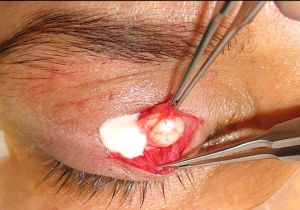 Intraoperative appearance of a ruptured IKC showing milky fluid material. Courtesy of Prof. Adel Alsuhaibani

Differential diagnosis
Chalazion- affects younger patients, typically accompanied by signs of inflammation and the margin of the lesion is less clear than that of an IKC with fluctuations in size.
Sebaceous cyst carcinoma- a highly malignant skin tumour, tends to masquerade as more common benign conditions such as blepharitis or chalazion, typically appears yellow with painless eyelash loss, effacement of the meibomian gland orifices and diffuse conjunctival inflammatory signs from intraepithelial growth.
Steatocystoma- affects young patients, arises from the skin dermis, not attached to the tarsus, and lined with a nonkeratinizing squamous epithelium without keratohyalin granules with sebaceous cell clusters, filled with yellow sebum and scattered hair shafts.
Epidermal inclusion cyst- freely movable, not fixed to the tarsus, and lined by focal acanthotic squamous epithelium and has a granular layer.
Wolfring gland ductal cysts-subconjunctival cysts attached to the proximal tarsal border and extending into the fornix. They have a double epithelial lining with a serpiginous cavity.[11]
Management
The intratarsal location of IKCs is the main factor contributing to a higher recurrence rate, compared to other eyelid cystic lesions. This position within the tarsal plate makes complete excision challenging, raising the possibility of leaving residual tissue. Therefore, IKCs tend to recur unless completely excised.
Surgery
Complete excision, with partial tarsectomy is the recommended treatment.[4] This can be accomplished through either the skin[9] or the conjunctiva.[6] Although the transconjunctival approach avoids skin incision , previous reports have shown comparable cosmetic outcomes and similar recurrence rates following both approaches. Simple incision and drainage with curettage is not effective, with a high recurrence rate.
Full-thickness eyelid resection can be used in selected cases.[1]

Complications
Recurrence often occurs after incomplete cyst excision.
Spontaneous rupture with transconjunctival leakage of the keratinous material may occur and cause eye irritation.[12]
References
- ↑ Jump up to: 1.0 1.1 1.2 1.3 1.4 1.5 Jakobiec FA, Mehta M, Iwamoto M, Hatton MP, Thakker M, Fay A. Intratarsal keratinous cysts of the Meibomian gland: distinctive clinicopathologic and immunohistochemical features in 6 cases. Am J Ophthalmol. 2010 Jan;149(1):82-94.
- ↑ Jump up to: 2.0 2.1 Tang T, Brownstein S, Chen H, Jordan DR, Iacob CE, Blanco P, Farmer J. Histopathological Study on the Proposed Pathogenesis of Intratarsal Keratinous Cysts. Ophthalmic Plast Reconstr Surg. 2019 Jul/Aug;35(4):365-368.
- ↑ Rajak SN, James C, Selva D. The clinical, histological and immunohistochemical characteristics and nomenclature of meibomian gland ductal cysts (intratarsal keratinous cysts) and eyelid steatocystomas. Eye (Lond). 2017 May;31(5):736-740.
- ↑ Jump up to: 4.0 4.1 Zhang ZD, Li X, Li M, Zhao J, Zhou KJ, Qu J. Clinicopathological features and surgical treatment of intratarsal keratinous cysts. Am J Dermatopathol. 2013 Feb;35(1):78-82.
- ↑ Patel VS, Meyer DR, Carlson JA (2011) Intratarsal kerati- nous cysts of the meibomian gland (a sebaceous duct cyst): report of 2 cases. Am J Dermatopathol 33(6):624–627.
- ↑ Jump up to: 6.0 6.1 Zhang ZD, Li X, Li M, Zhao J, Zhou KJ, Qu J. Clinicopathological features and surgical treatment of intratarsal keratinous cysts. Am J Dermatopathol. 2013;35(1):78-82. doi:10.1097/DAD.0b013e31825d9d0d
- ↑ Tang T, Brownstein S, Chen H, Jordan DR, Iacob CE, Blanco P, Farmer J (2018) Histopathological study on the proposed pathogenesis of intratarsal keratinous cysts. Ophthalmic Plast Reconstr Surg. https://doi.org/10.1097/iop. [[1]]
- ↑ Jump up to: 8.0 8.1 Jordan D, Stoica B, Brownstein S, Ali-Ridha A. Intratarsal keratinous cyst mimicking a large chalazion. Can J Ophthalmol. 2014 Dec;49(6):e149-51.
- ↑ Jump up to: 9.0 9.1 9.2 9.3 AlRubaian A, Alkatan HM, Al-Faky YH, Alsuhaibani AH. Clinical features differentiating intratarsal keratinous cyst from chalazion. Int Ophthalmol. 2020 Aug;40(8):2041-2045.
- ↑ Kim JA, Kim N, Choung HK, Lee MJ, Lee C, Khwarg SI. Clinical features of intratarsal keratinous cysts. Eye (Lond). 2016 Jan;30(1):59-63. doi: 10.1038/eye.2015.184.
- ↑ Diab MM, Allen RC, Mohammed KK, Saif ATS. Clinical characteristics and surgical outcomes of transcutaneous versus transconjunctival excision of Wolfring gland ductal cysts. BMC Ophthalmol. 2024;24(1):164. Published 2024 Apr 16. doi:10.1186/s12886-024-03420-x
- ↑ Gologorsky D, Jakobiec FA, Freitag SK. Transconjunctival elimination of keratin from an intratarsal meibomian cyst. Ophthalmic Plast Reconstr Surg. 2013 May-Jun;29(3):e88-91. 250332.