Hydro Manoeuvres in Cataract Surgery
All content on Eyewiki is protected by copyright law and the Terms of Service. This content may not be reproduced, copied, or put into any artificial intelligence program, including large language and generative AI models, without permission from the Academy.
Hydro manoeuvres are an integral part of modern cataract surgery, permitting nuclear rotation and facilitating epinuclear and cortical cleanup. Cortical cleaving hydro dissection and hydro delineation procedures if successfully performed, improves efficiency of phacoemulsification, reduces the risk of posterior capsular rupture and the rate of posterior capsule opacification.
Hydrodissection
The term Hydro dissection was coined by Faust in 1984.He described it as injection of fluid to separate the lens nucleus from cortex during a planned extra capsular cataract extraction, to facilitate mobilisation and easy removal of nucleus.[1] This procedure requires cortical removal by irrigation aspiration as a separate step.
Multi lamellar hydrodissection
Multilamellar hydro dissection, described in 1990, involves injection of fluid into multiple lamellae of lens, cleaving it into a small nucleus and multiple layers of cortex.[2] The advantages described were reduction phacoemulsification time as the nucleus is smaller, protection of the posterior capsule by a thick bed of cortex during emulsification and easier cortical clean up of multiple layers of loose cortex.
Cortical cleaving hydro dissection
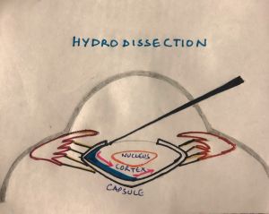
Although subcapsular injection of fluid in human cadaver eyes in a laboratory setting had been described earlier (D.J. Apple and co authors), a description of the procedure in a clinical setting was given by Fine in 1992 and he called it Cortical Cleaving Hydro dissection. This technique which is widely used, effectively cleaves the surrounding cortex from capsule by pre injection tenting of the anterior capsule and if successfully performed, does not require cortex removal as a separate step, eliminating the associated risk of a posterior capsule rupture(PCR).[3] [4]It ensures ease, safety and efficacy of nucleus emulsification, thereby reducing the time taken for phacoemulsification.[5] Another advantage is the removal of equatorial lens epithelial cells(LECs) from the adjacent capsule by the shearing effect of fluid during the procedure, thereby reducing the incidence of posterior capsule opacification(PCO).[6]
Instrumentation
Syringe with hydrodissection cannula is used. A small-diameter cannula (30- or 27-gauge) is preferred as it provides an increased flow resistance and maximizes the force that can be generated by a small volume of fluid.
A 3-cc syringe is recommended as it provides a fluid jet that is brief, sufficiently forceful and oriented in a radial direction. A tuberculin syringe contains insufficient volume and larger-volume syringes are bulky and do not provide enough tactile feedback as the plunger is advanced. Balanced salt solution (BSS), lidocaine, or ophthalmic viscosurgical device(0.5 -1 cc) can be injected.
Technique (Cortical Cleaving Hydrodissection)
Capsulorhexis size of 5-5.5mm with a large anterior capsular flap is optimum.A blunt hydrodissection cannula is passed inferiorly and is used to tent the anterior capsule away from the cortex and is advanced near the equator. Maintaining the tented up position, fluid is injected gently.This results in a posterior fluid wave between the capsule and the cortex, followed by bulging forward of the entire lens due to the fluid trapped by the firm equatorial cortical-capsular connections. This is the result of a temporary intraoperative capsular block syndrome and is characterised by an enlargement of capsulorhexis diameter. The central portion of the lens is then depressed with the side of the cannula, forcing fluid to come around the lens equator from behind by cleaving the cortical-capsular connections in the capsular fornix and under the anterior capsular flap, and exiting via capsulorhexis. This constricts the capsulorhexis to its original size, and mobilizes the lens.
Hydro dissection and capsular decompression may be repeated in the opposite distal quadrant and may be followed by hydrodelineation. Adequate hydro dissection at this point can be demonstrated by the ease with which the nuclear–cortical complex can be rotated by the cannula.
Surgical challenges
Separation of cannula
Separation of cannula from syringe during fluid ejection: This can be avoided by using Luer-lock syringe.
Incomplete Cortical Cleaving Hydro dissection
Inadequate fluid injection, injection into the lens or failure to create enough hydraulic pressure to separate the capsule and cortex can result in stressful rotation of nucleus. A repeat hydrodissection should be performed in such instances to avoid zonular stress.
Errant Placement of the Cannula
Inadvertent placement of the irrigating cannula beneath the iris, but on top of the anterior capsule may happen rarely in small pupils and can result in injection of fluid within the zonules, over the vitreous face, and into the posterior capsule. This can result in a shallowing of anterior chamber as occurs with fluid misdirection syndrome.
Prolapse of the nucleus partially or wholly in anterior chamber
This situation can be managed by gently repositioning the nucleus back into bag (if intact). Debulking the nucleus by chopping or sculpting may be required.
Anterior Capsular Block and Capsular Blow Out
Overzealous fluid injection can lead to excessive nucleus bulging blocking the anterior capsular opening, trapping fluid within the capsular bag and in extreme cases, posterior capsule rupture. Anterior capsular block is characterised by shallowing of anterior chamber not relieved by lens decompression. Attempting rotation can jeopardize zonular integrity. In such cases, creating a trench and initiating a chop or crack can relieve the capsular block. This is characterised by deepening of the anterior chamber and backward movement of lens. Capsular blow out can occur in cataracts with compromised posterior capsule (posterior polar cataract, posterior capsular injury from vitrectomy or intravitreal injection, or trauma) and femto second laser assisted cataract surgery(FLACS) cases with trapped intracapsular gas.[7] A brisk hydro dissection in small capsulorhexis in hard cataracts can also result in capsular blow out. It is characterised by a pupillary snap sign [8]or nucleus drop.[9] To avoid capsular block syndrome, the amount of fluid injection should be titrated and the anterior chamber should not be overfilled with visco elastic. Hydro dissection is also best avoided in cases with suspected or definitive posterior capsular dehiscence as in posterior polar cataracts to avoid build up of hydraulic pressure in the capsular bag and subsequent blow out.
Rare cases of anterior capsule tear through the posterior capsule, anterior hyaloid membrane tear and acute aqueous misdirection have been reported due to rapid elevation of intra ocular pressure during manual hydrodissection.
Modifications of Hydro dissection
Multi quadrant focal hydro dissection
Injection of small quantities of fluid focally in multiple quadrants. This is particularly useful in cases of corticocapsular adhesions which are unlikely to get cleaved by single quadrant conventional hydro dissection.[10]
Multi quadrant hydrodissection for posterior polar cataracts
Fine et described this technique where tiny quantities of fluid are injected gently in multiple quadrants, such that fluid wave is not allowed to spread across the entire posterior capsule thereby sparing the area of dehiscence.[11]
Minimal water-jet hydrodissection
Hydrodissection technique performed with high-speed pulse injection of only 0.1 cc liquid.[12]
Phaco sleeve irrigatiation assisted hydrodissection
This technique uses the phaco tip to intentionally vacuum the intraocular fluid in order to induce the irrigation dynamic pressure for cortical-capsular cleavage, there is a reduction in the intraocular pressure (IOP) from the bottle-height-dependent hydrostatic pressure thereby avoiding complications.[13]
Hydro delineation
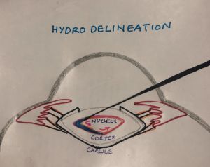
The procedure was first described by Anis (A. Anis, M. D., "Understanding Hydro delineation: The Term and Related Procedures," Ocular Surgery News, October 15, 1991, 134-137) as an injection of fluid into the body of the lens deep to the cortex resulting in separation of epinuclear shell or multiple shells from a more compact central nuclear mass. It is a useful technique in situations with compromised capsules.
The reduced overall size of the nucleus allows less deep and less peripheral grooving and smaller, more easily mobilized quadrants after cracking or chopping. The epinucleus acts as a protective cushion within which phacoemulsification forces can be confined. In addition, the epinucleus keeps the bag on stretch, making it unlikely for a knuckle of posterior capsule to occlude the phaco tip and rupture.
Technique
Instrumentation is same as that for hydrodissection. The cannula is placed in the nucleus, off centre to either side, and directed at an angle downward and forward towards the central plane of the nucleus. Endonucleus is reached when the nucleus starts to move.The cannula is then directed tangentially to the endonucleus, and a to-and-fro movement of the cannula is used to create a tract within the nucleus. The cannula is backed out of the tract approximately halfway and a gentle but steady pressure on the syringe allows fluid to enter the “empty” distal tract without resistance. Driven by the hydraulic force of the syringe, the fluid will find the path of least resistance, which is the junction between the endonucleus and the epinucleus, and flow circumferentially in this contour Most often, a circumferential golden ring will be seen outlining the cleavage between the epinucleus and the endonucleus.
Surgical challenges
Difficulty in finding the right plane in very soft and very hard cataracts. This may occasionally result in inadvertent hydrodissection.
Modifications of Hydrodelineation
Inside-Out Hydrodelineation
First described by Vasavada, in this technique fluid is injected from the inside of the nucleus to the outside, allowing precise hydrodeliniation.[14] The desired thickness of the nucleus–epinucleus bowl can be accomplished because the right-angled cannula can direct the fluid in the desired plane. This technique is useful in eyes with posterior polar cataracts and in eyes with dense cataracts in which aggressive phaco parameters are necessary for their removal.
References
- ↑ Faust KJ. Hydrodissection of soft nuclei. Am Intra-Ocular Implant Soc J 1984; 10:75–77
- ↑ Koch DD, Liu JF. Multilamellar hydrodissection in phacoemulsification and planned extracapsular surgery. J Cataract Refract Surg 1990; 16:559–562.
- ↑ Fine IH. Cortical cleaving hydrodissection. J Cataract Refract Surg 1992; 18:508–512.
- ↑ Fine IH. Cortical cleaving hydrodissection. (letter) J Cataract Refract Surg 2000; 26:943–944.
- ↑ Vasavada AR, Singh R, Apple DJ, Trivedi RH, Pandey SK, Werner L. Effect of hydrodissection on intraoperative performance: randomized study. J Cataract Refract Surg. 2002 Sep;28(9):1623-8.
- ↑ Peng Q, Apple DJ, Visessook N, et al. Surgical prevention of posterior capsule opacification: Part 2: enhancement of cortical cleanup by focusing on hydrodissection. J Cataract Refract Surg 2000; 26:188–197.
- ↑ Roberts TV, Sutton G, Lawless MA, et al. Capsular block syndrome associated with femtosecond laser–assisted cataract surgery. J Cataract Refract Surg. 2011;37(11):2068–2070. doi:10.1016/j.jcrs.2011.09.003
- ↑ Yeoh R. The‘pupil snap’ sign of posterior capsule rupture with hydrodissection in phacoemulsification. (letter) Br J Ophthalmol 1996; 80:486 23.
- ↑ Ota I, Miyake S, Miyake K. Dislocation of the lens nucleus into the vitreous cavity after standard hydrodissection. Am J Ophthalmol 1996; 121:706–708.
- ↑ Vasavada AR, Goyal D, Shastri L, Singh R. Corticocapsular adhesions and their effect during cataract surgery. J Cataract Refract Surg. 2003;29:309–14.
- ↑ Fine IH, Packer M, Hoffman RS. Management of posterior polar cataract. J Cataract Refract Surg. 2003;29:16–19.
- ↑ Taş A. Minimal water-jet hydrodissection. Clin Ophthalmol. 2017;12:1-5.
- ↑ Masuda Y, Iwaki H, Kato N, Takahashi G, Oki K, Tsuneoka H. Irrigation dynamic pressure-assisted hydrodissection during cataract surgery. Clin Ophthalmol. 2017;11:323–328.
- ↑ Vasavada AR, Raj SM. Inside-out delineation. J Cataract Refract Surg. 2004 Jun;30(6):1167-9.


