File:Malignant HTN RET.jpg
Malignant_HTN_RET.jpg (640 × 427 pixels, file size: 123 KB, MIME type: image/jpeg)
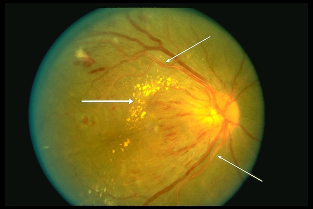
Common hypertensive retinal changes are flame-shaped hemorrhages in the superficial layers of the retina and cotton-wool patches caused by occlusion of the precapillary arterioles with ischemic infarction of the inner retina. Long-standing hypertension can produce arteriolar sclerotic vascular changes, such as copper or silver wiring of the arterioles, as shown by the two arrows in the figure, or arteriorvenous nicking. Another sign of chronic hypertension is lipid exudates resulting from abnormal vascular permeability, as shown by the arrow at left. More ominous in this photograph is swelling of the optic disc, seen here by the blurring of the temporal disc margins. This is the hallmark of malignant hypertension, which carries a poor prognosis for the patient’s health if left untreated. BP must be emergently controlled to decrease the risk of developing heart and renal failure and hypertensive encephalopathy as well as stroke and permanent vision loss. © 2019 American Academy of Ophthalmology
File history
Click on a date/time to view the file as it appeared at that time.
| Date/Time | Thumbnail | Dimensions | User | Comment | |
|---|---|---|---|---|---|
| current | 12:23, January 5, 2019 | | 640 × 427 (123 KB) | Daniel.kiernan (talk | contribs) | Common hypertensive retinal changes are flame-shaped hemorrhages in the superficial layers of the retina and cotton-wool patches caused by occlusion of the precapillary arterioles with ischemic infarction of the inner retina. Long-standing hypertension... |
You cannot overwrite this file.
File usage
The following 2 pages use this file: