File:Conjunctival RLH - Figure 3.png
From EyeWiki
No higher resolution available.
Conjunctival_RLH_-_Figure_3.png (517 × 232 pixels, file size: 113 KB, MIME type: image/png)
Summary
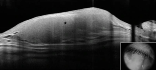
Figure 3. Anterior segment optical coherence tomography scan showing homogeneous, hyporeflective lesion with thin overlying epithelium.[2] Open access under terms of the Creative Commons License: http://creativecommons.org/licenses/by/4.0/. Original figures available at: https://eandv.biomedcentral.com/articles/10.1186/s40662-019-0151-4.
File history
Click on a date/time to view the file as it appeared at that time.
| Date/Time | Thumbnail | Dimensions | User | Comment | |
|---|---|---|---|---|---|
| current | 20:13, January 11, 2023 | | 517 × 232 (113 KB) | Michael.Yen (talk | contribs) | Figure 3. Anterior segment optical coherence tomography scan showing homogeneous, hyporeflective lesion with thin overlying epithelium.[2] Open access under terms of the Creative Commons License: http://creativecommons.org/licenses/by/4.0/. Original figures available at: https://eandv.biomedcentral.com/articles/10.1186/s40662-019-0151-4. |
You cannot overwrite this file.
File usage
The following page uses this file: