Eyelid Burns
All content on Eyewiki is protected by copyright law and the Terms of Service. This content may not be reproduced, copied, or put into any artificial intelligence program, including large language and generative AI models, without permission from the Academy.
Disease Entity
General Pathology
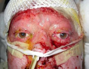
Burns may be caused by thermal, chemical or electrical sources which can all be both life threatening and sight threatening. Thermal burns are the most common, often affecting the eyelids, the periocularregion & the face. Ophthalmic involvement occurs between 7.5% and 27% of patients admitted to burn units. However, the loss of an eye and vision primarily from a thermal injury is uncommon, primarily due to a significant number of inherent protective mechanisms such as the blink reflex, Bell's phenomenon, and protective movements of the head and arms to avoid the source of a burn.
The most common causes of thermal burn injury are fire/flame (46%) and scald (32%), with scald burns being a particular problem in children. Of those admitted to burn units, eyelid burns were more common than other burn/injury to the eye(s). While chemical burns, accidental and intentional, although uncommon also inflict serious harm , the focus of this article will be on thermal burns.[1][2]
Pathophysiology
Burn injury results in the release of multiple inflammatory mediators that result in vasodilatation, pain, and edema.
The depth of burn depends on the intensity of heat exposure, the duration of exposure, and the thickness of epidermis and dermis. Eyelid skin is thin without subcutaneous fat, leading to deeper burns compared to similar exposure to skin elsewhere.
The three zones of a burn were described by Jackson in 1947.
- Zone of coagulation—This occurs at the point of maximum damage. In this zone there is irreversible tissue loss due to coagulation of the constituent proteins.
- Zone of stasis—The surrounding zone of stasis is characterized by decreased tissue perfusion. The tissue in this zone is potentially salvageable. Thus the main goal of burn resuscitation is to increase tissue perfusion and minimize irreversible damage. Additional insults, such as prolonged hypotension, infection, or edema, can convert this zone into an area of complete tissue loss.
- Zone of hyperemia—In this outermost zone tissue perfusion is increased. The tissue here will invariably recover unless there is severe sepsis or prolonged hypoperfusion.[3]
Diagnosis
Diagnosis is clinically obvious, especially when combined with a typical and reliable history. Occasionally, the exact mechanism of injury may not be available - hot water, chemical(s), fire, contact thermal burn etc and hence may be misleading. Eyelid burns are classified depending on how deep and severe they penetrate the skin's surface.
Classification
Ocular adnexal burns may be classified depending on the extent, depth and severity of the injury including underlying tissue damage.
First degree burns / Epidermal burns
This corresponds to the zone of hyperemia in Jackson’s model. Severe sunburn is the most common example of first-degree burn. By definition, this affects only the epidermis, and blistering is not common. Pain is due to local vasodilator prostaglandins, and healing is usually complete within a week.[1]
Second degree burns / Partial-thickness burns
Partial-thickness burns involve the dermis and epidermis. This corresponds to the zone of stasis in Jackson’s model. It is commonly divided into superficial and deep dermal injury.
- SUPERFICIAL PARTIAL-THICKNESS BURNS
- Injury to the epidermis and superficial papillary dermis results in thin-walled, fluid-filled blisters with a moist red base. The exposure of superficial nerves makes these injuries painful. A burn of this depth usually heals within 2 weeks by regeneration of epidermis from keratinocytes within sweat glands and hair follicles, with minimal scarring.
- DEEP PARTIAL-THICKNESS BURNS
Third degree burns / Full-thickness burns
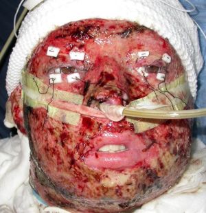
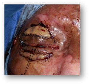
These destroy epidermis, dermis, and all regenerative elements and correspond to the zone of coagulation in Jackson’s burn wound model. The skin is dry, leathery, and as a result of heat coagulation of dermal blood vessels, the affected tissue is avascular and white. Such burns are typically painless due to loss of sensation in the involved area. Healing only occurs from the edges and is associated with significant contraction. Early excision of affected tissue and skin grafting is almost always required to resurface the burnt area and prevent secondary severe corneal complications from exposure and secondary infection.[6][7][8]
Fourth degree burns / Deep burns
These are full-thickness burns with destruction of the underlying muscle, bone and vital structures. Such burns require extensive and complex multidisciplinary management and often result in severe contracture and prolonged disability.
Based on these definitions and the likelihood of surgery, eyelid burns may be classified as mild, moderate, or major. Mild or minimal burns describe superficial partial-thickness burns that usually heal without the need for surgery. Moderate burns refer to deeper partial-thickness burns that are delayed in healing but may not require surgery. Major eyelid burns are deep-partial or full-thickness and invariably require early surgery with skin grafts.[1]
Physical Examination
Patients with facial burns should be seen by an ophthalmologist as soon as possible to assess the extent of eyelid, eyelid margin and ocular surface injury. Ocular surface examination should include not only the cornea, but also the bulbar, forniceal and tarsal conjunctiva. Particulate material and foreign bodies on the ocular surface and when suspected intraocular or even intraorbital - with high velocity or blast injuries should be suspected when appropriate and excluded by imaging if indicated. The presence of a contact lens should not be overlooked. This assessment should be made, if possible, in the emergency room if necessary under topical anesthetic (after ruling out open globe injury) before significant conjunctival and eyelid edema prevents a thorough exam.
The most critical issue for the ophthalmologist is the integrity of the corneal surface. Therefore, a proper corneal evaluation should be performed using fluorescein strips and a cobalt blue light in the emergency room or burn unit. Even with no initial corneal damage, sequelae from eyelid retraction can occur later and monitoring is mandatory.
The depth and extent of burns in the eyelid and facial area should be initially assessed, and documented with photography if possible. In the presence of a partial-thickness burn, eyelid contracture producing progressive lagophthalmos and corneal exposure is likely. Therefore, the presence or absence of a Bell’s phenomenon should be documented.[1][9]
Intraocular pressure (IOP) monitoring may be important in the patient who receives intravenous fluid resuscitation, as elevation may occur within 72 hours of large shifts from the intravascular compartment to the extravascular compartment. Third spacing of intravascular volume peaks at 6-12 hours and increases with aggressive fluid resuscitation. Positive pressure ventilation can also exacerbate the edema and potential lagophthalmos.[1][10][11]
Management
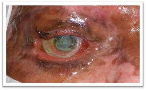
Immediate priorities following burns including assessment of the extent of the burns and presence of orofacial involvement. Securing airway, skin protection, hydration and antibiotics are essential. Ophthalmic management following assessment, includes adequate lubricating antibiotic eyedrops and ointments, moisture chambers, and frequent evaluation of both the globes and the eyelids. All efforts to minimize scarring of the ocular surface and periocular area should be taken. Once cicatricial changes begin in the eyelids and ocular surface, a relentless and rapid deterioration often ensues with secondary cicatricial eyelid retraction, ectropion, lagophthalmos, and corneal exposure. Chemical injuries should be treated based on the extent of injury after the ocular surface pH is normalized post-irrigation, details on chemical injuries can be seen under that topic. In the acute setting, head elevation may be helpful in preventing further swelling around the eyelids. Topical steroids should be avoided due to the risk of secondary infection.[1][4][9][12]
General treatment
- Lubrication within 24 hours of admission, with copious application as required, is particularly important as burn patients often have reduced tear production, blink reflex, and eyelid mobility or excursion.
- Singed or scorched eyelashes are usually present in a thermal eyelid burn and should be removed to avoid the possibility of char falling into the eye and prolonging ocular surface discomfort.
- The principles of wound management for eyelid burns are those for any burn: assessment, cleansing, and protection followed by resurfacing for deeper burns.
- Cicatricial lagophthalmos can develop early and requires aggressive treatment until skin grafting can be performed.
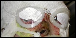
- In the presence of lagophthalmos with a severe burn of the eyelid skin, clear occlusive dressing with adequate lubricant gels ('cling wrap or Saran Wrap' or TegaDerm(R)) is generally useful. Measures than can be used to protect the corneal surface until skin grafting can be performed include amniotic membrane and suture tarsorrhaphy.
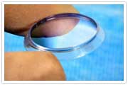 [13]Prokera
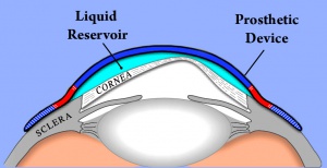
 - In cases where amniotic membrane is not available and tarsorrhapy is not feasible due to extensive tissue loss a Boston Ocular Surface Prosthesis can also be use to protect the cornea.[12]
- As extreme injury with secondary orbtial edema may present as an orbital compartment syndrome (OCS), in such cases a canthotomy, cantholysis & septolysis may be necessary to control the IOP and prevent permanent visual loss.
- In the past, skin grafting was usually delayed until the cicatricial changes stabilized, but the early use of full-thickness skin grafts, amniotic membrane, and various types of flaps can effectively reduce ocular morbidity in selected patients.[7][8][14]
Long term management includes retraction repair with skin grafts, repair of medial canthal deformities, trichiasis, palpebral aperture stenosis, eyebrow deformities, canalicular obstruction, and surgical or laser scar revision.
References
- ↑ Jump up to: 1.0 1.1 1.2 1.3 1.4 1.5 1.6 Raman M., Ijaz S, and Baljit D. The management of Eyelid Burns. Survey of ophthalmology. Volume 54. June 2009
- ↑ Kaplan AT, Yalcin SO, Günaydın NT, Kaymak NZ, Gün RD. Ocular-periocular burns in a tertiary hospital: Epidemiologic characteristics. J Plast Reconstr Aesthet Surg. 2023 Jan;76:208-215. doi: 10.1016/j.bjps.2022.10.049. Epub 2022 Nov 2. PMID: 36527902.
- ↑ JACKSON DM. [The diagnosis of the depth of burning]. Br J Surg. 1953 May;40(164):588-96. Undetermined Language. doi: 10.1002/bjs.18004016413. PMID: 13059343.
- ↑ Jump up to: 4.0 4.1 Achauer BM, Adair SR. Acute and reconstructive management of the burned eyelid. Clin Plast Surg. 2000;7:87-95
- ↑ Papini R. Management of burn injuries of various depths. BMJ. 2004 Jul 17;329(7458):158-60. doi: 10.1136/bmj.329.7458.158. PMID: 15258073; PMCID: PMC478230.
- ↑ Bouchard CS, Morno K, Jeffrey P, et al. Ocular complications of thermal injury: a 3-year retrospective. J Trauma. 2001; 50(1):79-82
- ↑ Jump up to: 7.0 7.1 Cole JK, Engrav LH, Heimbach DM, et al: Early excision and grafting of face and neck burns in patients over 20 years. Plast Reconstr Surg 2002; 109: 1266-1273
- ↑ Jump up to: 8.0 8.1 Fallah LY, Ahmadi A, Ruche AB, Taremiha A, Soltani N, Mafi M. The effect of early change of skin graft dressing on pain and anxiety among burn patients: a two-group randomized controlled clinical trial. Int J Burns Trauma. 2019 Feb 15;9(1):13-18. PMID: 30911431; PMCID: PMC6420706.
- ↑ Jump up to: 9.0 9.1 Orbit, eyelids, and lacrimal system. (2021). San Francisco, CA: American Academy of Ophthalmology.
- ↑ Rachel Dahl, MD, MPH, MS and others, Regional Burn Review: Neither Parkland Nor Brooke Formulas Reach 85% Accuracy Mark for Burn Resuscitation, Journal of Burn Care & Research, 2023;, irad047,
- ↑ Brundridge, WL. Adnexal Burns. Ch 68. In: Servat, JJ et al. eds. Smith and Nesi's Ophthalmic Plastic and Reconstructive Surgery. 4th ed. Springer, 2021: 1223-1230.
- ↑ Jump up to: 12.0 12.1 Kalwerisky K, Davies B, Mihora L, Czyz CN, Foster JA, DeMartelaere S. Use of the Boston Ocular Surface Prosthesis in the management of severe periorbital thermal injuries: a case series of 10 patients. Ophthalmology. 2012 Mar;119(3):516-21. doi: 10.1016/j.ophtha.2011.08.027. Epub 2011 Nov 30. PMID: 22133791.
- ↑ lorieanngrover.blogspot.com/2018/01/dry-eyes-solutions-part-five-amniotic.html
- ↑ Reed DS, Plaster AL, Mehta A, Hill MD, Zanganeh TS, Soeken TA, DeMartelaere SL, Davies BW. Acute And Sub-Acute Reconstruction Of Periorbital Thermal Burns Involving The Anterior Lamella Of The Eyelid With Simultaneous Fullthickness Skin Grafting And Amniotic Membrane Grafting. Ann Burns Fire Disasters. 2020 Dec 31;33(4):323-328. PMID: 33708023; PMCID: PMC7894844.


