Dacryoadenoma
All content on Eyewiki is protected by copyright law and the Terms of Service. This content may not be reproduced, copied, or put into any artificial intelligence program, including large language and generative AI models, without permission from the Academy.
Disease Entity
Dacryoadenoma is a rare conjunctival tumor usually seen in children or young adults.
Clinical Findings
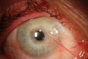
This tumor presents as mass on the bulbar or palpebral conjunctiva. According to case reports, dacryoadenoma presents as a pink, soft, and lobulated mass. It is usually small, measuring 7mm x 4mm in one case report.[1]
While the exact presentation varies from case to case, it generally involves features typical of benign conjunctival lesions. This includes having a size of less than 15 mm in basal dimension and lacking elevation, vascularity, or corneal extension.[2] The mass may grow slowly over time, but can also remain stable for many years (one case reported stable size for fifteen years).[1] Like most conjunctival tumors, dacryoadenomas do not usually cause symptoms.
Diagnosis
The diagnosis of a conjunctival neoplasm such as dacryoadenoma begins with a comprehensive history of the mass, followed by external examination and examination under slit-lamp biomicroscopy.
Due to the tumor’s small size and benign features, further diagnostic evaluation is usually not indicated. Incisional biopsy is reserved for larger lesions or lesions suspicious for malignancy. Excisional biopsy can be done for smaller lesions that are symptomatic, or to rule out other diagnoses.
Differential Diagnosis
Differential diagnosis includes other growths and neoplasms of the conjunctival epithelium:
Benign
- Squamous papilloma

- Hereditary Benign Intraepithelial Dyskeratosis
- Epithelial Inclusion Cyst
- Keratotic Plaque
- Pterygium

Malignant
- Conjunctival Intraepithelial Neoplasia
- Squamous Cell Carcinoma
Histology
This tumor arises from bulbar or palpebral conjunctival epithelium. Metaplastic cuboidal or columnar epithelial cells penetrate underlying connective tissue, forming tubular and glandular structures. Electron microscopy will show lacrimal secretory granules in the metaplastic cells. Dacryoadenoma is distinct from complex choristoma histologically.[3]
Images for dacryoadenoma not available on AAO. For external photographs and histopathological images, see referenced publication.[1]
Management
Because of the benign nature of this disease, serial observation is generally the first step in management. Careful documentation of slit-lamp findings and serial photographs are recommended to track progression. Excisional biopsy can be considered to exclude other diagnoses. Excision can also be performed as per patient preference.[1] [2]
Prognosis
Excellent.
References
- ↑ Jump up to: 1.0 1.1 1.2 1.3 Jakobiec FA, Perry HD, Harrison W, et al. Dacryoadenoma. A unique tumor of the conjunctival epithelium. Ophthalmology 1989; 96:1014-1020
- ↑ Jump up to: 2.0 2.1 Carol L Shields, Jerry A Shields, Tumors of the conjunctiva and cornea, Survey of Ophthalmology, Volume 49, Issue 1, 2004, Pages 3-24, ISSN 0039-6257, https://doi.org/10.1016/j.survophthal.2003.10.008.
- ↑ Milman T, Jakobiec FA, Lally SE, Shields JA, Shields CL, Eagle RC Jr. Lacrimal Gland Hamartoma (Formerly Termed Dacryoadenoma). Am J Ophthalmol. 2020 Sep;217:189-197. doi: 10.1016/j.ajo.2020.04.015. Epub 2020 Apr 29. PMID: 32360334.
- Shields JA, Shields CL. Eyelid, Conjunctival, and Orbital Tumors: An Atlas and Textbook. Lippincott Williams & Wilkins, 2008; 2:18:280.
- Smolin G, Foster CS, Azar DT, Dohlman CH. Smolin and Thoft’s The Cornea: Scientific Foundations and Clinical Practice. Lippincott Williams & Wilkins, 2005; 40:745.
- Yanoff M, Sassani, J Ocular Pathology 6th edition Mosby, 2008; 7: 244.

