Conjunctival Concretions
All content on Eyewiki is protected by copyright law and the Terms of Service. This content may not be reproduced, copied, or put into any artificial intelligence program, including large language and generative AI models, without permission from the Academy.

Disease Entity
Disease
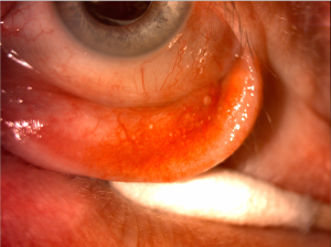
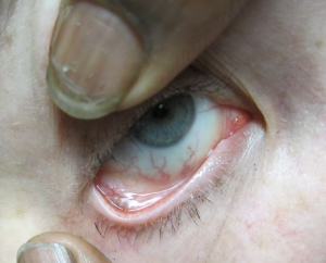
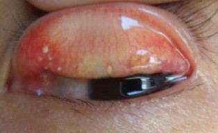
Conjunctival concretions are small, typically multiple, yellow-white lesions commonly found on palpebral conjunctiva of elderly individuals and those with chronic inflammation. They are thought to be widespread and typically asymptomatic, occurring in 40-50% of studied populations.
Etiology
The cause for these concretions is variable but most commonly associated with aging and chronic inflammation. Long standing inflammation, such as chronic conjunctivitis (e.g., trachoma or vernal), severe atopic conjunctivitis, and meibomian gland disease, have been associated with the formation of these concretions. Conjunctival concretions have also been associated with recrystallization of certain eye drops (e.g., sulfadiazine).
General Pathology
The primary components of conjunctival concretions are degenerating epithelial cells and proteinaceous secretions from conjunctival glands. Following inflammation, the debris becomes trapped in the subconjunctival depressions and recesses, and they often undergo calcification. Although the concretions may contain substantial calcification, they are not typically hard and have no crystalline pattern under electron microscopy, and therefore, the term "lithiasis" should not be applied. Occasionally epithelial cysts are located on top of these concretions. Goblet cells are not thought to play a major role in the pathogenesis of these lesions.
Diagnosis
Typically, patients are asymptomatic and the concretions are noted incidentally on ocular evaluation. Some patients may complain of irritation or foreign body sensation. Patients may or may not have history of chronic conjunctivitis.
Physical Exam
On exam, there are small 1-2 mm, chalky, yellow-white lesions typically on palpebral conjunctiva. They may be singular, multiple, or rarely confluent. They tend to be located on inferior tarsal and forniceal conjunctiva but may be superiorly located as well.
Rarely, the degenerated substances will become secondarily inflamed or may erode through overlying epithelium resulting in local irritation and foreign body sensation.
Differential diagnosis
- Epidermal inclusion cysts
- Lymphoid follicles
Management
Treatment is usually unnecessary because the concretions are asymptomatic and located in the subepithelial space. If they erode though epithelium, they can often be removed with needlepoint forceps or a 30 gauge needle after instillation of topical anesthetic. In cases of foreign body sensation or persistent irritation, they may be removed surgically.
References
- Haicl P, Jankova H. "[Prevalence of conjunctival concretions]." Cesk Slov Oftalmol. 2005 Jul;61(4):260-4.
- Kulshrestha MK, Thaller VT. "Prevalence of conjunctival concretions." Eye. 1995(9):797–798.
- Boettner EA, Fralick FB, Wolter JR. Conjunctival concretions of sulfadiazine. Arch Ophthalmol 1974;92:446.
- Chin, GN, Chi EY, Bunt AH. "Ultrastructural and histochemical studies of conjunctival concretions." Arch Ophthalmol. 1980 Apr;98(4):720-4.
- Chang SW, Hou PK, Chen MS. "Conjunctival concretions. Polarized microscopic, histopathologic, and ultrastructural studies." Arch Ophthalmol. 1990 Mar;108(3):405-7.


