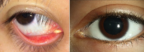
Caruncular Dermoid Cyst
All content on Eyewiki is protected by copyright law and the Terms of Service. This content may not be reproduced, copied, or put into any artificial intelligence program, including large language and generative AI models, without permission from the Academy.
Disease Entity
Dermoid Cyst are congenital, benign neoplasms of the orbit that are usually located superotemporally near the frontozygomatic suture, or superonasally.[1] They are composed of keratinized stratified squamous epithelium and other dermal derivatives, including hair follicles and sebaceous glands. They are one of the most common lesions of the orbit in children[2]; however, dermoid cysts of the caruncle are rarely reported.[3]
Etiology
When fetal suture lines close during embryogenesis, embryonic epithelial nests may become entrapped and form a cyst.[1]
Risk Factors
No known risk factors
Pathophysiology
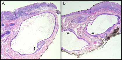
Dermoid cysts are common encapsulated cystic lesions composed of keratinized stratified squamous epithelium with dermal appendages and adnexal structures, including hair follicles, sebaceous glands, sweat glands, smooth muscle, and fibroadipose tissue. The lumen of the cyst contains keratin and hair.
Dermoid cysts are typically thought of as choristomas, which are lesions that consist of normal tissue arising in an atypical location, but when they are localized to the caruncle they are better classified as hamartomas because dermal appendages can be found at the caruncle. On histology, these cystic lesions are epithelial-lined with dermal appendages, and while the appendages are native to the caruncle, keratinizing epithelium is not.
Diagnosis
This diagnosis can be suspected by the focal, circumscribed clinical appearance, but more often confirmed histologically.
History
Similar to other superficial dermoids, these in the caruncle may present as a slowly progressive and nontender mass.[1] Patients often notice them more easily when they are yellow and begin to protrude between the medial palpebral fissure. The lesions can also be found bilaterally on both caruncles.
Physical Examination
These superficial lesions present as a smooth painless mass. If the cysts leaks or ruptures, granulomatous inflammation may be present.[2]
Signs
- Palpable mass
- Often yellow in color
- Inflammation (redness, swelling)
Symptoms
Typically asymptomatic and nontender
Clinical Diagnosis
Caruncular dermoid cysts are often not a clinical diagnosis because of the infrequency with which it is reported.
Diagnostic procedures
Excisional biopsy is the preferred method due to the possible differential diagnoses
Differential diagnosis
The majority of caruncular lesions, including dermoid cysts, are benign in nature.[3] The most common of these benign lesions include nevi, papillomas and epithelial cysts.[3] In evaluating caruncular lesions, however, providers should be aware that malignant lesions such as melanoma can occur, and can be fatal.[3] Early studies estimated malignancy to be as high as 27% of these lesions[3]; however, more recent data have reported much lower rates (range: 2.7-5.4%).[3] Differential diagnosis of dermoid cysts of the caruncle vary. Sebaceous gland hyperplasia and sebaceous gland adenoma appear similar to dermoid cysts clinically, but differ pathologically.[4] While steatocystomas closely resemble dermoid cysts both clinically and pathologically, steatocystomas typically have a more corrugated eosinophilic cuticle.[5] They also typically arise in puberty and are not present from birth.
Management
The mainstay of treatment is complete surgical excision.
Surgery
Care should be taken to avoid excising excess caruncle or surrounding plica semilunaris, that could lead to scarring and medial scar tissue.
Complications
Typically, if a dermoid cyst ruptures during surgery, copious irrigation of the area can avoid a lipogranulomatous inflammatory reaction. However, these lesions in the caruncle are much smaller and can usually be completely removed with ease. If the capsule is incompletely excised, the cyst may recur.
Prognosis
Prognosis is excellent after surgical excision.
References
- ↑ 1.0 1.1 1.2 Shields J, Shields C (2004) Orbital Cysts of Childhood – Classification, Clinical Features and Management. Surv Ophthalmol 49: 281-299. [Pubmed]
- ↑ 2.0 2.1 Cavazza S, Laffi GL, Lodi L, Gasparrini E, Tassinari G (2011) Orbital Dermoid Cyst of Childhood: Clinical Pathologic Findings, Classification and Management. Int Ophthalmol 31: 93-97. [Pubmed]
- ↑ 3.0 3.1 3.2 3.3 3.4 3.5 Levy J, Ilsar M, Deckel Y, Maly A, Pe’er J (2009) Lesions of the Caruncle: A Description of 42 Cases and a Review of the Literature. Eye 23:1004–18. [Pubmed]
- ↑ Ishida Y, Takahashi Y, Takahashi E, Kitaguchi Y, Kakizaki H (2016) Steatocystoma simplex of the lacrimal caruncle: a case report. BMC Ophthalmology 16: 183 [Pubmed]
- ↑ Elston DM, Ferringer T, Ko C, Peckham S, High WA et al. (2019) Dermatopathology (3rd edtn). Elsevier. [Elsevier]


