Aflibercept
All content on Eyewiki is protected by copyright law and the Terms of Service. This content may not be reproduced, copied, or put into any artificial intelligence program, including large language and generative AI models, without permission from the Academy.
Original Author: Christian Swinney B.A. and Peter A Karth, M.D., M.B.A.
Aflibercept is a soluble decoy receptor that binds vascular endothelial growth factor-A (VEGF-A) and placental growth factor (PIGF) with a greater affinity than the body’s native receptors. VEGF-A is a biochemical signal protein that promotes angiogenesis throughout the body and in the eye. By decreasing VEGF-A’s activation of its native receptors, aflibercept reduces subsequent growth of new blood vessels.1,2
Overview of VEGF and its Role in Retinal Disease
VEGF is a member of the platelet-derived growth factor (PDGF) family. The VEGF gene family consists of VEGF-A, VEGF-B, VEGF-C, VEGF-D and placental growth factor (PlGF), located on chromosome 6p12.3 The binding of VEGF with its receptors leads to endothelial cell proliferation and new blood vessel growth and therefore plays a key role in angiogenesis. New blood vessel growth and development are extremely complex and coordinated processes that require a cascade of receptor activations. In this cascade, VEGF represents an initial and critical rate-limiting step in physiological angiogenesis. 4,5,6 The critical role of VEGF in angiogenesis can be seen in the fact that a loss of a single VEGF allele can result in defective vascularization.7
There are nine VEGF-A isoforms: VEGF121, VEGF145, VEGF148, VEGF162, VEGF165, VEGF165b, VEGF183, VEGF189 and VEGF206. 8 The most abundant isoform found in the eye is VEGF165. 9,10 VEGF165 is a secreted heparin-binding homodimeric 45-kDa glycoprotein with a significant fraction bound to the cell surface. 11 VEGF activates endothelial cells by binding VEGFR-1 (Flt-1) and VEGFR-2 (KDR) endothelial cell receptors, which in turn activate intracellular signal transduction cascades. 11 VEGFR-2 is thought to be principally responsible for VEGF signaling in angiogenesis.11
VEGF-A levels have been found to be elevated in the vitreous of patients with neovascular (wet) age-related macular degeneration (AMD),12 diabetic macular edema, and retinal vein occlusion. Choroidal neovascularization (CNV) in AMD may be instigated by several events, such as accumulation of lipid metabolic byproducts, oxidative stress, reduction in choriocapillaris blood flow, and alterations in Bruch’s membrane. 13-15 Hypoxia has been shown to induce VEGF gene transcription. As a response to metabolic distress, the retinal pigment epithelium (RPE) and retinal tissue produce various factors, particularly VEGF, which induce CNV proliferation. VEGF has been shown to be a chemo-attractant for endothelial cell precursors, causing CNV in mouse models. 16 VEGF also prevents endothelial cell apoptosis. 17 Additionally, VEGF promotes metalloproteinase production by endothelial cells, causing tissue degradation that facilitates invasion by new vessels. 18,19
VEGF is a powerful agonist of vascular permeability, which causes vascular leakage and macular edema. 20 Placental Growth Factor (PIGF) may work synergistically with VEGF, contributing to vascular inflammation and leukocyte infiltration.1,2 VEGF is thought to cause increased vascular permeability by formation of fenestrations in microvascular endothelium. 21,22 Furthermore, VEGF was shown to up-regulate leukocyte adhesion to ICAM-1 in mice, thereby promoting vascular permeability and capillary non-perfusion. 23 On this basis, inhibition of VEGF activity is central to the treatment of macular edema and prevention of progressive capillary non-perfusion, especially in diabetic retinopathy and retinal vein occlusion.
Mechanism of Action
Aflibercept is a 115 kDa fusion protein. It consists of an IgG backbone fused to extracellular VEGF receptor sequences of the human VEGFR1 and VEGFR2. 1,2 As a soluble decoy receptor, it binds VEGF-A with a greater affinity than its natural receptors. In an experimental model, aflibercept’s equiibrium disassociation constant (Kd, inversely related to binding affinity) for VEGF-A165 was 0.49 pM, compared with 9.33 pM and 88.8 pM for experimental native VEGFR1 and VEGFR2, respectively.1 Aflibercept’s high affinity for VEGF prevents the subsequent binding and activation of native VEGF receptors. Reduced VEGF activity leads to decreased angiogenesis and vascular permeability. 1,2 Inhibition of PIGF and VEGF-B may also aid the treatment of angiogenic conditions.1 PIGF has been associated with angiogenesis and can be elevated in multiple conditions, such as wet AMD.1,24,25,26,27 VEGF-B overexpression has recently been connected with breakdown of the blood-retinal battier retinal angiogenesis.28 Thus, inhibition of VEGF-A, VEGF-B, and PIGF may all contribute to the efficacy of aflibercept.
Aflibercept has a unique binding action and binds to both sides of the VEGF dimer, forming an inert 1:1 complex, also termed a VEGF trap. Additionally, aflibercept is the only drug in its class to bind to PIGF-2.1,2
Indications for Use
Ophthalmology
Intravitreal aflibercept injection (EYLEA®; Regeneron Pharmaceuticals, Inc) was approved by the FDA in 2011 for the treatment of neovascular (wet) age-related macular degeneration (AMD) following two large clinical trials.29,30 Since then, it has also been approved for macular edema following retinal vein occlusion (RVO), diabetic macular edema (DME), and, most recently for diabetic retinopathy (DR) in patients with DME.29,31
Oncology
Ziv-aflibercept (Zaltrap®, Sanofi, developed in collaboration with RegeneronPharmaceuticals, Inc.) in combination with 5-flourouracil, leucovorin, irinotecan-(FOLFIRI), is indicated for patients with metastatic colorectal cancer (mCRC) that is resistant to or has progressed following an oxaliplatin‑containing regimen. Ziv-aflibercept contains the same protein (active drug) as aflibercept, but is specifically formulated for injection as an intravenous infusion. Ziv-aflibercept is not intended for ophthalmic use, as the osmolarity of the ziv-aflibercept preparation is significantly higher than that of intravitreal aflibercept injection.32
Intravitreal Aflibercept Injection: Dosing, Administration, and Preparation
The approved dose of intravitreal aflibercept injection (IAI) is 2.0mg in 0.05ml. Recommended dosing regimen and frequency vary according to disease indication:29
- Neovascular (Wet) Age-Related Macular Degeneration (AMD) - The recommended dose for aflibercept is 2 mg (0.05 mL or 50 microliters) administered by intravitreal injection every 4 weeks (monthly) for the first 12 weeks (3 months), followed by 2 mg (0.05 mL) via intravitreal injection once every 8 weeks (2 months).
- Macular Edema Following Retinal Vein Occlusion (RVO) - The recommended dose for aflibercept is 2 mg (0.05 mL or 50 microliters) administered by intravitreal injection once every 4 weeks (monthly)
- Diabetic Macular Edema (DME) and Diabetic Retinopathy (DR) in Patients with DME- The recommended dose for aflibercept is 2 mg (0.05 mL or 50 microliters) administered by intravitreal injection every 4 weeks (monthly) for the first 5 injections, followed by 2 mg (0.05 mL) via intravitreal injection once every 8 weeks (2 months).
Aflibercept is typically given by transconjunctival intravitreal injections into the posterior segment via the pars plana.
Aflibercept is supplied as one single-use, 3-mL, sterile, glass vial. It is designed to deliver .05 mL of 40 mg/mL. It should be refrigerated at 2-8 degrees C. It should not be frozen or used beyond the date on the carton and container label.29
Review of Pivotal Trial Data in Ophthalmology
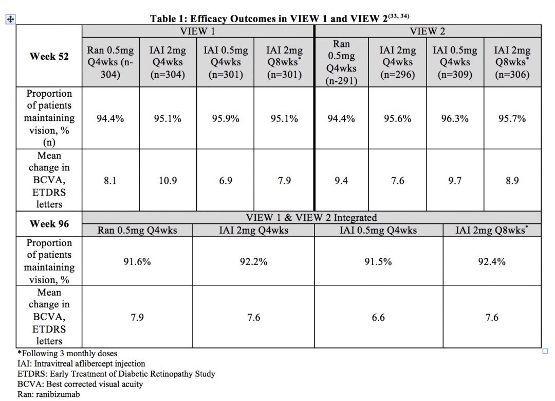
Age-related Macular Degeneration (neovascular with CNV) - VIEW 1 and VIEW 2 were two prospective, multicenter, double-masked, randomized, parallel-group, active-controlled phase 3 studies that evaluated the efficacy and safety of various doses and dosing regimens of IAI compared with ranibizumab in a non-inferiority paradigm in wet AMD. In total, 2,457 patients were randomized in the two trials. The primary endpoint was the proportion of patients who maintained vision (defined as loss of less than 15 ETDRS letters) from baseline at 52 weeks. Patients were followed for 96 weeks. Table 1 summarizes key efficacy results from VIEW 1 and VIEW 2.33,34
Adverse Events
Through week 96 of the VIEW 1 and VIEW 2 studies, the most frequent ocular adverse events (>10% of patients for the overall study population) were conjunctival hemorrhage, eye pain, retinal hemorrhage, and reduced visual acuity. The most frequent serious non-ocular adverse events (>1% of patients for the overall study population) were falls, pneumonia, myocardial infarction, and atrial fibrillation. The incidence of arterial thromboembolic events (ATEs) as defined by the Antiplatelet Trialists’ Collaboration (APTC) criteria was 3.2% for patients receiving ranibizumab and 3.3% for patients receiving IAI. Overall, adverse events were infrequent and occurred with similar rates across all treatment groups.33,34
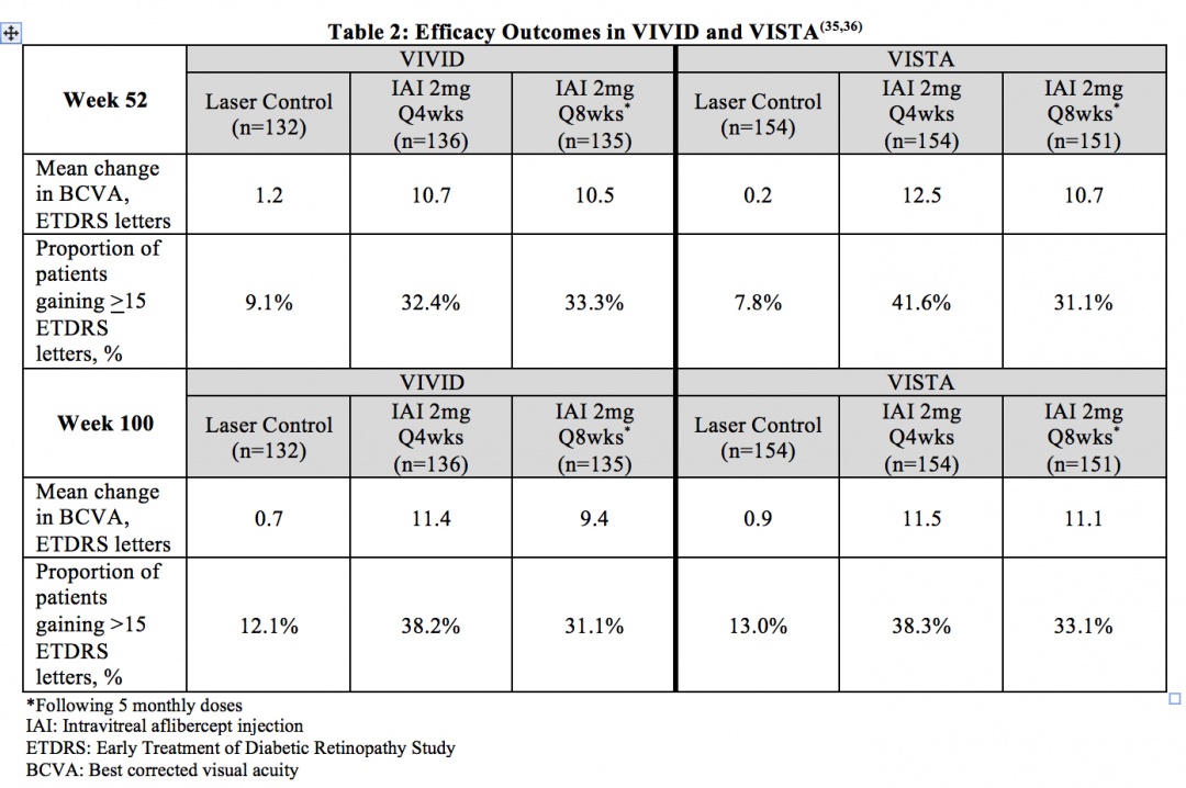
Diabetic Macular Edema (DME) – VIVID and VISTA were two prospective, multicenter, double-masked, randomized, laser-controlled phase 3 studies that evaluated the efficacy and safety of IAI compared with laser in patients with DME. In total, 872 patients were randomized in the two trials. The primary endpoint of VIVID and VISTA was the mean change from baseline of BCVA at week 52 as measured by ETDRS letter score. Patients were followed for a total of 148 weeks. Table 2 summarizes key efficacy results of the two studies.35,36
Adverse Events
The most common adverse reactions, occuring in > 10% of patients receiving IAI through week 100, were conjunctival hemorrhage, cataract, and eye pain. The incidence of non-ocular SAEs was slightly higher for some events in the combined IAI group (e.g. anemia and cerebrovascular accident), and for others in the laser group (e.g. acute myocardial infarction and acute cardiac failure) with no apparent general trend. Arterial thromboembolic events as defined by APTC criteria occurred at similar rates across the IAI treatment groups and the laser control group.35,36
Diabetic Retinopathy (DR) in Patients with DME – In the VIVID and VISTA trials, patients’ Diabetic Retinopathy Severity Score (DRSS) were assessed at baseline and approximately every 6 months thereafter for the duration of the study. The proportion of patients who experienced a > 2 step improvement in DRSS at 100 weeks was included as a secondary endpoint of the studies. In VIVID, 29.3% of patients improved at least 2 steps on ETDRS-DRSS from baseline with IAI 2mg q4 weeks administration and 32.6% with IAI 2 mg q8 weeks administration, compared to 8.2% in the laser control group. In VISTA, 37.1% of patients improved at least 2 steps on ETDRS-DRSS from baseline with Q4 weeks administration and 37.1% with Q8 weeks administration, compared to 15.6% in the laser control group.36
Macular Edema due Retinal Vein Occlusion (RVO)
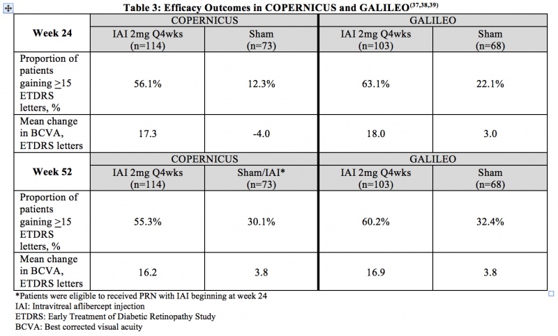
Macular edema due to Central RVO (CRVO) - COPERNICUS and GALILEO were two prospective, multicenter, double-masked, randomized, sham-controlled phase 3 studies that evaluated the efficacy and safety of monthly (2mg q4 weeks) IAI compared with sham in patients with CRVO. In total, 361 patients were randomized in the two trials. The primary endpoint was the proportion of patients who gained at least 15 letters in BCVA at week 24. Table 3 summarizes key efficacy outcomes from the two trials.37,38,39
Adverse Events
Through week 52 in COPERNICUS, the most common ocular adverse events in the IAI arm were conjunctival hemorrhage, eye pain, and maculopathy. The most common ocular adverse event in the sham/IAI arm through week 52 was increased intraocular pressure. Through week 24 in GALILEO, the most common ocular adverse events reported for IAI-treated eyes were eye pain, increased IOP, and conjunctival hemorrhage. From week 24 to week 52 in GALILEO, worsening of macular edema, increased IOP, and reduced visual acuity were the most common ocular adverse events. At week 52 in COPERNICUS, hypertension and upper respiratory tract infection were the most common non-ocular adverse events in both study arms, while in GALILEO, nasopharyngitis was the most common non-ocular adverse event at week 52 in both study arms. APTC-defined arteriothromboembolic events were rare and occurred with similar frequencies in the IAI and sham arms through week 52 in both COPERNICUS and GALILEO.37,38,39
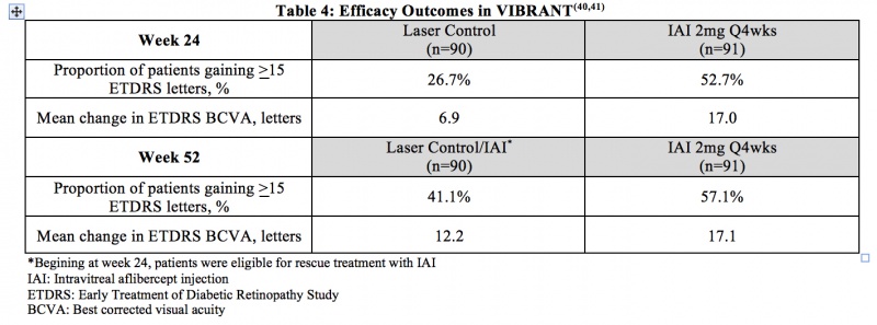
Macular Edema due to Branch RVO (BRVO) – VIBRANT was a prospective, multicenter, double-masked, randomized, laser-controlled phase 3 study that evaluated the efficacy and safety of monthly (2 mg q4 week) IAI compared with laser in patients with BRVO. In total, 183 patients were randomized. The primary endpoint of VIBRANT was the proportion of patients who gained at least 15 ETDRS letters in BCVA at week 24. Table 4 summarizes key efficacy results at week 24 and week 52 from the VIBRANT trial.40,41
lih
Diagnosis
Add text here
History
Add text here
Physical examination
Add text here
Signs
Add text here
Symptoms
Add text here
Clinical diagnosis
Add text here
Diagnostic procedures
Add text here
Laboratory test
Add text here
Differential diagnosis
Add text here
Management
Add text here
General treatment
Add text here
Medical therapy
Add text here
Medical follow up
Add text here
Surgery
Add text here
Surgical follow up
Add text here
Complications
Add text here
Prognosis
Add text here
Additional Resources
Add text here
References
Add text here


