Strabismus Surgery, Horizontal: Difference between revisions
No edit summary |
No edit summary |
||
| Line 113: | Line 113: | ||
== Surgery == | == Surgery == | ||
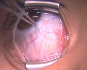
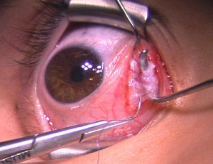
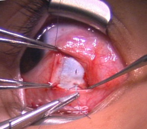
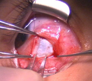
This section outlines the common strabismus surgical techniques and instrumentation, but the same results can be achieved with other techniques.<br>'''Anesthesia and Prep'''<br> Prior to surgery a decision is made as to the type of anesthesia to be performed. All children and many adults are placed under general anesthesia. If general anesthesia is not desired, retrobulbar injections can produce adequate muscle anesthesia. On a cooperative adult, surgery can also be performed under topical anesthesia with IV or oral sedation. <br>Once in the operating room, the periorbital skin is cleaned with povidone-iodine or hexachlorophene, after which the ocular surface is rinsed with half-strength povidone-iodine solution, followed by balanced salt solution. Draping of the face and head is performed to maintain sterility.<br>'''Isolating the muscle'''<br> A lid speculum is used to retract the lids as well as keep the lashes out of the field. If local anesthesia is to be used, a drop of topical anesthetic is applied. With the patient under general anesthesia, the globe requires fixation to prevent rotation and to maintain the field of view. This can be done with either a traction suture, or forceps. Depending on the type of surgery to be performed and the surgeon preference, the first incision through conjunctiva is made either in the fornix or at the limbus. The limbal incision provides better exposure and is thought to cause less scarring because tenon’s capsule is not as traumatized.22 However, the limbal incision is thought to affect the anterior segment blood supply more and can be more irritating secondary to suture placement. A small incision is made through conjunctiva and tenon’s capsule through which local anesthetic can be applied with a cannula in the peribular space. A surgical plane is created down to bare sclera, with both the conjunctiva and tenon’s retracted. A Stevens hook is then passed between tenon’s and sclera, with the plane of the hook parallel to the insertion of the muscle. Traction is placed on the hook towards the limbus to isolate the fibers of the muscle insertion. A larger muscle hook, such as a Jameson or Greene hook, is then passed behind the Stevens hook to ensure all muscle fibers are obtained. The insertion of the muscle is then exposed, by removing any tenon’s attachments if limbal incision or by freeing up and retracting conjunctiva and draping it over the toe of the hook if a fornix incision is used. The intermuscular septum at the toe of the hook is incised and special attention is made to ensure all muscle fibers were obtained. If a resection or adjustable suture procedure is to be performed then it is useful to isolate the superior and inferior aspects of the muscle for dissection of the fascial attachments. By placing a hooks at the superior and inferior borders, the falciform folds of Guerin are exposed and can easily be freed by sharp dissection. | This section outlines the common strabismus surgical techniques and instrumentation, but the same results can be achieved with other techniques.<br>'''Anesthesia and Prep'''<br> Prior to surgery a decision is made as to the type of anesthesia to be performed. All children and many adults are placed under general anesthesia. If general anesthesia is not desired, retrobulbar injections can produce adequate muscle anesthesia. On a cooperative adult, surgery can also be performed under topical anesthesia with IV or oral sedation. <br>Once in the operating room, the periorbital skin is cleaned with povidone-iodine or hexachlorophene, after which the ocular surface is rinsed with half-strength povidone-iodine solution, followed by balanced salt solution. Draping of the face and head is performed to maintain sterility.<br>'''Isolating the muscle'''<br> A lid speculum is used to retract the lids as well as keep the lashes out of the field. If local anesthesia is to be used, a drop of topical anesthetic is applied. With the patient under general anesthesia, the globe requires fixation to prevent rotation and to maintain the field of view. This can be done with either a traction suture, or forceps(Figure 1.1). Depending on the type of surgery to be performed and the surgeon preference, the first incision through conjunctiva is made either in the fornix or at the limbus. The limbal incision provides better exposure and is thought to cause less scarring because tenon’s capsule is not as traumatized.22 However, the limbal incision is thought to affect the anterior segment blood supply more and can be more irritating secondary to suture placement. A small incision is made through conjunctiva and tenon’s capsule through which local anesthetic can be applied with a cannula in the peribular space. A surgical plane is created down to bare sclera, with both the conjunctiva and tenon’s retracted. A Stevens hook is then passed between tenon’s and sclera, with the plane of the hook parallel to the insertion of the muscle(Figure 1.2). Traction is placed on the hook towards the limbus to isolate the fibers of the muscle insertion. A larger muscle hook, such as a Jameson or Greene hook, is then passed behind the Stevens hook to ensure all muscle fibers are obtained(Figure 1.3). The insertion of the muscle is then exposed, by removing any tenon’s attachments if limbal incision or by freeing up and retracting conjunctiva and draping it over the toe of the hook if a fornix incision is used. The intermuscular septum at the toe of the hook is incised and special attention is made to ensure all muscle fibers were obtained. If a resection or adjustable suture procedure is to be performed then it is useful to isolate the superior and inferior aspects of the muscle for dissection of the fascial attachments. By placing a hooks at the superior and inferior borders, the falciform folds of Guerin are exposed and can easily be freed by sharp dissection. | ||
[[Image:Recession 1.jpg|thumb|left | [[Image:Recession 1.jpg|thumb|left]][[Image:Recession 4 copy.jpg|thumb|right]][[Image:Recession 2 copy.jpg|thumb|center|Figure 1.2: Hooking the muscle with Stevens hook]]<br> | ||
<br>'''Recession'''<br> For a recession, the anterior insertion of the muscle is cleaned and adequately exposed. A double armed 6-0 suture on a spatulated needle is used to secure the muscle insertion. Absorbable sutures such as Vicryl( Ethicon) or Polysorb (Covidien) are commonly used. The spatulated needle has a curvature that makes it safer and easier to create scleral passes. Depending on the surgeon and their preferred technique the fibers of the muscle insertion are sutured. One technique is a partial thickness muscle pass exiting at the muscle margin. A full thickness locking whip stitch is then placed at the superior and inferior poles of the muscle. <br> The muscle is disinserted from the globe by placing traction on the muscle hook and the sutures to keep them away from the insertion. Westcott or other scissors are then used to disinsert the muscle from the sclera. The cut stump of the insertion remaining on the sclera is grasped with locking forceps at both the superior and inferior aspects to maintain orientation of the globe and will be used to measure the amount of recession to be performed. Calipers set at a predetermined amount are used to mark the distance posterior from the original insertion for placement of the muscle. The posterior sclera is marked to correspond with both the superior and inferior aspects of the insertion. The needles attached to the previously disinserted muscle are passed partial thickness through the sclera with visualization of the needle through the superficial sclera lamellae. The direction of the pass is angled slightly anterior to parallel with the insertion. Special caution is taken to ensure that a perforation or penetration of the globe does not occur. This is repeated with the second arm of the suture which is passed at the second marked site posterior to the insertion. The scleral exit sites of the needles usually are closely approximated and a popular technique is the “cross swords” to allow for a more secure knot. For a straight recession the sutures are then pulled to bring the muscle up to the new recessed position on the sclera. The sutures are then tied and trimmed. <br> Based on the surgical plan, a hang-back suture can be performed which is done similar to a straight recession except for the placement of the knot is not directly on sclera. For a hang-back suture, the muscle is brought up to the sclera and the sutures are held taught above the sclera. Calipers are used to measure the amount of hang-back along the length of the suture. Locking needle driver is then clamped across the sutures at the premeasured length. The sutures are then tied over the needle driver. The needle driver is removed allowing the muscle to hang-back the predetermined amount. <br> For adjustable sutures, multiple techniques have been described. This builds on the hang-back technique but uses a suture remnant to create a sliding knot around the previously passed scleral sutures. The knot can be slid along the sutures and is brought down to a premeasured distance from the sclera. This same knot can be adjusted later for modification of the ocular alignment, intraoperatively if the patient is under local anesthesia, in the outpatient setting the same day, or up to 1-2 weeks post-op if additional sensorimotor adaptation is desired. In the delayed adjustable technique the overlying conjunctiva is reapproximated, giving patients additional time to develop sensorimotor adaptation. If no adjustment is to be performed all sutures can be left in place and absorbed.23 | <br>'''Recession'''<br> For a recession, the anterior insertion of the muscle is cleaned and adequately exposed. A double armed 6-0 suture on a spatulated needle is used to secure the muscle insertion. Absorbable sutures such as Vicryl( Ethicon) or Polysorb (Covidien) are commonly used. The spatulated needle has a curvature that makes it safer and easier to create scleral passes. Depending on the surgeon and their preferred technique the fibers of the muscle insertion are sutured. One technique is a partial thickness muscle pass exiting at the muscle margin. A full thickness locking whip stitch is then placed at the superior and inferior poles of the muscle. <br> The muscle is disinserted from the globe by placing traction on the muscle hook and the sutures to keep them away from the insertion. Westcott or other scissors are then used to disinsert the muscle from the sclera. The cut stump of the insertion remaining on the sclera is grasped with locking forceps at both the superior and inferior aspects to maintain orientation of the globe and will be used to measure the amount of recession to be performed. Calipers set at a predetermined amount are used to mark the distance posterior from the original insertion for placement of the muscle. The posterior sclera is marked to correspond with both the superior and inferior aspects of the insertion. The needles attached to the previously disinserted muscle are passed partial thickness through the sclera with visualization of the needle through the superficial sclera lamellae. The direction of the pass is angled slightly anterior to parallel with the insertion. Special caution is taken to ensure that a perforation or penetration of the globe does not occur. This is repeated with the second arm of the suture which is passed at the second marked site posterior to the insertion. The scleral exit sites of the needles usually are closely approximated and a popular technique is the “cross swords” to allow for a more secure knot. For a straight recession the sutures are then pulled to bring the muscle up to the new recessed position on the sclera. The sutures are then tied and trimmed. <br> Based on the surgical plan, a hang-back suture can be performed which is done similar to a straight recession except for the placement of the knot is not directly on sclera. For a hang-back suture, the muscle is brought up to the sclera and the sutures are held taught above the sclera. Calipers are used to measure the amount of hang-back along the length of the suture. Locking needle driver is then clamped across the sutures at the premeasured length. The sutures are then tied over the needle driver. The needle driver is removed allowing the muscle to hang-back the predetermined amount. <br> For adjustable sutures, multiple techniques have been described. This builds on the hang-back technique but uses a suture remnant to create a sliding knot around the previously passed scleral sutures. The knot can be slid along the sutures and is brought down to a premeasured distance from the sclera. This same knot can be adjusted later for modification of the ocular alignment, intraoperatively if the patient is under local anesthesia, in the outpatient setting the same day, or up to 1-2 weeks post-op if additional sensorimotor adaptation is desired. In the delayed adjustable technique the overlying conjunctiva is reapproximated, giving patients additional time to develop sensorimotor adaptation. If no adjustment is to be performed all sutures can be left in place and absorbed.23 | ||
Revision as of 13:33, June 28, 2010
Strabismus, abnormal ocular alignment, is one of the most common ocular problems in children, affecting 5% of the preschool population.1 It also occurs in adults de novo or as a result of a lifelong condition. The ocular misalignment can be vertical, horizontal, torsional or a combination of the three. Strabismus can be treated with conservative therapy such as glasses, prisms, patching and/or orthoptic exercises, with a majority of the cases eventually requiring correction with eye muscle surgery. Horizontal eye muscle surgery is performed on patients with exodeviations (out turning), esodeviations (in turning), dissociated vertical deviations, abnormal head postures and/or nystagmus, and is the most common strabismus surgery performed.
Disease Entity
Add text here
Disease
Horizontal strabismus is an ocular misalignment of one or both eyes. The two main forms are esodeviation or exodeviation.
Add text here
Risk Factors
Risk factors for strabismus include:
Prematurity
Amblyopia
Refractive error in children younger than 6 years old
Family history of accommodative esotropia
Family history of infantile esotropia
Craniofacial disorder
Cranial nerve palsies
Non-correctable causes of poor vision in one or both eyes
Thyroid Eye Disease
Genetic Syndromes: Moebius syndrome, Prader-Willi syndrome, mitochondrial myopathies, Down’s Syndrome and many others
Demyelinating or other cerebral disorder
Neuromuscular disorder 1, 2
General Pathology
Add text here
Pathophysiology
Add text here
Primary prevention
Add text here
Diagnosis
Strabismus can be confirmed with a comprehensive ocular evaluation. A thorough history is obtained regarding; age of onset, symptoms, percentage of time the eye(s) is deviated, whether misalignment occurs when fixating on distance or near objects, other health problems, recent trauma and family history. A comprehensive evaluation of a patient with strabismus includes; visual acuity, stereoacuity, 10 prism base down test for ocular preference, measurement of ocular alignment, duction and version movements, slit lamp examination, cycloplegic refraction and fundus ophthalmoscopy.
The amount of misalignment can be measured by a variety of methods based on the age of the patient. In infants and young children who are not cooperative with fixating on an object, misalignment is measured based on the pupillary light reflex using Hirschberg (corneal light reflex alone) or Krimsky (corneal light reflex and prism) methods. If patients are able to fixate on a 20/70 object, cross-cover testing is used to measure the amount of deviation in prism diopters.3 This is done by covering one eye and switching the occluder to the contralateral eye. A deviated eye will show a re-fixation movement when uncovered. Prisms are then held in front of the eye until there is no longer a re-fixation movement. Other methods can be used to evaluate strabismus such as prism cover test and the major amblyoscope (synoptophore). Measurements are taken in the nine cardinal positions of gaze in addition to right head tilt, left head tilt and near. Assessment of ocular torsion is performed using double Maddox rods, the major amblyoscope, Bagolini striated lenses, Lancaster Red-Green test or examination for fundus torsion.
History
Important aspects of patient history are age of onset, whether diplopia is appreciated, percentage of time eye(s) is deviated, misalignment present with distance viewing and/or near viewing, anomalous head positions, family history, medications, health problems, history of trauma and previous ocular treatments used.
Physical examination
- Visual acuity with current correction
- Best corrected visual acuity
- Stereoacuity
- 10 base down test for ocular preference.
- 4 prism base out test or Bagolini lenses to test for suppression
- Ocular motility
- Pupillary examination for afferent pupillary defect
- Worth four-dot test
- Pupillary light reflex (Hirschberg and Krimsky)
- Cover test / alternate cover test/ prism and alternate cover test / simultaneous
- prism cover test.
- Double Maddox Rods / Major Amblyoscope (Synoptophore) / Bagolini Lenses
- Lancaster Red-Green Test/ fundus examination - to evaluate for ocular torsion.
- Near point of convergence
- Fusional vergences
- External exam – to evaluate upper and lower eyelid height and pupillary light
- reflex
- Slit lamp examination
- Cycloplegic Retinoscopy
- Ophthalmoscopy
- Exophthalmometry – when orbital disease is suspected along with other
- measures of optic nerve health (i.e. color vision)
Symptoms
Strabismus Signs and Symptoms:
- Ocular misalignment
- Asymmetry in location of corneal light reflex
- Abnormal head position
- Diplopia
- Closing one eye in bright sunlight
- Asthenopia
- Inability to perceive 3-D
- Visual confusion
Clinical diagnosis
Add text here
Diagnostic procedures
Add text here
Laboratory test
Add text here
Differential diagnosis
Add text here
Management
Add text here
General treatment
The primary goal of strabismus surgery is to improve the alignment of the visual axis of the eyes. Surgery is used to help re-establish binocular single vision (decrease diplopia), increase stereoacuity, improve ocular strain related to intermittent deviations, decrease visual confusion, improve abnormal head posture and restore peripheral visual field.4 Strabismus surgery can also help to restore normal facial appearance by straightening the eyes which can have a large socioeconomic impact. In a study performed by Olitsky et al., patients without strabismus had their photos digitally altered to create the appearance of strabismus. Study subjects were then asked to judge the photos and rate personality characteristics and employment capability of the strabismic and non-strabismic patients. The study found, “Overall, the strabismic faces were judged significantly more negatively, than the nonstrabismic face . . . The effect of esotropia was worse than exotropia.”5
Pre-operative Evaluation
Once it is decided to proceed with strabismus surgery, a determination is made by the ophthalmologist as to which muscles will be operated on. A muscle can be surgically weakened (recession or myotomy) or its antagonistic muscle tightened (resection). A muscle’s insertion can be transposed to change the direction of its force. The number of muscles, type of surgery, amount of surgery and which muscles will be operated on is dependent on the type and amount of deviation.
The versions, or ability of the eyes to move together, are evaluated to assess for paresis, restriction, or clinical evidence of excessive movement in the direction of action of a given muscle. Prism measurements of the angle of deviation are performed in multiple gaze positions which may include the 9 cardinal gaze positions, right tilt, left tilt and near gaze. These measurements help to determine the muscles involved, type of surgery and amount of surgery to be performed. Other tests which can assist surgical planning include; stereoacuity, worth four-dot, test for binocular single vision, evaluation for post-operative diplopia, double Maddox rod or Lancaster red/green test for torsion and amblyoscope / synoptophore testing. Forced duction testing is usually performed intraoperatively to evaluate for mechanical restriction. and may affect the surgical planning. Certain anesthetics, most commonly, Succinylcholine, can affect forced duction testing. It is a short acting depolarizing muscle relaxant used with general anesthesia that causes sustained muscle contraction for up to 20 minutes and should be avoiding when planning to perform forced ductions.
Surgical Decision
The planned amount of resection (strengthening) or recession (weakening) of muscles is derived from retrospective analyses of dose response curves. There are multiple surgical tables published which list the millimeters of resection or recession of a muscle based on prism diopters of deviation. The data is based on deviation in primary gaze and is a general guideline and not a precise predictor of final results. The variability in surgical results is dependent not only on the surgeon’s technique but also on the anatomy and pathology of the patient’s disease.
In infants with congenital esotropia, the success rate of alignment at one year was, 62%.6 In those with a re-occurrence of strabismus, repeat surgical intervention can be performed in most cases increasing the percentage of successful re-alignment. Several large case series report successful surgical realignment rates of 63% to 81% in adults, up to one year after surgery.7 In adult patients with strabismus, an important determination for post-operative success is the resolution of diplopia. Patients younger than age 10 typically can suppress the deviated eye, thus not appreciate post-operative diplopia. It is important, prior to surgery, to have clearly defined goals which are agreed upon by the physician and patient. Post operative goals can be to restore binocular single vision, improve stereopsis, expand peripheral vision, decrease abnormal head position and / or improve the appearance of ocular alignment.
There are some who advocate the use of Fresnel prisms pre-operatively in esotropic patients to determine the surgical dosage. This is thought to help identify those patients in who the deviation increases after surgical correction and also to allow for a time period of fusion pre-operatively so that the patient might be better at maintaining fusion post operatively.8,9 The Prism Adaptation Study, showed 1-year after surgery that those who were operated on based on the prism adapted angle had a satisfactory alignment outcome in 89% of patients versus those operated on without prism adaptation at 79%.10 Arguments against prism adaptation are the fact that children may adapt to greater prism correction than needed thus resulting consecutive deviations. There are also some patients who never prism adapt, as well as the added cost, time and patient compliance with prism glasses are additional factors in this treatment.
To affect horizontal alignment a decision must then be made whether to perform recessions, resections or both. Muscle recessions have a greater effect per millimeter than muscle resections. In addition, post-operative discomfort is less in those with recessions. The disadvantage of recession is that large recessions of muscles can create limitation in the field of action of that muscle. The disadvantage of resections is that discomfort is greater and there is a limit to the amount of resection of a muscle that can be done. A medial rectus resection is best not to exceed 7 mm and lateral rectus resections not more than 10 mm.11 These numbers are chosen to help keep the portion of resected tissue mostly to tendon and also to help prevent globe retraction. The combined resection-recession operation is predominantly used in cases of comitant strabismus in which the deviation is unchanged in different directions of gaze and is the same at distance and near.12 According to E. Helveston, “Weakening of an agonist combined with strengthening the action of the antagonist in the same operative session adds greatly to the effectiveness of each procedure and tends to stabilize the surgical result.”3 Performing bilateral eye muscle recession or resections are thought to be better at correcting incomitant deviations. There is no definitive answer as to which is “better” a resection or a recession, thus is it dependent on the patient’s underlying deviation as well as the surgeon’s experience.
There are modifications to performing straight recessions or resections, which can help to correct secondary deviations. Muscles can be vertically transposed to change the vector force of contraction helping with vertical deviations.13 McClatchy, reported large recessions of the lateral rectus to help decrease, dissociated horizontal deviations (DHD).14 Kestenbaum developed a nomogram for recession and resection of horizontal muscles to correct head turn. A and V patterns of esotropia and vertical deviations can be corrected by vertical transposition of the horizontal muscles. In patients with limitation of motility in a certain direction, the duction deficit can be matched by placing posterior fixation suture “faden procedure” on the contralateral yolk muscle. 15,16,17 Spielmann, suggested slanting the rectus muscle insertions to treat cyclotropia or head tilt.18 Hertle, has found tenotomy and replacement of horizontal muscles to dampen horizontal nystagmus and improve foveation time.19 For deviations that are too small to correct with a recession or resection, a marginal myotomy or central tenotomy can be performed.20,21
Medical therapy
Add text here
Medical follow up
Add text here
Surgery
This section outlines the common strabismus surgical techniques and instrumentation, but the same results can be achieved with other techniques.
Anesthesia and Prep
Prior to surgery a decision is made as to the type of anesthesia to be performed. All children and many adults are placed under general anesthesia. If general anesthesia is not desired, retrobulbar injections can produce adequate muscle anesthesia. On a cooperative adult, surgery can also be performed under topical anesthesia with IV or oral sedation.
Once in the operating room, the periorbital skin is cleaned with povidone-iodine or hexachlorophene, after which the ocular surface is rinsed with half-strength povidone-iodine solution, followed by balanced salt solution. Draping of the face and head is performed to maintain sterility.
Isolating the muscle
A lid speculum is used to retract the lids as well as keep the lashes out of the field. If local anesthesia is to be used, a drop of topical anesthetic is applied. With the patient under general anesthesia, the globe requires fixation to prevent rotation and to maintain the field of view. This can be done with either a traction suture, or forceps(Figure 1.1). Depending on the type of surgery to be performed and the surgeon preference, the first incision through conjunctiva is made either in the fornix or at the limbus. The limbal incision provides better exposure and is thought to cause less scarring because tenon’s capsule is not as traumatized.22 However, the limbal incision is thought to affect the anterior segment blood supply more and can be more irritating secondary to suture placement. A small incision is made through conjunctiva and tenon’s capsule through which local anesthetic can be applied with a cannula in the peribular space. A surgical plane is created down to bare sclera, with both the conjunctiva and tenon’s retracted. A Stevens hook is then passed between tenon’s and sclera, with the plane of the hook parallel to the insertion of the muscle(Figure 1.2). Traction is placed on the hook towards the limbus to isolate the fibers of the muscle insertion. A larger muscle hook, such as a Jameson or Greene hook, is then passed behind the Stevens hook to ensure all muscle fibers are obtained(Figure 1.3). The insertion of the muscle is then exposed, by removing any tenon’s attachments if limbal incision or by freeing up and retracting conjunctiva and draping it over the toe of the hook if a fornix incision is used. The intermuscular septum at the toe of the hook is incised and special attention is made to ensure all muscle fibers were obtained. If a resection or adjustable suture procedure is to be performed then it is useful to isolate the superior and inferior aspects of the muscle for dissection of the fascial attachments. By placing a hooks at the superior and inferior borders, the falciform folds of Guerin are exposed and can easily be freed by sharp dissection.
Recession
For a recession, the anterior insertion of the muscle is cleaned and adequately exposed. A double armed 6-0 suture on a spatulated needle is used to secure the muscle insertion. Absorbable sutures such as Vicryl( Ethicon) or Polysorb (Covidien) are commonly used. The spatulated needle has a curvature that makes it safer and easier to create scleral passes. Depending on the surgeon and their preferred technique the fibers of the muscle insertion are sutured. One technique is a partial thickness muscle pass exiting at the muscle margin. A full thickness locking whip stitch is then placed at the superior and inferior poles of the muscle.
The muscle is disinserted from the globe by placing traction on the muscle hook and the sutures to keep them away from the insertion. Westcott or other scissors are then used to disinsert the muscle from the sclera. The cut stump of the insertion remaining on the sclera is grasped with locking forceps at both the superior and inferior aspects to maintain orientation of the globe and will be used to measure the amount of recession to be performed. Calipers set at a predetermined amount are used to mark the distance posterior from the original insertion for placement of the muscle. The posterior sclera is marked to correspond with both the superior and inferior aspects of the insertion. The needles attached to the previously disinserted muscle are passed partial thickness through the sclera with visualization of the needle through the superficial sclera lamellae. The direction of the pass is angled slightly anterior to parallel with the insertion. Special caution is taken to ensure that a perforation or penetration of the globe does not occur. This is repeated with the second arm of the suture which is passed at the second marked site posterior to the insertion. The scleral exit sites of the needles usually are closely approximated and a popular technique is the “cross swords” to allow for a more secure knot. For a straight recession the sutures are then pulled to bring the muscle up to the new recessed position on the sclera. The sutures are then tied and trimmed.
Based on the surgical plan, a hang-back suture can be performed which is done similar to a straight recession except for the placement of the knot is not directly on sclera. For a hang-back suture, the muscle is brought up to the sclera and the sutures are held taught above the sclera. Calipers are used to measure the amount of hang-back along the length of the suture. Locking needle driver is then clamped across the sutures at the premeasured length. The sutures are then tied over the needle driver. The needle driver is removed allowing the muscle to hang-back the predetermined amount.
For adjustable sutures, multiple techniques have been described. This builds on the hang-back technique but uses a suture remnant to create a sliding knot around the previously passed scleral sutures. The knot can be slid along the sutures and is brought down to a premeasured distance from the sclera. This same knot can be adjusted later for modification of the ocular alignment, intraoperatively if the patient is under local anesthesia, in the outpatient setting the same day, or up to 1-2 weeks post-op if additional sensorimotor adaptation is desired. In the delayed adjustable technique the overlying conjunctiva is reapproximated, giving patients additional time to develop sensorimotor adaptation. If no adjustment is to be performed all sutures can be left in place and absorbed.23
Resection
For resection of a muscle, once the insertion is isolated and cleaned, calipers are used to measure posterior to the insertion the amount of muscle to be resected or removed. A double armed 6-0 absorbable suture is then used to secure the muscle with a central locking bite, partial thickness muscle passes and full thickness locking whip stitches placed at the superior and inferior edges. A straight clamp is placed anterior to the previously passed sutures. The muscle as above is disinserted from the globe and the stump of insertion is grasped with locking forceps to stabilize the globe. The portion of muscle to be resected is excised from the clamp. The cut muscle edge is then cauterized with low temp cautery. After the clamp is removed the sutures are secured to the sclera at the original insertion site through partial thickness scleral passes as described above. Some physicians are proponents of multiple sclera passes due to the greater tension that is created with a resected muscle.
Upon completion of a horizontal muscle surgery, the conjunctival cut edges are approximated and closed. The type of suture varies on physician from Vicryl to chromic gut. The surgeon may choose to perform sub-tenon’s injections of local anesthetic, steroids, antibiotics, and/or viscoelastic material.
Surgical follow up
The postoperative care of patients usually involves examination within 1-2 weeks after surgery, with subsequent visits varying in time from a month to 3 months. Postoperative regimens of topical and/or oral antibiotics, corticosteroids and NSAIDs vary among practitioners. The ophthalmologist who performs the surgery is also responsible for informing the patient of possible complications and concerning signs for which the patient is to notify the physician if they should occur.
If adjustable strabismus surgery is performed, the patient is seen anywhere from the same day as surgery to as late as 5-7 days post-operatively depending on the adjustable technique used. Most adjustments that are done after post-operative day 1 are done in clinic with topical anesthetic medication. Subsequent follow-up is usually at 1-3 months to allow time for stabilization.
Complications
While not a true surgical complication, misalignment of the eye post-operatively is the most common cause for patient dissatisfaction. In almost all cases, the post-operative misalignment can be corrected with time, prisms or additional surgery.
True complications of strabismus surgery occur rarely. Patients are instructed to stop any blood thinners and anti-coagulants, under the supervision of their primary physician, to decrease the risk of intraoperative bleeding. Local anesthetic containing epinephrine can also be used at the time of surgery to help maintain hemostasis. While blood loss during strabismus surgery is minimal, subconjunctival blood looks cosmetically unsatisfactory and can also create increased scarring. Other intraoperative risks are scleral perforation during scleral suture passes or with excising the muscle off the globe.24 In most cases, the perforation does not create a problem other than a chorioretinal scar, but in some cases can trigger endophthalmitis, vitreous hemorrhage or retinal detachment.25 Depending on the depth and location of the perforation, some are treated with prophylactic laser to prevent retinal detachment. The sclera is thinnest right behind the muscle insertion making small recession surgery, more concerning for perforation. While there is a risk to a deep sclera pass, there is also a risk of a slipped muscle, if sutures capture only the superficial muscular capsule and not the muscle tissue or if an adequate depth sclera pass is not made. A slipped muscle is usually not truly “lost” but has slipped back in its muscle capsule. Clinically, patients with a slipped muscle present with a limitation of motility in the field of action of that muscle. Patients thus require further surgical exploration to identify the muscle and re-secure it to the globe, which is often challenging and time consuming.26,27
Infection rate for strabismus surgery is low and is limited by the use of standard surgical technique, dilute betadine applied to the ocular surface as part of surgical prepping and postoperative antibiotic injections subconjunctivally or antibiotic topical drops applied during recovery.28 Infection when it occurs, is most commonly seen 2-3 days after surgery. Patients may present with redness, pain, discharge and/or lid swelling. Significantly less common but more serious is the risk of the infection spreading into the deeper orbital tissues, creating an orbital cellulitis which requires aggressive antibiotic therapy. The conjunctiva when closed can form an epithelial inclusion cyst or granuloma requiring steroid treatment or surgical resection. Also in areas where there is irregularity of the conjunctiva, a dellen can form on the cornea that is treated with close monitoring and aggressive lubrication. Some patients also experience mild refractive changes after strabismus surgery. Lastly in those patients who have had previous muscle surgery and have had 3 ipsilateral rectus muscles operated on are at risk for anterior segment ischemia. The anterior segment blood supply is via the ciliary arteries within the four rectus muscles as well as from conjunctival vessels. In excising a rectus muscle off the globe the ciliary vessels are compromised, affecting the anterior segment circulation. This process although rare, can ultimately result in iritis, corneal edema, anterior segment necrosis, loss of vision or phthysis.29 Some advocate an iris angiogram to ensure adequate anterior segment blood supply in patients with multiple previous strabismus surgeries who are considering undergoing additional strabismus surgery.30
Prognosis
Add text here
Additional Resources
Add text here
References
- Robaei R, Rose KA, Kifley A, Cosstick M, Ip JM, Mitchell P. Factors associated with childhood strabismus: findings from a population-based study. Ophthalmol. 7:113, 2006.
- Chew E, Rernaley N, Tamboli A, Jialiang Z, Podgor M, Klebanoff M. Risk factors for esotropia and exotropia. Arch Ophtahlmol. 10:112, 1994.
- Von Noorden GK, Campos EC. Binocular Vision and Ocular Motility: Theory and management of strabismus. Orbis Cyber-Sight. http://telemedicine.orbis.org/bins/content_page.asp?cid=1-2193-2194
- Mills MD, Coats DK, Donahue SP, Wheeler DT; American Academy of Ophthalmology. Strabismus surgery for adults: a report by the American Academy of Ophthalmology. Ophthalmol. 6:111, 2004.
- Olitsky SE, Sudesh S, Graziano A, Hamblen J, Brooks SE, Shaha SH. The negative psychosocial impact of strabismus in adults. J AAPOS. 4:3, 1999.
- Kampanartsanyakorn S, Surachatkumtonekul T, Dulayajinda D, Jumroendararasmee M, Tongsae S. The outcomes of horizontal strabismus surgery and influencing factors of the surgical success. J Med Assoc Thai, 9:88, 2005.
- Beauchamp GR, Black BC, Coats DK, Enzenauer RW, Hutchinson AK, Saunders RA, Simon JW, Stager DR, Stager DR Jr, Wilson ME, Zobal-Ratner J, Felius J. The management of strabismus in adults--I. Clinical characteristics and treatment. J AAPOS. 2003 ;4:233-40.
- Repka MX, Connet JE, Scott WE: The one-year surgical outcome after prism adaptation for the management of acquired esotropia. Ophthalmology 103:922,1996.
- Scott WE, Thalacker A: Preoperative prism adaptation in acquired esotropia. Ophthalmologica 189:49, 1984.
- Efficacy of prism adaptation in the surgical management of acquired esotropia. Prism Adaptation Study Research Group. Arch Ophthalmol. 9:108, 1990.
- Wright K: Color Atlas of Strabimus Surgery. Philadelphia, JB Lippincott, 1991.
- Bock CJ, Buckley E, Freedman S: Combined resection and recession of a single rectus muscle for the treatment of incomitant strabismus. J Am Assoc Pediatr Ophthalmol Strabismus 3:263, 1999.
- Jones ST: Treatment of hypertropia by vertical displacement of horizontal recti. Am Orthopt J 27:107, 1977.
- Wilson ME, McClatchey SK. Dissociated horizontal deviation. J Pediatric Ophtahlmol Strabismus. 2:28, 1991.
- Paliaga GP, Braga M: Passive limitation of adduction after Cüppers’s "Fadenoperation" on medial recti. Br J Ophthalmol 73:633, 1989.
- Noorden GK von: Posterior fixation suture in strabismus surgery. In Symposium on Strabismus: Transactions of the New Orleans Academy of Ophthalmology. St Louis, Mosby–Year Book, 1978, p 307.
- Scott AB. The faden operation: mechanical effects. Am Orthopt J. 27, 1977.
- Spielmann A: The "oblique Kestenbaum" procedure revisited (sloped recessions of the recti). In Lenk-Schäfer M, ed: Orthoptic Horizons. Transactions of the Sixth International Orthoptic Congress, Harrogate, UK, 1987, p 433.
- Hertle RW. Does eye muscle surgery improve vision in patients with infantile Nystagmus syndrome? Ophthalmol. 10:116, 2009.
- Helveston EM, Cofield DD.: Indications for marginal myotomy and technique. Am J Ophthalmol 70:574, 1970.
- L. Faber JTHN de, Noorden GK von: Medial rectus muscle marginal myotomies for persistent esotropia. Am J Ophthalmol 112:702, 1991.
- Noorden GK von: The limbal approach to surgery of the rectus muscles. Arch Ophthalmol 80:94, 1968.
- Robbins SL, D B Granet DB, Burns C, Freeman RS, Eustis HS, Yafai S, Cruz F, Danylyshyn-Adams K, Langham K. Delayed adjustable sutures: a multicentred clinical review, pending publication in BJO 2010.
- Awad AH, Mullaney PB, Al-Hazmi A, et al. Recognized globe perforation during strabismus surgery: incidence, risk factors, and sequelae. J AAPOS 2000;4:150-3.
- Salamon SM, Friberg TR, Luxenburg MN: Endophthalmitis after strabismus surgery. Am J Ophthalmol 93:39, 1982.
- Plager DA, Parks MM. Recognition and repair of the "lost" rectus muscle. A report of 25 cases. Ophthalmology 1990;97:131-6.
- Lenart TD, Lambert SR. Slipped and lost extraocular muscles. Ophthal Clin North Am 2001;14:433-42.
- Berger RW, Haase W: Complications in strabismus surgery. Strabismus 5:67, 1997.
- Saunders RA, Bluestein EC, Wilson ME, Berland JE. Anterior segment ischemia after strabismus surgery. Surv Ophthalmol 1994;38:456-66.
- Saunders R, Phillips MS: Anterior segment ischemia after three rectus muscle surgery. Ophthalmology 95:533, 1988.
All content on Eyewiki is protected by copyright law and the Terms of Service. This content may not be reproduced, copied, or put into any artificial intelligence program, including large language and generative AI models, without permission from the Academy.


