Phlyctenular Keratoconjunctivitis: Difference between revisions
(Text copyedits and formatting.) |
(Reference formatting to AMA style.) |
||
| Line 20: | Line 20: | ||
= Disease = | = Disease = | ||
Phlyctenular keratoconjunctivitis is a nodular inflammation of the cornea or conjunctiva that results from a hypersensitivity reaction to a foreign antigen. Prior to the 1950s, phlyctenular keratoconjunctivitis often presented as a consequence of a hypersensitivity reaction to tuberculin protein due to high prevalence of tuberculosis. It was typically seen in poor, malnourished children with a positive tuberculin skin test.<ref name=":3">Thygeson P. The etiology and treatment of phlyctenular keratoconjunctivitis. '' | Phlyctenular keratoconjunctivitis is a nodular inflammation of the cornea or conjunctiva that results from a hypersensitivity reaction to a foreign antigen. Prior to the 1950s, phlyctenular keratoconjunctivitis often presented as a consequence of a hypersensitivity reaction to tuberculin protein due to high prevalence of tuberculosis. It was typically seen in poor, malnourished children with a positive tuberculin skin test.<ref name=":3">Thygeson P. The etiology and treatment of phlyctenular keratoconjunctivitis. ''Am J Ophthalmol. ''1951;34(9):1217-1236. | ||
</ref><ref name=":0">Culbertson WW, Huang AJ, Mandelbaum SH, Pflugfelder SC, Boozalis GT, Miller D. Effective treatment of phlyctenular keratoconjunctivitis with oral tetracycline. ''Ophthalmology. ''1993;100(9):1358-1366.</ref><ref name=":1">Rohatgi J, Dhaliwal U. Phlyctenular eye disease: a reappraisal. '' | </ref><ref name=":0">Culbertson WW, Huang AJ, Mandelbaum SH, Pflugfelder SC, Boozalis GT, Miller D. Effective treatment of phlyctenular keratoconjunctivitis with oral tetracycline. ''Ophthalmology. ''1993;100(9):1358-1366.</ref><ref name=":1">Rohatgi J, Dhaliwal U. Phlyctenular eye disease: a reappraisal. ''Jpn J Ophthalmol''. 2000;44(22):146-150.</ref> Following improvements in public health efforts and decreasing rates of tuberculosis, there was a decline in phlyctenular keratoconjunctivitis and subsequent patients were found to have negative tuberculin tests.<ref name=":1" /><ref name=":4">Thygeson P. Observations on nontuberculous phlyctenular keratoconjunctivitis. ''Trans Am Acad Ophthalmol Otolaryngol.'' 1954;58(1):128-132.</ref><ref>Thygeson P. Nontuberculous phlyctenular keratoconjunctivitis. In: Golden B, ed. ''Ocular Inflammatory Disease''. Charles C Thomas Publisher Ltd; 1974.</ref> Currently in the United States, microbial proteins of ''Staphylococcus aureus'' are the most common causative antigens in phlyctenular keratoconjunctivitis.<ref name=":1" /><ref name=":2">Beauchamp GR, Gillette TE, Friendly DS. Phlyctenular keratoconjunctivitis. ''J Pediatr Ophthalmol Strabismus.'' 1981;18(3):22-28.</ref> Risk factors for ''S aureus'' exposure include chronic blepharitis and suppurative keratitis.<ref name=":2" /><ref name=":5">Ostler HB, Lanier JD. Phlyctenular keratoconjunctivitis with special reference to the staphylococcal type. ''Trans Pac Coast Otoophthalmol Soc Annu Meet.'' 1974;55:237-252.</ref> Phlyctenular keratoconjunctivitis is a common cause of pediatric referrals as it occurs primarily in children from 6 months to 16 years old. There is a higher prevalence in females and higher incidence during spring.<ref name=":0" /><ref name=":2" /><ref name=":6">Abu El Asrar AM, Geboes K, Maudgal PC, Emarah MH, Missotten L, Desmet V. Immunocytological study of phlyctenular eye disease. ''Int Ophthalmol''. 1987;10(1):33-39.</ref> | ||
= General Pathophysiology = | = General Pathophysiology = | ||
Phlyctenular keratoconjunctivitis is postulated to occur secondary to an allergic, hypersensitivity reaction at the cornea or conjunctiva, following re-exposure to an infectious antigen that the host has been previously sensitized to.<ref name=":3" /><ref name=":2" /><ref name=":5" /> Antigens of ''S aureus'' and ''Mycobacterium tuberculosis'' are most commonly associated; however, herpes simplex, ''Chlamydia'', ''Streptococcus viridans'', ''Dolosigranulum pigrum'', and intestinal parasites including ''Hymenolepis nana'' have also been reported as causative agents.<ref name=":0" /><ref name=":4" /><ref>Juberias RJ, Calonge M, Montero J, Herreras JM, Saornil AM. Phlyctenular keratoconjunctivitis a potentially blinding disorder. '' | Phlyctenular keratoconjunctivitis is postulated to occur secondary to an allergic, hypersensitivity reaction at the cornea or conjunctiva, following re-exposure to an infectious antigen that the host has been previously sensitized to.<ref name=":3" /><ref name=":2" /><ref name=":5" /> Antigens of ''S aureus'' and ''Mycobacterium tuberculosis'' are most commonly associated; however, herpes simplex, ''Chlamydia'', ''Streptococcus viridans'', ''Dolosigranulum pigrum'', and intestinal parasites including ''Hymenolepis nana'' have also been reported as causative agents.<ref name=":0" /><ref name=":4" /><ref>Juberias RJ, Calonge M, Montero J, Herreras JM, Saornil AM. Phlyctenular keratoconjunctivitis a potentially blinding disorder. ''Ocul Immunol Inflammation. ''1996;4(2):119-123.</ref><ref name=":7">Venkateswaran N, Kalsow CM, Hindman HB. Phlyctenular keratoconjunctivitis associated with Dolosigranulum pigrum. ''Ocul Immunol Inflammation. ''2014;22(3):242-245.</ref><ref>Al-Amry MA, Al-Amri A, Khan AO. Resolution of childhood recurrent corneal phlyctenulosis following eradication of an intestinal parasite. ''J AAPOS.'' 2008;12(1):89-90. </ref><ref>Holland EJ, Mahanti RL, Belongia EA, et al. Ocular involvement in an outbreak of herpes gladiatorum. ''Am J Ophthalmol.'' 1992;114(6):680-684.</ref> | ||
Histologically, scrapings from affected eyes with phlyctenular keratoconjunctivitis infiltrates show predominantly helper T cells, as well as suppressor/cytotoxic T lymphocytes, monocytes, and Langerhans cells.<ref name=":6" /><ref name=":8">Neiberg MN, Sowka J. Phlyctenular keratoconjunctivitis in a patient with Staphylococcal blepharitis and ocular rosacea. ''Optometry. ''2008;79(3):133-137.</ref> The majority of cell scrapings were HLA-DR positive.<ref name=":6" /><ref name=":8" /> The presence of antigen-presenting cells (Langerhans cells), monocytes, and T cells supports the rationale that phlyctenular keratoconjunctivitis is likely due to a delayed cell-mediated reaction. Phlyctenular keratoconjunctivitis may have an association with ocular rosacea, a skin condition that may have a similar underlying type IV hypersensitivity origin.<ref name=":0" /><ref name=":8" /><ref name=":9">Zaidman GW, Brown SI. Orally administered tetracycline for phlyctenular keratoconjunctivitis. '' | Histologically, scrapings from affected eyes with phlyctenular keratoconjunctivitis infiltrates show predominantly helper T cells, as well as suppressor/cytotoxic T lymphocytes, monocytes, and Langerhans cells.<ref name=":6" /><ref name=":8">Neiberg MN, Sowka J. Phlyctenular keratoconjunctivitis in a patient with Staphylococcal blepharitis and ocular rosacea. ''Optometry. ''2008;79(3):133-137.</ref> The majority of cell scrapings were HLA-DR positive.<ref name=":6" /><ref name=":8" /> The presence of antigen-presenting cells (Langerhans cells), monocytes, and T cells supports the rationale that phlyctenular keratoconjunctivitis is likely due to a delayed cell-mediated reaction. Phlyctenular keratoconjunctivitis may have an association with ocular rosacea, a skin condition that may have a similar underlying type IV hypersensitivity origin.<ref name=":0" /><ref name=":8" /><ref name=":9">Zaidman GW, Brown SI. Orally administered tetracycline for phlyctenular keratoconjunctivitis. ''Am J Ophthalmol''. 1981;92(2):187-182.</ref> Previous reports of phlyctenular keratoconjunctivitis with associated asthma and allergies also support the notion of an altered immune mechanism contributing to the pathogenesis.<ref name=":7" /> | ||
=Clinical Presentation and Diagnosis = | =Clinical Presentation and Diagnosis = | ||
| Line 52: | Line 52: | ||
= Complications = | = Complications = | ||
Phlyctenular nodules can lead to ulceration, scarring, and mild to moderate vision loss.<ref name=":0" /><ref name=":9" /> Although rare, corneal perforation is possible as well.<ref name=":0" /><ref>Ostler HB. Corneal | Phlyctenular nodules can lead to ulceration, scarring, and mild to moderate vision loss.<ref name=":0" /><ref name=":9" /> Although rare, corneal perforation is possible as well.<ref name=":0" /><ref>Ostler HB. Corneal perforation in nontuberculosis (staphylococcal) phlyctenular keratoconjunctivitis. ''Am J Ophthalmol''. 1975;79(3):446-448.</ref> | ||
= General Treatment = | = General Treatment = | ||
The first line of treatment for phlyctenular keratoconjunctivitis is to decrease the inflammatory response. Phlyctenulosis is generally responsive to topical steroids. However, risk of increased intraocular pressure has to be kept in mind. In cases with multiple recurrences or those which become steroid dependent, topical cyclosporine A is an effective treatment option.<ref>Doan S, Gabison E, Gatinel D, Duong MH, Abitbol O, Hoang-Xuan T. Topical cyclosporine A in severe steroid-dependent childhood phlyctenular keratoconjunctivitis. '' | The first line of treatment for phlyctenular keratoconjunctivitis is to decrease the inflammatory response. Phlyctenulosis is generally responsive to topical steroids. However, risk of increased intraocular pressure has to be kept in mind. In cases with multiple recurrences or those which become steroid dependent, topical cyclosporine A is an effective treatment option.<ref>Doan S, Gabison E, Gatinel D, Duong MH, Abitbol O, Hoang-Xuan T. Topical cyclosporine A in severe steroid-dependent childhood phlyctenular keratoconjunctivitis. ''Am J Ophthalmol. ''2006;141(1):62-66.</ref> The use of cyclosporine A may reduce the sequelae of long-term steroid use such as cataracts, ocular hypertension, and decreased wound healing. In cases with corneal ulceration, pretreatment or concurrent use of an antibiotic is recommended. Corneal cultures may also be considered prior to starting treatment. | ||
In addition to treating the inflammatory response, it is important to decrease the source of antigens inciting the inflammation. This usually requires treating the associated blepharitis or underlying infectious process. In cases of blepharitis, lid hygiene with warm compresses and lid scrubs should be started. One study found that 1.5% topical azithromycin was effective in treating phlyctenular keratoconjunctivitis with underlying ocular rosacea.<ref>Doan S, Gabison E, Chiambaretta F, Touati M, Cochereau I. Efficacy of azithromycin 1.5% eye drops in childhood ocular rosacea with phlyctenular blepharokeratoconjunctivitis. '' | In addition to treating the inflammatory response, it is important to decrease the source of antigens inciting the inflammation. This usually requires treating the associated blepharitis or underlying infectious process. In cases of blepharitis, lid hygiene with warm compresses and lid scrubs should be started. One study found that 1.5% topical azithromycin was effective in treating phlyctenular keratoconjunctivitis with underlying ocular rosacea.<ref>Doan S, Gabison E, Chiambaretta F, Touati M, Cochereau I. Efficacy of azithromycin 1.5% eye drops in childhood ocular rosacea with phlyctenular blepharokeratoconjunctivitis. ''J Ophthalmic Inflamm Infect''. 2013;3(1):38.</ref> Adjunctive treatment with oral doxycycline may also be of benefit. In children under the age of 8, erythromycin is preferred to prevent dental discoloration from tetracycline use.<ref name=":0" /> | ||
In patients with communicable diseases such as tuberculosis and chlamydia, the underlying infections should be properly addressed and treated appropriately. Chlamydia-induced phlyctenular keratoconjunctivitis should be treated with azithromycin or doxycycline. Patients with positive tuberculin tests should be referred to receive proper systemic treatment of tuberculosis. Close contacts should also be evaluated and treated fittingly. | In patients with communicable diseases such as tuberculosis and chlamydia, the underlying infections should be properly addressed and treated appropriately. Chlamydia-induced phlyctenular keratoconjunctivitis should be treated with azithromycin or doxycycline. Patients with positive tuberculin tests should be referred to receive proper systemic treatment of tuberculosis. Close contacts should also be evaluated and treated fittingly. | ||
Latest revision as of 10:28, March 25, 2025
All content on Eyewiki is protected by copyright law and the Terms of Service. This content may not be reproduced, copied, or put into any artificial intelligence program, including large language and generative AI models, without permission from the Academy.
Disease
Phlyctenular keratoconjunctivitis is a nodular inflammation of the cornea or conjunctiva that results from a hypersensitivity reaction to a foreign antigen. Prior to the 1950s, phlyctenular keratoconjunctivitis often presented as a consequence of a hypersensitivity reaction to tuberculin protein due to high prevalence of tuberculosis. It was typically seen in poor, malnourished children with a positive tuberculin skin test.[2][3][4] Following improvements in public health efforts and decreasing rates of tuberculosis, there was a decline in phlyctenular keratoconjunctivitis and subsequent patients were found to have negative tuberculin tests.[4][5][6] Currently in the United States, microbial proteins of Staphylococcus aureus are the most common causative antigens in phlyctenular keratoconjunctivitis.[4][7] Risk factors for S aureus exposure include chronic blepharitis and suppurative keratitis.[7][8] Phlyctenular keratoconjunctivitis is a common cause of pediatric referrals as it occurs primarily in children from 6 months to 16 years old. There is a higher prevalence in females and higher incidence during spring.[3][7][9]
General Pathophysiology
Phlyctenular keratoconjunctivitis is postulated to occur secondary to an allergic, hypersensitivity reaction at the cornea or conjunctiva, following re-exposure to an infectious antigen that the host has been previously sensitized to.[2][7][8] Antigens of S aureus and Mycobacterium tuberculosis are most commonly associated; however, herpes simplex, Chlamydia, Streptococcus viridans, Dolosigranulum pigrum, and intestinal parasites including Hymenolepis nana have also been reported as causative agents.[3][5][10][11][12][13]
Histologically, scrapings from affected eyes with phlyctenular keratoconjunctivitis infiltrates show predominantly helper T cells, as well as suppressor/cytotoxic T lymphocytes, monocytes, and Langerhans cells.[9][14] The majority of cell scrapings were HLA-DR positive.[9][14] The presence of antigen-presenting cells (Langerhans cells), monocytes, and T cells supports the rationale that phlyctenular keratoconjunctivitis is likely due to a delayed cell-mediated reaction. Phlyctenular keratoconjunctivitis may have an association with ocular rosacea, a skin condition that may have a similar underlying type IV hypersensitivity origin.[3][14][15] Previous reports of phlyctenular keratoconjunctivitis with associated asthma and allergies also support the notion of an altered immune mechanism contributing to the pathogenesis.[11]
Clinical Presentation and Diagnosis
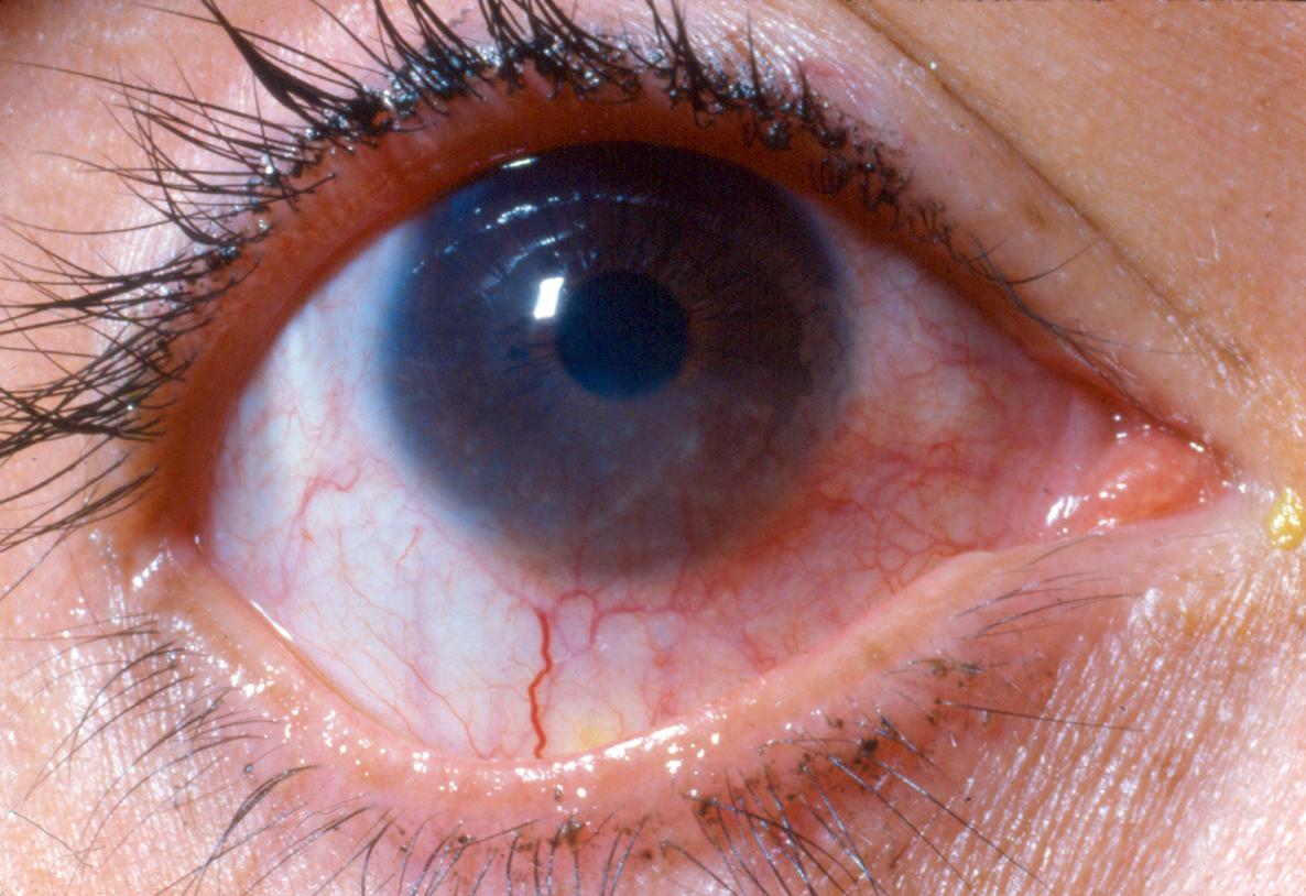
The clinical presentation of phlyctenulosis is dependent on the location of the lesion as well as the underlying etiology. Conjunctival lesions may cause only mild to moderate irritation of the eye, while corneal lesions typically may have more severe pain and photophobia. More severe light sensitivity can also be associated with tuberculosis-related phlyctenules compared to S aureus–related phlyctenules.[8] Phlyctenules can occur anywhere on the conjunctiva but are more common in the interpalpebral fissure and are frequently noted along the limbal region. They commonly present with a gelatinous, nodular lesion with marked injection of the surrounding conjunctival vessels. The lesions may show some degree of ulceration and staining with fluorescein as they progress. In some cases, multiple 1- to 2-mm nodules may be present along the limbal surface.
Corneal phlyctenules similarly begin along the limbal region and frequently degenerate to corneal ulceration and neovascularization. In some instances, the phlyctenule will progress across the corneal surface due to repeated episodes of inflammation along the central edge of the lesion. These “marching phlyctenules” demonstrate an elevated leading edge trailed by a leash of vessels.
The diagnosis of phlyctenular keratoconjunctivitis is made based on history and clinical exam findings. The underlying infectious etiology requires further investigation when the possibility of tuberculosis or chlamydia is suspected. Chest radiographs, purified protein derivative skin testing, or QuantiFERON-TB Gold testing should be ordered for patients with a history of travel to tuberculosis-endemic regions or symptoms consistent with tuberculosis infection. For patients suspected of having chlamydia, immunofluorescent antibody testing and PCR of conjunctival swabs provide quick and accurate screening. If positive, appropriate systemic treatment of these infections is required as well as screening and possible treatment of close contacts.
Differential Diagnosis
- Acne rosacea keratitis
- Rosacea keratoconjunctivitis
- Staphylococcal marginal keratitis
- Nodular episcleritis
- Salzmann nodules
- Trachoma
- Inflamed pingueculum/pterygium
- Vernal keratoconjunctivitis
- Infectious corneal ulcer with vascularization
- Catarrhal ulcer
- Peripheral ulcerative keratitis
- Herpes simplex keratitis or keratoconjunctivitis
Complications
Phlyctenular nodules can lead to ulceration, scarring, and mild to moderate vision loss.[3][15] Although rare, corneal perforation is possible as well.[3][16]
General Treatment
The first line of treatment for phlyctenular keratoconjunctivitis is to decrease the inflammatory response. Phlyctenulosis is generally responsive to topical steroids. However, risk of increased intraocular pressure has to be kept in mind. In cases with multiple recurrences or those which become steroid dependent, topical cyclosporine A is an effective treatment option.[17] The use of cyclosporine A may reduce the sequelae of long-term steroid use such as cataracts, ocular hypertension, and decreased wound healing. In cases with corneal ulceration, pretreatment or concurrent use of an antibiotic is recommended. Corneal cultures may also be considered prior to starting treatment.
In addition to treating the inflammatory response, it is important to decrease the source of antigens inciting the inflammation. This usually requires treating the associated blepharitis or underlying infectious process. In cases of blepharitis, lid hygiene with warm compresses and lid scrubs should be started. One study found that 1.5% topical azithromycin was effective in treating phlyctenular keratoconjunctivitis with underlying ocular rosacea.[18] Adjunctive treatment with oral doxycycline may also be of benefit. In children under the age of 8, erythromycin is preferred to prevent dental discoloration from tetracycline use.[3]
In patients with communicable diseases such as tuberculosis and chlamydia, the underlying infections should be properly addressed and treated appropriately. Chlamydia-induced phlyctenular keratoconjunctivitis should be treated with azithromycin or doxycycline. Patients with positive tuberculin tests should be referred to receive proper systemic treatment of tuberculosis. Close contacts should also be evaluated and treated fittingly.
In rare instances of corneal perforation, surgical treatment may be required. Options for treating peripheral perforations include corneal gluing, amniotic membrane grafting, or corneal patch grafts.
References
- ↑ American Academy of Ophthalmology. Staph phlyctenular keratoconjunctivitis. https://www.aao.org/image/staph-phlyctenular-keratoconjunctivitis-2 Accessed October 10, 2017.
- ↑ Jump up to: 2.0 2.1 Thygeson P. The etiology and treatment of phlyctenular keratoconjunctivitis. Am J Ophthalmol. 1951;34(9):1217-1236.
- ↑ Jump up to: 3.0 3.1 3.2 3.3 3.4 3.5 3.6 Culbertson WW, Huang AJ, Mandelbaum SH, Pflugfelder SC, Boozalis GT, Miller D. Effective treatment of phlyctenular keratoconjunctivitis with oral tetracycline. Ophthalmology. 1993;100(9):1358-1366.
- ↑ Jump up to: 4.0 4.1 4.2 Rohatgi J, Dhaliwal U. Phlyctenular eye disease: a reappraisal. Jpn J Ophthalmol. 2000;44(22):146-150.
- ↑ Jump up to: 5.0 5.1 Thygeson P. Observations on nontuberculous phlyctenular keratoconjunctivitis. Trans Am Acad Ophthalmol Otolaryngol. 1954;58(1):128-132.
- ↑ Thygeson P. Nontuberculous phlyctenular keratoconjunctivitis. In: Golden B, ed. Ocular Inflammatory Disease. Charles C Thomas Publisher Ltd; 1974.
- ↑ Jump up to: 7.0 7.1 7.2 7.3 Beauchamp GR, Gillette TE, Friendly DS. Phlyctenular keratoconjunctivitis. J Pediatr Ophthalmol Strabismus. 1981;18(3):22-28.
- ↑ Jump up to: 8.0 8.1 8.2 Ostler HB, Lanier JD. Phlyctenular keratoconjunctivitis with special reference to the staphylococcal type. Trans Pac Coast Otoophthalmol Soc Annu Meet. 1974;55:237-252.
- ↑ Jump up to: 9.0 9.1 9.2 Abu El Asrar AM, Geboes K, Maudgal PC, Emarah MH, Missotten L, Desmet V. Immunocytological study of phlyctenular eye disease. Int Ophthalmol. 1987;10(1):33-39.
- ↑ Juberias RJ, Calonge M, Montero J, Herreras JM, Saornil AM. Phlyctenular keratoconjunctivitis a potentially blinding disorder. Ocul Immunol Inflammation. 1996;4(2):119-123.
- ↑ Jump up to: 11.0 11.1 Venkateswaran N, Kalsow CM, Hindman HB. Phlyctenular keratoconjunctivitis associated with Dolosigranulum pigrum. Ocul Immunol Inflammation. 2014;22(3):242-245.
- ↑ Al-Amry MA, Al-Amri A, Khan AO. Resolution of childhood recurrent corneal phlyctenulosis following eradication of an intestinal parasite. J AAPOS. 2008;12(1):89-90.
- ↑ Holland EJ, Mahanti RL, Belongia EA, et al. Ocular involvement in an outbreak of herpes gladiatorum. Am J Ophthalmol. 1992;114(6):680-684.
- ↑ Jump up to: 14.0 14.1 14.2 Neiberg MN, Sowka J. Phlyctenular keratoconjunctivitis in a patient with Staphylococcal blepharitis and ocular rosacea. Optometry. 2008;79(3):133-137.
- ↑ Jump up to: 15.0 15.1 Zaidman GW, Brown SI. Orally administered tetracycline for phlyctenular keratoconjunctivitis. Am J Ophthalmol. 1981;92(2):187-182.
- ↑ Ostler HB. Corneal perforation in nontuberculosis (staphylococcal) phlyctenular keratoconjunctivitis. Am J Ophthalmol. 1975;79(3):446-448.
- ↑ Doan S, Gabison E, Gatinel D, Duong MH, Abitbol O, Hoang-Xuan T. Topical cyclosporine A in severe steroid-dependent childhood phlyctenular keratoconjunctivitis. Am J Ophthalmol. 2006;141(1):62-66.
- ↑ Doan S, Gabison E, Chiambaretta F, Touati M, Cochereau I. Efficacy of azithromycin 1.5% eye drops in childhood ocular rosacea with phlyctenular blepharokeratoconjunctivitis. J Ophthalmic Inflamm Infect. 2013;3(1):38.


