Gonioscopic Angle Imaging: Difference between revisions
No edit summary |
|||
| Line 17: | Line 17: | ||
<youtube>XOr-X-JfxCU</youtube> | <youtube>XOr-X-JfxCU</youtube> | ||
(Video recorded with smartphone showing gonioscopy) | (Video recorded with smartphone showing gonioscopy)[[File:GonioPen.png|alt=Goniopen angle imaging device from Singapore|thumb|188x188px|Goniopen angle imaging device from Singapore]]EyeCam (Clarity Medical Systems, Pleasanton, CA, USA), designed for widefield pediatric fundus imaging, can also be used for Angle Photography by addition of a 130 degree wide-field lens.<ref>Quek DTL, Nongpiur ME, Perera SA, Aung T. Angle imaging: Advances and challenges. ''Indian J Ophthalmol''. 2011;59(Suppl1):S69-S75. doi:10.4103/0301-4738.73699</ref> | ||
EyeCam (Clarity Medical Systems, Pleasanton, CA, USA), designed for widefield pediatric fundus imaging, can also be used for Angle Photography by addition of a 130 degree wide-field lens.<ref>Quek DTL, Nongpiur ME, Perera SA, Aung T. Angle imaging: Advances and challenges. ''Indian J Ophthalmol''. 2011;59(Suppl1):S69-S75. doi:10.4103/0301-4738.73699</ref> | |||
A team from Singapore is working on GonioPen,<ref>Shinoj VK, Murukeshan VM, Baskaran M, Aung T. Integrated flexible handheld probe for imaging and evaluation of iridocorneal angle. ''J Biomed Opt''. 2015;20(1):016014. doi:10.1117/1.JBO.20.1.016014</ref> a handheld angle photography device, which provides high resolution iridocorneal angle photographs. It is a small, compact device that can be used by a technician with minimal training.<ref>Hong XJJ, Shinoj VK, Murukeshan VM, Baskaran M, Aung T. Preclinical imaging of iridocorneal angle and fundus using a modified integrated flexible handheld probe. ''J Med Imaging (Bellingham)''. 2017;4(2):026001. doi:10.1117/1.JMI.4.2.026001</ref> | A team from Singapore is working on GonioPen,<ref>Shinoj VK, Murukeshan VM, Baskaran M, Aung T. Integrated flexible handheld probe for imaging and evaluation of iridocorneal angle. ''J Biomed Opt''. 2015;20(1):016014. doi:10.1117/1.JBO.20.1.016014</ref> a handheld angle photography device, which provides high resolution iridocorneal angle photographs. It is a small, compact device that can be used by a technician with minimal training.<ref>Hong XJJ, Shinoj VK, Murukeshan VM, Baskaran M, Aung T. Preclinical imaging of iridocorneal angle and fundus using a modified integrated flexible handheld probe. ''J Med Imaging (Bellingham)''. 2017;4(2):026001. doi:10.1117/1.JMI.4.2.026001</ref> | ||
| Line 28: | Line 25: | ||
= Indications = | = Indications = | ||
All indications of gonioscopy are indications of gonioscopic imaging | * All indications of gonioscopy are indications of gonioscopic imaging | ||
* Objective documentation of gonioscopic findings | |||
Objective documentation of gonioscopic findings | |||
= Limitations = | = Limitations = | ||
Imaging resolution is limited based on the quality of the camera and the lighting | * Imaging resolution is limited based on the quality of the camera and the lighting | ||
Nothing currently compares to the resolution of direct visualization of gonioscopy | * Nothing currently compares to the resolution of direct visualization of gonioscopy | ||
<youtube>9LFeeNX8JL8</youtube> | <youtube>9LFeeNX8JL8</youtube> | ||
= References = | = References = | ||
<references /> | <references /> | ||
Revision as of 10:10, June 1, 2020
All content on Eyewiki is protected by copyright law and the Terms of Service. This content may not be reproduced, copied, or put into any artificial intelligence program, including large language and generative AI models, without permission from the Academy.
Diagnostic Intervention
Gonioscopic Angle Imaging
Overview
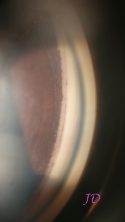
Gonioscopy is a very important part of glaucoma evaluation, but is often underrated. In addition to differentiating between open and closed angle glaucoma, other clues in the angle also may help in diagnosis. Documenting the gonioscopic photographs would help in objective assessment. This article covers the latest techniques in gonioscopic imaging.
In addition to the conventional single-mirror, two-mirror, three-mirror and four-mirror gonioprisms, there is also a six-mirror Gonio Lens named Volk G-6 goniolens (Volk Optical, Inc., Mentor, OH) which helps to do a more complete angle examination without rotating the goniolens. Angle photography can be done with slitlamp cameras using a goniolens, but is difficult to get correct focus and exposure. Smartphone cameras can also be used with a slitlamp adapter to take gonioscopic photographs and videos.
(Video recorded with smartphone showing gonioscopy)
EyeCam (Clarity Medical Systems, Pleasanton, CA, USA), designed for widefield pediatric fundus imaging, can also be used for Angle Photography by addition of a 130 degree wide-field lens.[1]
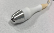
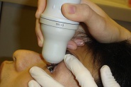
A team from Singapore is working on GonioPen,[2] a handheld angle photography device, which provides high resolution iridocorneal angle photographs. It is a small, compact device that can be used by a technician with minimal training.[3]
The new GS-1 Gonioscope (Nidek, Aichi, Japan) released in September 2018,[4] is a table-top, contact-based, angle photography device that takes gonioscopic photographs and stitiches them together in 360 degrees. It captures using a 16 mirror goniolens and takes images at multiple focal points so that focus can be adjusted on to different angle structures later.[5]
Direct Smartphone Imaging of Goniscopy can be done without a slitlamp as shown by Dr Nilesh Kumar et al.[6]
Indications
- All indications of gonioscopy are indications of gonioscopic imaging
- Objective documentation of gonioscopic findings
Limitations
- Imaging resolution is limited based on the quality of the camera and the lighting
- Nothing currently compares to the resolution of direct visualization of gonioscopy
References
- ↑ Quek DTL, Nongpiur ME, Perera SA, Aung T. Angle imaging: Advances and challenges. Indian J Ophthalmol. 2011;59(Suppl1):S69-S75. doi:10.4103/0301-4738.73699
- ↑ Shinoj VK, Murukeshan VM, Baskaran M, Aung T. Integrated flexible handheld probe for imaging and evaluation of iridocorneal angle. J Biomed Opt. 2015;20(1):016014. doi:10.1117/1.JBO.20.1.016014
- ↑ Hong XJJ, Shinoj VK, Murukeshan VM, Baskaran M, Aung T. Preclinical imaging of iridocorneal angle and fundus using a modified integrated flexible handheld probe. J Med Imaging (Bellingham). 2017;4(2):026001. doi:10.1117/1.JMI.4.2.026001
- ↑ NIDEK launches the GS-1 Gonioscope | News | NIDEK CO.,LTD. Accessed May 3, 2020. https://www.nidek-intl.com/news-event/news/entry-3367.html
- ↑ Gonioscope GS-1 | Retina & Glaucoma | NIDEK CO.,LTD. Accessed May 3, 2020. https://www.nidek-intl.com/product/ophthaloptom/diagnostic/dia_retina/gs-1.html
- ↑ Kumar N, Francesco B, Sharma A. Smartphone-based Gonio-Imaging: A Novel Addition to Glaucoma Screening Tools. J Glaucoma. 2019;28(9):e149-e150. doi:10.1097/IJG.0000000000001306


