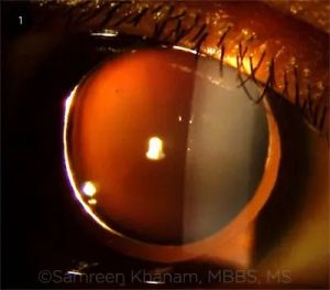
Weill-Marchesani Syndrome
All content on Eyewiki is protected by copyright law and the Terms of Service. This content may not be reproduced, copied, or put into any artificial intelligence program, including large language and generative AI models, without permission from the Academy.
Disease Entity
Disease
Weill-Marchesani syndrome, also known as Spherophakia-Brachymorphia syndrome and Mesodermal dysmorphodystrophy, is an inherited connective tissue disorder characterized by abnormalities of the lens of the eye, secondary glaucoma, short stature, brachydactyly, joint stiffness, and cardiovascular defects.[1]
Weill-Marchesani syndrome may be inherited in Autosomal Dominant or Autosomal Recessive patterns.
Autosomal Recessive Weill-Marchesani syndrome (WMS) often presents with microspherophakia and cardiac anomalies.[2]
Autosomal Dominant Weill-Marchesani syndrome often presents with ectopia lentis and joint limitations.[2]
However, autosomal dominant WMS cannot be distinguished from autosomal recessive WMS based on clinical findings alone and at this time there is consensus on clinical diagnostic criteria.
Genetics
Weill-Marchesani syndrome can present either as a sporadic mutation or via an autosomal recessive or autosomal dominant inheritance pattern, as mentioned. Four genes have been found to be associated with Weill-Marchesani syndrome. Most of those affected display similar clinical manifestations despite genetic heterogeneity. Intra and interfamilial variable expressivity is observed in Weill-Marchesani syndrome.[3] There are four subtypes based on the genes affected in the disease.
Autosomal Dominant Weill-Marchesani Syndrome Gene: FBN-1 (WMS 2)
Autosomal Recessive Weill-Marchesani Syndrome Genes: ADAMTS10 (WMS 1), ADAMTS17 (WMS 4), and LTPBP2 (WMS 3)
Autosomal recessive and autosomal dominant inheritance account for roughly 45% and 39% of Weill-Marchesani syndrome cases, respectively, while the remaining cases are sporadic.[2]
Generally, genes associated with Weill-Marchesani syndrome provide instructions for the production of enzymes and extracellular matrix proteins responsible for connective tissue integrity.
Epidemiology
Weill-Marchesani syndrome is a rare disease in the general population. The prevalence is estimated to be 1 case in 100,000 in the population.[3]
Pathophysiology
Fibrillin, a glycoprotein, is implicated directly in the pathogenesis of Weill-Marchesani syndrome by defects in the FBN-1 gene. The ADAMTS superfamily of proteins act as zinc metalloproteases that appear to be involved in proteoglycan processing. ADAMTS proteins partner with Fibrillin-1 to fulfill its structural and regulatory role and biogenesis. The exact mechanism is unknown, but clinical presentation resembling Weill-Marchesani syndrome with defective ADAMTS and normal FBN-1 suggests an association between the two sets of genes.[4]
Fibrillin is the principal component of the ciliary zonule which exists as a structural scaffold of extensible microfibrils. Zonular weakness, due to impairment in extracellular matrix proteins and enzymes involved with the production of fibrillin, allows for a highly mobile lens which can lead to pupillary block with anterior dislocation of the lens.[5] Mobility of the lens can lead to reduced vision due to refractive shift as well as dysregulation of intraocular pressure. Decreased visual acuity also results from an abnormally shaped, hyper-spherical, lens in Weill-Marchesani syndrome. This is due to increased refractive power of the hyper-spherical lens. This spherical change results in myopia.
Fibrillin is also present in many connective tissues including blood vessels, lung, ligaments, and dermis.[6] Dysfunctional fibrillin can explain the systemic disease manifestations of WMS. Individuals with certain FBN-1 variants have been shown to be of particularly short stature.[7] A specific variant of ADAMTS10 has been found in association with heart-developed membranes in the supra-pulmonic, supramitral, and subaortic areas, creating stenosis that recurred after surgical resection.[8] The heterogeneous nature of this syndrome is quite apparent.
Diagnosis
Definitive diagnosis can be made in a proband by identification of biallelic pathogenic variants in ADAMTS10, ADAMTS17, and LTPBP2 or by a heterozygous pathogenic variant in FBN-1 by molecular genetic testing if clinical presentation is inconclusive. Genetic testing can be performed via serial single-gene testing in individuals with a high clinical suspicion, or via comprehensive genomic testing when the phenotype is indistinguishable from other connective tissue abnormalities. Exome sequencing is the most commonly used comprehensive genomic testing approach.[3]
Ophthalmoscopic examination and anterior segment Optical Coherence Tomography (OCT) can aid in identifying lens shape and position abnormalities.[9]
A comprehensive physical exam is essential identifying other clinical manifestations like brachydactyly, short stature, and cardiac abnormalities.
Clinical Presentation
Ocular abnormalities are typically recognized in childhood (mean age 7.5 years) with routine eye examination. Findings may include microspherophakia (small spherical lens), myopia secondary to an abnormally shaped lens, lens dislocation/abnormal positioning, secondary glaucoma, and increased corneal thickness.[10] Individuals may present with severe myopia with narrow anterior chambers but absent myopic retinopathy.[3]
Systemic presenting features may include short stature, brachydactyly (short fingers or toes), progressive joint stiffness, thickened skin, pseudomuscular build, cardiovascular defects, and mild intellectual disability.
Cardiovascular defects may include a patent ductus arteriosis, pulmonic stenosis, thoracic aortic aneurysm, cervical artery dissection, or prolonged QT interval.[3]
Radiographic findings may include shortened long tubular bones, delayed bone age, and broad proximal phalanges.[3]
Differential diagnosis
- Ectopia Lentis
- Homocystinuria
- Marfan syndrome
- Sulfite Oxidase Deficiency
- Acromelic Dysplasia
- Geleophysic Dysplasia
- Acromicric Dysplasia
- Myhre Syndrome
Management
There is no cure for Weill-Marchesani syndrome. Management is focused on treating the visual and systemic manifestations.
Ophthalmic Management
Annual ocular examinations of affected patients and family members of patients with Weill-Marchesani syndrome allow for early diagnosis of Weill-Marchesani syndrome. Early detection and treatment of disease manifestations can help optimize and preserve visual function. Additionally, genetic evaluation can be considered for family members of patients with Weill-Marchesani syndrome to determine genetic risk and carrier status.
Ophthalmic miotics and mydriatics should be avoided to decrease the risk of pupillary block in these patients. Early removal of the microspherophakic lens can be considered to improve visual acuity, control intraocular pressure, and to prevent pupillary block and glaucoma. Glaucoma can also be managed with peripheral iridectomy and trabeculectomy. [3] Increased corneal thickness in patients with Weill-Marchesani syndrome needs to be accounted for in glaucoma monitoring and treatment as it can affect intraocular pressure readings. Individuals should be counseled on the risk of potential ocular complications with contact sports.
Systemic Management
Regular medical examinations are recommended to assess growth and mobility issues. Systemic symptoms may be managed with physical therapy for joint issues. Regular cardiac follow-up to screen for anomalies, with potential echocardiogram and electrocardiography, are important as cardiac issues can be life threatening.
General anesthesia may be difficult in these individuals due to stiff joints and maxillary hypoplasia. [11]Extra considerations and appropriate patient counseling are important in surgical planning.
Prognosis
Weill-Marchesani syndrome has a variable prognosis but with early detection and timely management outcomes can be favorable.
Conclusion
Weill-Marchesani syndrome is a rare genetic condition that involves ocular and systemic abnormalities. It is classified into four subtypes according to the affected genes. There is no cure for Weill- Marchesani syndrome. Management is focused on treating the active disease processes.
References
- ↑ Weill-Marchesani Syndrome. Genetic and Rare Diseases Information Center (GARD). 2017. https://rarediseases.info.nih.gov/diseases/4936/weill-marchesani-syndrome. Accessed March 1 2021.
- ↑ Jump up to: 2.0 2.1 2.2 Faivre L, Dollfus H, Lyonnet S, et al. Clinical homogeneity and genetic heterogeneity in Weill–Marchesani syndrome. Am J Me Genet A. 2003:123(2), 204-207.
- ↑ Jump up to: 3.0 3.1 3.2 3.3 3.4 3.5 3.6 Marzin P, Cormier-Daire V, Tsilou E. Weill-Marchesani Syndrome. 2007 Nov 1 [Updated 2020 Dec 10]. In: Adam MP, Ardinger HH, Pagon RA, et al., editors. GeneReviews® [Internet]. Seattle (WA): University of Washington, Seattle; 1993-2021. Available from: https://www.ncbi.nlm.nih.gov/books/NBK1114/.
- ↑ Hubmacher D, Apte SS. Genetic and functional linkage between ADAMTS superfamily proteins and fibrillin-1: a novel mechanism influencing microfibril assembly and function. Cell Mol Life Sci. 2011:68(19), 3137-3148.
- ↑ Yazgan S, Çelik T, Çelik E. Insufficiency of YAG laser iridotomy to prevent pupillary Block glaucoma in a microspherophakic patient with Weill-Marchesani syndrome. İstanbul Med J. 2018:19, 73-5.
- ↑ Ashworth JL, Kielty CM, McLeod D. (2000). Fibrillin and the eye. Br J Ophthalmol. 2000:84(11), 1312-1317.
- ↑ Marzin P, Rondeau S, Alessandri JL, Dieterich K, le Goff C, Mahaut C, Mercier S, Michot C, Moldovan O, Miolo G, Rossi M, Van-Gils J, Francannet C, Robert MP, Jaïs JP, Huber C, Cormier-Daire V. Weill-Marchesani syndrome: natural history and genotype-phenotype correlations from 18 news cases and review of literature. J Med Genet. 2024 Jan 19;61(2):109-116.
- ↑ Levitas A, Aspit L, Lowenthal N, Shaki D, Krymko H, Slanovic L, Yagev R, Parvari R. A Novel Mutation in the ADAMTS10 Associated with Weil-Marchesani Syndrome with a Unique Presentation of Developed Membranes Causing Severe Stenosis of the Supra Pulmonic, Supramitral, and Subaortic Areas in the Heart. Int J Mol Sci. 2023 May 16;24(10):8864.
- ↑ Guo H, Wu X, Cai K, Qiao Z. Weill-Marchesani syndrome with advanced glaucoma and corneal endothelial dysfunction: a case report and literature review. BMC ophthalmol, 2015:15(1), 1-4.
- ↑ Roszkowska AM, Aragona, P. Corneal microstructural analysis in Weill-Marchesani syndrome by in vivo confocal microscopy. Open Access J Ophthalmol, 2011(5)48.
- ↑ Dal D, Sahin A, Aypar U. Anesthetic management of a patient with Weill-Marchesani syndrome. Acta anaesthesiol Scand. 2003:47(3), 369–370. https://doi.org/10.1034/j.1399-6576.2003.00069.x


