Virtual Reality Perimetry
All content on Eyewiki is protected by copyright law and the Terms of Service. This content may not be reproduced, copied, or put into any artificial intelligence program, including large language and generative AI models, without permission from the Academy.
Diagnostic Intervention
Description/Overview
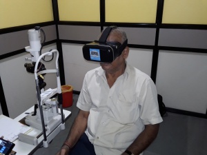
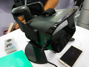
Visual Field Perimetry, even though a cornerstone of glaucoma diagnosis, has always been a cumbersome and difficult test to administer as well as undergo. It was the only standard test to assess the functional damage due to glaucoma for several decades and had not evolved much despite rapid advancements in technology. This is now the era of Virtual Reality, and several innovators started to develop head-mounted visual field testing devices derived from gaming headsets including the extremely low-cost Google Cardboard.
Virtual Reality is an upcoming field and Perimetry is only one of the aspects of Ophthalmology being advanced by it. The Periscreener[1] developed out of a collaboration between Aravind Eye Hospita[2]l and Vellore Institute of Technology, essentially consists of a low-cost Google Cardboard headset, two ordinary android smartphones, and a Bluetooth clicker. It was crude and did only suprathreshold testing, but worked as a prototype.
VirtualEye[3] is a head-mounted, eye-tracking perimeter, that does the equivalent of a full threshold 24-2 visual field. In addition to manual patient response with a click, it can also do an innovative technique called Visual Grasp, where the eye tracker detects change in gaze as detection of stimulus. This mode utilizes the natural tendency to look at a new, moving, or transient visual stimulus. The hypothesis is that the M-cell system inputs to a reflex that, unsuppressed, drives an eye movement to acquire the novel target to the fovea. This mode does not require a manual input from the patient and does not require the cortical processing and decision making required for manual mode.
There are a few other virtual reality perimeters like VIP Visual Fields by Arieh Solomon[4] from Tel Aviv, Israel, Vivid Vision Perimetry by Vivid Vision Inc, imo[5] by Matsumoto et al, Virtual Field by Virtual Field Inc, VR eCloud Perimeter by Ashvin Agarwal et al, Visual Field Visualizer[6] by Nguyen et al, and so on. Tsapakis et al[7] developed low cost Virtual Reality headset based fast-threshold 3 dB step staircase algorithm for the central 24° of visual field (52 points).[8] Karam Alawa et al developed a version with frequency doubling technology[9] in the virtual reality headset.
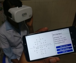
In India, C3 Field Analyzer(C3FA)(Alfaleus Technology Pvt Ltd, India) is developed as a commercial product by Indian Startup company Alfaleus in collaboration with Remidio[10] and is currently capable of glaucoma threshold field testing.[11] The advantages of a compact, portable, head-mounted perimeter are numerous. The patient need not sit in a fixed position and can freely move their head. In fact, they be standing, sitting, lying down or even moving while doing the test. Bedridden patients and wheelchair patients can easily do this virtual reality perimetry. The machine can be carried in hand and taken to the patient if they are difficult to mobilize. They work on a rechargeable battery pack and does not need an external power supply. It does not require an internet connection to connect wirelessly to the operator’s smartphone which displays the real-time stimuli and report while the test is ongoing.
Going beyond subjective perimetry where the patient response is a factor, is the Visual Grasp technique of VirtualEye.[3] Another objective method is Pupil Perimetry, which measured amplitude or latency of pupillary responses using infrared pupillometry. Horn et al realized that using VEPs can give an objective patient response for perimetry testing.[12] Thus, the nGoggle(NGoggle, Inc, San Diego, California, USA)[13] Virtual Reality Perimeter, uses a Brain Computer Interface(BCI) using ElectroEnchephaloGram(EEG).[14] It uses multifocal Steady State Visual Evoked Potentials(mfSSVEP) to detect patient response.[15] This would eliminate patient errors, like false positives and false negatives and make the test faster and more reliable.
Indications
- Screening Visual Fields
- Perimetry in bedridden patients
- Portable Visual Fields for those who cannottravel to a center with full scale visual fields
- Home based Visual Field Charting
- Tele-Glaucoma
Procedure
The subject wears the Virtual Perimetry headset while seated comfortably. In case the subject is more comfortable lying down or even standing up, that is also acceptable.
The subject also holds a wireless clicker to respond to the visual stimuli.
The operator activates the headset from their controls on the Tablet computer.
This displays a visual acuity test and to which they respond verbally.
Then the appropriate visual field test is started.
Subject keeps both eyes open and looks at the fixation point, while stimuli are displayed in the periphery.
When the subject sees the stimuli, they are supposed to press the button on the wireless clicker.
Live result of the test will be continuously be displayed on the Tablet computer.
Once the tests for both eyes are completed, the final report is displayed and can be exported as a PDF file.
Headset can be removed as soon as the test is completed, and can also be removed during the test if the subject wants to take a pause.
Risks/Benefits
Risks/Limitations
- Sensitivity of this test is not as good as Standard Automated Perimetry, so early glaucoma may be missed
- Can be uncomfortable for claustrophobic patients
- Gaze Tracking by camera is not there in most models
- Normative database is still new and may need to be updated
Benefits are
- Portable
- Affordable
- Convenient
- Can be done in bedridden or wheelchair patients
References
- ↑ Akkara JD, Kuriakose A. Review of recent innovations in ophthalmology. Kerala Journal of Ophthalmology. 2018;30(1):54. doi:10.4103/kjo.kjo_24_18
- ↑ Aravind Eye Hospital Pondicherry - AUROTUBE. Peri-Screener. Accessed April 19, 2020. https://www.youtube.com/watch?v=Vink4PCBfLI
- ↑ Jump up to: 3.0 3.1 Wroblewski D, Francis BA, Sadun A, Vakili G, Chopra V. Testing of visual field with virtual reality goggles in manual and visual grasp modes. Biomed Res Int. 2014;2014:206082. doi:10.1155/2014/206082
- ↑ Solomon A. Method and apparatus for evaluating and mapping visual field. Published online March 9, 1999. Accessed April 19, 2020. https://patents.google.com/patent/US5880812A/en
- ↑ Matsumoto C, Yamao S, Nomoto H, et al. Visual Field Testing with Head-Mounted Perimeter ‘imo.’ PLOS ONE. 2016;11(8):e0161974. doi:10.1371/journal.pone.0161974
- ↑ Nguyen NT, Nanayakkara S, Lee H. Visual Field Visualizer: Easier & Scalable Way to Be Aware of the Visual Field. In: Proceedings of the 9th Augmented Human International Conference. AH ’18. ACM; 2018:31:1–31:3. doi:10.1145/3174910.3174931
- ↑ Tsapakis S, Papaconstantinou D, Diagourtas A, et al. Home-based visual field test for glaucoma screening comparison with Humphrey perimeter. Clin Ophthalmol. 2018;12:2597-2606. doi:10.2147/OPTH.S187832
- ↑ Tsapakis S, Papaconstantinou D, Diagourtas A, et al. Visual field examination method using virtual reality glasses compared with the Humphrey perimeter. Clin Ophthalmol. 2017;11:1431-1443. doi:10.2147/OPTH.S131160
- ↑ Alawa KA, Sayed M, Arboleda A, Durkee HA, Aguilar MC, Lee RK. Low-cost, smartphone based frequency doubling technology visual field testing using virtual reality (Conference Presentation). In: Ophthalmic Technologies XXVII. Vol 10045. International Society for Optics and Photonics; 2017:100451K. doi:10.1117/12.2256535
- ↑ C3 Field Analyzer - Alfaleus - Remidio. Accessed May 1, 2020. https://www.remidio.com/c3fa.php
- ↑ Mees L, Upadhyaya S, Kumar P, et al. Validation of a Head-mounted Virtual Reality Visual Field Screening Device. J Glaucoma. 2020;29(2):86-91. doi:10.1097/IJG.0000000000001415
- ↑ Horn FK, Kaltwasser C, Jünemann AG, Kremers J, Tornow RP. Objective perimetry using a four-channel multifocal VEP system: correlation with conventional perimetry and thickness of the retinal nerve fibre layer. Br J Ophthalmol. 2012;96(4):554-559. doi:10.1136/bjophthalmol-2011-300844
- ↑ NGoggle. NGoggle. Accessed April 19, 2020. http://ngoggle.com/
- ↑ Nakanishi M, Wang Y-T, Jung T-P, et al. Detecting Glaucoma With a Portable Brain-Computer Interface for Objective Assessment of Visual Function Loss. JAMA Ophthalmol. 2017;135(6):550-557. doi:10.1001/jamaophthalmol.2017.0738
- ↑ Nakanishi M, Wang Y-T, Daga FB, et al. Detecting Preperimetric Glaucoma with the nGoggle, a Portable Brain-Computer Interface for Assessing Neural Damage. INVESTIGATIVE OPHTHALMOLOGY & VISUAL SCIENCE. 2017;58.


