Uveitis Cataract
All content on Eyewiki is protected by copyright law and the Terms of Service. This content may not be reproduced, copied, or put into any artificial intelligence program, including large language and generative AI models, without permission from the Academy.
Introduction and Epidemiology
Cataracts are one of the most common complications in patients with chronic uveitis. Its incidence varies from 57% in pars planitis to 78% in Fuchs heterochromic iridocyclitis.[1][2]
Cataract formation in patients with uveitis is usually caused by uncontrolled and sustained inflammation, but also by prolonged use of high-dose topical, periocular and/or systemic corticosteroids.[3]
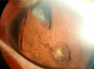
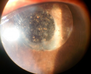
Cataract extraction in patients with uveitis has a different surgical approach compared to conventional phacoemulsification procedure. This is attributed to specific factors to be taken into consideration before surgery. The age of the patient, the etiology of uveitis, its clinical course and response to medical therapy, and the past experience accumulated on previous surgical results described in the literature are very important aspects that need to be considered before performing cataract phacoemulsification. From the stand point of surgical preparation, it is key to have absolute control of intraocular inflammation, this means no anterior chamber cells for at least three months before surgery. Secondly, the surgery needs to be considered and planned according to pre-existing corneal, anterior, and posterior segment pathologic changes such as corneal scarring and leukoma, band keratopathy; iris atrophy and vascular fragility; peripheral anterior and posterior synechiae; pupillary and cyclic membrane formation; anterior and posterior vitreous haze, cystoid macular edema, epiretinal membrane and choroidal neovascularization; macular scarring and atrophy; optic nerve inflammation, ischemia and atrophy; retinal vascular disease, among others. Last, but equally important, pre, intra, and postoperative anti-inflammatory medications (mainly steroids and NSAIDs) need to be administered to the patient in order to avoid severe and abrupt post-operative inflammation that may affect the visual outcome of the patient.[4]
This surgical procedure is considered complex, and represents a major challenge for the anterior segment surgeon. Intraoperative maneuvers such as synechiolysis, membranectomy, and/or pupillary sphincterotomy, use of iris retractor hooks, among others, are required to improve cataract visualization. Management of intraocular bleeding and iris pigment dispersion also need to be taken into consideration on such cases. If significant vitreous haze, or hemorrhage present, possible combined pars plana posterior vitrectomy needs to be considered.[5][6]
Postoperative management requires a close and special monitoring to abrupt inflammation, particularly in children. While in adult patients, uveitis cataract extraction is considered a relatively safe and effective procedure, in children postoperative intraocular inflammation represents a serious threat and therefore the prognosis is reserved.[7][8][9]
Knowledge of the etiology and clinical course of uveitis; analyzing pre-existing pathological changes in the anterior and posterior segment; understanding intraoperative risks; surgical techniques and the importance of the pre and postoperative management are crucial to reduce potential complications of surgery in these cases.
Clinical Characteristics of Different Forms of Cataracts in Uveitis
The etiology and type of uveitis influence the clinical course, treatment response and associated complications. An accurate diagnosis should be the guide for therapeutic and surgical planning. Therefore, it is very important to have a detailed medical history with the support of appropriate diagnostic studies, including investigation of potential primary or systemic causes (infectious or autoimmune). The importance of these efforts is evidenced in cases such as juvenile idiopathic arthritis, in which ocular complications and severe inflammation are frequently observed after surgery, compared to disorders like Fuchs´ heterochromic iridocyclitis in which postoperative inflammation is usually mild and the visual outcome is good.[10][11]
Thus, postoperative visual prognosis is influenced significantly by the age of the patient, the etiology of uveitis, the clinical course of the disease and previous ocular complications, and the appropriate control of inflammation before and after surgery.
Preoperative Evaluation
Prior to surgery, careful eye examination is essential to establish the pathologic changes produced by the inflammatory process. The presence of corneal opacities, condensation and vitreous opacity, cystic macular edema, macular scarring or atrophy of the optic nerve, among others, may limit the visual outcome. Therefore the patient must be informed truthfully and objectively on the visual expectations of the surgical procedure. [12][13]
Auxiliary diagnostic tests are necessary to obtain an accurate postoperative visual potential outcome. In some cases, it is useful to take potential visual acuity using the PAM ("Potential Acuity Meter") or laser interferometry (Lambda, Heine®).[14][15]
In other instances, A, and B ultrasound scan helps identify vitreous condensations and opacities, along with posterior segment structures. Fluorescein angiogram allows detection of macular edema, optic nerve and vasculature inflammation, particularly when significant media opacity is present. The optical coherence tomography (OCT) may be helpful in detecting and analyzing macular pathology (edema, epiretinal membrane, holes, choroidal neovascularization, among others) in cases where media is clear enough. [16][17][18]
With a good preoperative evaluation, the visual expectative and prognosis will be more accurate and objective, and the surgical approach may be planned in advance to avoid surprises and complications, optimizing the surgical result.
Preoperative Management
The key to improve the visual outcome of the surgery is the complete control of the inflammatory process before the procedure is performed. The general consensus suggests that for the majority of cases of uveitis, at least three months of absolute inflammatory control is required (no cells in the anterior chamber and keeping the vitreous inflammation to a minimum) before scheduling surgery. However in some cases, including Fuchs heterochromic iridocyclitis, the presence of inflammatory cells in the anterior chamber cannot be abolished completely despite intensive therapy with steroids. Therefore, in such cases surgery can be performed even in the presence of a low grade of cells. [3][4]
Preoperative management specifically depends on the type of uveitis. In anterior non granulomatous uveitis and in Fuchs heterochromic iridocyclitis, topical administration of prednisolone acetate 1% (1 drop every 6 h) starting three to seven days prior to surgery may be enough. By contrast, patients with uveitis associated with juvenile idiopathic arthritis, granulomatous anterior uveitis, intermediate uveitis including pars planitis, posterior uveitis, panuveitis, or in patients with history of cystoid macular edema, topical therapy should be complemented with systemic corticosteroids or in some cases periocular or intraocular steroid injections. Administration of prednisone (0.5 to 1.0 mg/kg/day), starting three to seven days before surgery should be added to the actual regimen of classical immunosuppressive therapy (IMT) and/or biologic agents that the patient may be already receiving for long-term control of inflammation. If systemic prednisone is contraindicated (diabetes mellitus, acid-peptic disease, obesity, or osteoporosis), the periocular route should be considered. Either sub-Tenon´s , sub-conjunctival, or transeptal injections of 40mg triamcinolone acetone or methyl-prednisolone are the most frequently used steroids for this purpose. [19][20][21]
Topical non-steroidal anti-inflammatory drugs (NSAIDs) is recommended, (nepafenac 0.1%, ketorolac tromethamine 0.4% or bromfenac 0.9%) starting at least three days before surgery, and extending at least six to eight weeks after surgery are usually administered to all uveitis patients. Topical NSAIDs help to prevent cystoid macular edema secondary to surgery, which is common in patients with uveitis.[22]This is weighed against the risk of causing sometimes severe epitheliopathy in certain patients.
Additionally, cataract surgery may exacerbate pre-existing cystoid macular edema therefore, topical NSAIDs should be used to try to reduce the edema before surgery, and periocular or intravitreal corticosteroids may also be administered to reduce or resolve macular inflammation. Despite the general consensus among specialists on these therapeutic strategies nowadays, there are not well-controlled, prospective, randomized clinical trials evaluating the efficacy of topical and systemic prophylactic therapy for patients with uveitis who will undergo cataract surgery.[23][24]
Other special considerations in particular uveitis conditions should be taken into account before conducting cataract surgery. Such is the case of patients with herpetic (HSV-1, VZV) uveitis, in whom it is very important to start, or maintain antiviral treatment at full dose (2g/day of acyclovir or 1-3g/day of valacyclovir orally) at least one week prior to surgery, and up to 4 weeks or more after surgery at prophylactic dosage (600-800mg/day of acyclovir; and 500-1000mg/day of valacyclovir in order to avoid recurrence of inflammation due to viral reactivation.[25]
Central and dense band keratopathy that interferes with visualization of the cataract must be treated previously by 1-2% EDTA-chelation and debridement or by using the excimer laser in photo therapeutic keratectomy (PTK) mode to remove calcium deposits on Bowman´s membrane. Once the corneal epithelium has healed, then cataract surgery may be performed.[26][27]
Some uveitis cataract eyes are complicated by glaucoma or retinal disease that may limit the visual outcome after surgery. For eyes with concomitant uveitis glaucoma, combined surgery (phacoemulsification with IOL implantation plus trabeculectomy or valve implantation) has shown an increased risk of failure of the filtering procedure[AG1] . In these cases, it may be better to do first the cataract extraction through a corneal incision to prevent damage to the conjunctiva. Subsequently, in a second event, either trabeculectomy with antimetabolites (mitomycin-c or 5-fluorouracil) or implantation of a filter valve may be performed.[28] [29] [30]
Regarding retinal complications, such as epiretinal membranes or coexisting retinal detachment, cataract surgery may be combined with vitreo-retinal surgery.
Surgical Technique
The goal of cataract extraction is to improve vision. The key to improve the visual prognosis of cataract surgery in patients with uveitis is the absolute control of the inflammatory process before surgery.
There is still controversy over how to proceed in children with uveitis. Historically, cataract surgery in pediatric patients with uveitis has been associated with a higher rate of complications due to the technical difficulty of the surgery, and the occurrence of excessive postoperative inflammation. Previous or especially case techniques for cataract extraction of pediatric uveitis patients include, extra capsular surgery, lensectomy, and pars plana vitrectomy with phacofragmentation.[27]
Recent advances in surgical techniques and development of new instruments, and polymers for intraocular lenses, have significantly changed the surgical results in adult patients with uveitis. The most successful surgical technique is micro-incisions (MICS) and clear cornea phacoemulsification. The first intraoperative challenge is the proper exposure and visualization of the cataract. To achieve this, in many cases it is necessary to perform synechiolysis, remove pupillary membranes, perform pupillary relaxing incisions or sphyncterotomies, even using iris retractor hooks to maintain adequate pupil size during surgery . However, an effort must be done to try to keep the anatomy of the anterior segment structures intact, avoid cornea, iris and anterior chamber angle trauma, pigment dispersion, or excessive bleeding.[9][10][11][12][13]
Curvilinear continuous capsulorrhexis should be kept between 5-6mm. in diameter, smaller openings often lead to capsular contraction, phimosis, and increased possibility of IOL displacement, decantation, and posterior synechiae. On the other hand, very large capsulorrhexis lead to instability of the IOL.[10]
Ultrasound energy and time may vary depending on the size, hardness of the cataract, and status of the zone, however those parameters should be kept to a minimum to avoid excessive inflammation, corneal endothelium trauma, risk of posterior capsule rupture, loss of nucleus fragments and vitreous exposure. In small pupils, the safest technique is the vertical in situ chop. Chopping of fragments is done within the pupillary aperture with the phase tip kept in view at all times and minimal risk of engaging and traumatizing the iris.[10]
The need of anterior vitrectomy and placement of the lenticular haptic in the sulcus frequently produce severe and persistent inflammatory reaction in the immediate and late postoperative period, resulting in fibrosis, scarring, decantation, and in some cases the need to remove the IOL in a second surgery . Therefore, keeping the posterior capsule intact, and placing the IOL within the capsular bag are key to success in uveitis cataract surgery.
After closure of the corneal wounds, you may apply an intra-cameral injection of 400mcg unpreserved dexamethasone phosphate. Finally, except in cases of secondary glaucoma or steroid-responders, a periocular steroid injection, preferably 40 mg of triamcinolone acetone is very helpful for immediate and long-term control of inflammation.[31]
Intraocular Lenses Considerations
The decision of implanting or not an IOL during cataract surgery in patients with intraocular inflammation is more dependent on the etiology of the uveitis, the age of the patient, and the integrity of the posterior capsule.
Several studies have investigated what is the optimal IOL for patients with uveitis. In adults, it has been shown that various materials such as acrylic and hydrogel are relatively safe with a favorable outcome however, seen with with acrylic IOLs . It has been shown that acrylic lenses have lower levels of postoperative inflammation and lower rate of posterior capsule opacity in a period of six months after surgery in uveitis patients.[32]It is important to consider that this study excluded patients with a history of juvenile idiopathic arthritis. Foster, and Kansai, have the largest series of cataract patients with JIA associated iridocyclitis, which describe some severe postoperative complications, including secondary glaucoma, extensive lenticular fibrosis and cyclic membrane formation related to IOL implantation. One must carefully consider the risks and benefits of placing an IOL at time of cataract surgery in JIA patients vs leaving them aphakic.
In a prospective randomized study of two acrylic lenses, one hydrophobic (AcrySof®) and the other hydrophilic (Akreos®) sharp-edged intraocular lenses were compared on patients with noninfectious uveitis. The study found that visual outcome and capsular opacification were similar between both IOLs. Advancements in lens materials and design continue to offer potential improvement in surgical outcomes.[33] AcrySof® lenses have a history of developing glistenings and sub-surface nanoglistenings (SSNGs) over time and therefore may not be the best choice in a younger patient.
There have been no randomized controlled trials to delineate the optimal lens material in adult patients with uveitis. On the other hand, despite the fact that the implantation of intraocular lenses in the pediatric population is now better accepted, we still have a long way to go to improve the postoperative outcome in such patients.
Postoperative Management
Postoperative treatment in patients with uveitis is as important as preoperative preparation and surgical procedure. From the immediate postoperative period, topical corticosteroids should be administered intensively (prednisolone acetate 1% hourly), along with topical NSAIDs (nepafenac 0.1% every 8h, bromfenac, and wide spectrum topical antibiotics every 6h. During the night, it is useful to administer a corticosteroid ointment. It is also recommended to administer a short-action mydriatic (1% tropicamide every 6h) for a period of 10 to 14 days to avoid pupil synechiae formation.[10]
If oral corticosteroids were given to the patient, these should be maintained at the target dose for a week before tapering to maintenance levels (ideally ≤10 mg/day). Reducing topical steroids depends on the level of intraocular inflammation, and its reduction should be done as soon as possible to prevent the development of ocular hypertension or secondary glaucoma.[19]In the case of systemic antivirals, after a 10-14 days post-operative period, these should be kept at prophylactic doses (acyclovir 600-800 mg/day) by the time predetermined.[25]
Complications and Management
The risk of ocular complications after cataract surgery in patients with uveitis depends on the type, etiology, previous clinical course, degree of previous ocular damage, and therapeutic compliance of the patient.
Despite the preventive measures mentioned above, the most common and most feared complication is post-operative intraocular inflammation. Inflammation may be severe, manifesting by significant inflammatory cells (≥2 +) in the anterior chamber, protein exudation with formation of fibrin membranes or plasmid bodies aqueous obstructing the pupil, and the presence of hypopyon. Pupillary membranes are especially common in conditions such as juvenile idiopathic arthritis and Vogt-Koyanagi-Harada disease. In these cases, it may be necessary to consider the surgical removal of the membrane once the inflammation in the anterior chamber has been controlled. This can be reached increasing the dose of oral prednisone and application of 1% atropine every 12h.[34] While published data is lacking, there is anecdotal evidence that intraocular enoxaparin (lovenox) may be useful in preventing recurrent pupillary membranes.
Excessive inflammation after cataract surgery can also produce vitreous opacity due to acute vitritis. Once the possibility of acute infectious endophthalmitis has been ruled out, this condition should be treated aggressively with oral steroids and even consider intravitreal injections. Furthermore posterior vitrectomy in case of vitreous tensile membranes or organization may be necessary.[35]
Another common complication causing significant visual loss is cystoid macular edema (CME). Its incidence has been reported between 33-56% of cases. Treatment of CME includes periocular injections (subtenant or trans-septal) of 40 mg triamcinolone and intravitreal steroids. In addition, NSAIDs (nepafenac every 8 h) or systemic acetazolamide (250 mg every 12 h) for one to three months may be helpful in some cases.[18][31]Other chronic macular related complication is epiretinal membrane formation, which has been reported to occur in 15 to 56% of cases. Treatment involves delimitation of the membrane through a pars plana posterior vitrectomy.[36]
Posterior capsule opacity is also a frequent problem (approximately 48% of cases) in patients with uveitis. It is more commonly seen than in patients undergoing conventional cataract extraction. Nd-Yag laser capsulotomy may be performed under absolute control of inflammation and considering the higher risk of CME after treatment.[37]
The intraocular pressure (IOP) may rise transiently during the early postoperative period in eyes with compromised trabecular meshwork or angle closure. This complication can often be managed with topical and systemic anti-glaucoma therapy, avoiding when possible, the use of prostaglandin analogues (latanoprost, travoprost and bimatoprost), as well as alpha-adrenergic (brimonidine), as these drugs may favor the inflammatory process.[28]Hypotony can be particularly troublesome and once wound leakage has been ruled out, the next step is to increase the anti-inflammatory therapy topically and systemically. This is often effective in increasing the IOP, but the topical steroids may be difficult to taper or withdraw, and patients may require long-term topical steroids to maintain the IOP. [38] Hypotony maculopathy may not resolve even after IOP increases after prolonged hypotony.
Conclusion
It is possible to achieve successful visual outcomes after cataract surgery in patients with uveitis if special attention is paid to details before, during and after the surgical procedure is performed. The visual prognosis has improved mainly because of a higher level of understanding of the pathogenesis, etiology, clinical course and response to therapy of different forms of uveitis. But also because of better preoperative preparation of the patient, advanced surgical techniques with biocompatible IOL materials, and finally a close and aggressive post-operative care of the patient.
The use of immunosuppressive chemotherapy other than steroids and more recently, the administration of biologic agents have changed enormously the efficacy of control of intraocular inflammation. It has also enabled a longer time of therapy, especially as a steroid-sparing strategy.
Management of the uveitis cataract requires careful case selection and proper timing for surgery, meticulous and careful surgical maneuvers, as well as aggressive anti-inflammatory therapy and close monitoring with appropriate handling of postoperative complications that may occur.
References
- ↑ S. M. Malinowski, J. S. Pulido, and J. C. Folk, “Long-term visual outcome and complications associated with pars planitis,” Ophthalmology, vol. 100, no. 6, pp. 818–825, 1993
- ↑ S. Velilla, E. Dios, J. M. Herreras, and M. Calonge, “Fuchs' heterochromic iridocyclitis: a review of 26 cases,” Ocular Immunology and Inflammation, vol. 9, no. 3, pp. 169–175, 2001.
- ↑ Jump up to: 3.0 3.1 Urban Jr. Rc, Cotlier E. Corticosteroid-induced cataracts. Surv Ophthalmol 1986; 31:102-110
- ↑ Jump up to: 4.0 4.1 Foster CS. Cataract surgery in patients with uveitis. Am Acad Ophthalmol. Focal Points 1994; 12:1-6.
- ↑ Foster CS, Vitale at. Cataract surgery in uveitis. Ophthalmic Clin North Am 1993; 6:139-146.
- ↑ Foster RE, Loader CY, Meisler DM, et al. Combined extracapsu- lar extraction, posterior chamber intraocular lens implantation and pars plana vitrectomy. Ophthalmic Surg 1993; 24:446- 454.
- ↑ Benezra d, cohen e. Cataract surgery in children with uveitis. Ophthalmology 2000; 107:1255–1260.
- ↑ Foster CS, Barrett f. Cataract development and cataract surgery in patients with juvenile rheumatoid arthritis-associated uveitis. Ophthalmology 1993; 100:809-817.
- ↑ Jump up to: 9.0 9.1 Skarin A, Elborgh R, Edlund E, Bengtsson-Stigmar E. Long-term follow-up of patients with uveitis associated with juvenile idiopathic arthritis: a cohort study. Ocul Immune In amm 2009; 17:104-108.
- ↑ Jump up to: 10.0 10.1 10.2 10.3 10.4 Foster CS, Rashid S. Management of coincident cataract and uveitis. Curr Opin Ophthalmic 2003; 14:1-6.
- ↑ Jump up to: 11.0 11.1 Rojas B, Foster CS. Cataract surgery in patients with uveitis. Curr Opin Ophthalmol 1996; 7:11-16.
- ↑ Jump up to: 12.0 12.1 Sabri K, Saurenmann RK, Silverman Ed, Levin AV. Course, complications, and outcome of juvenile arthritis-related uveitis. JAAPOS 2008; 12:539-545.
- ↑ Jump up to: 13.0 13.1 Vidovic-Valentincic NN Kraut a, Hawlina M, et al. Intermediate uveitis: long term course and visual outcome. Br J Ophthalmic 2009; 3:477-480.
- ↑ Joschpe P, Kwitko i, roe d, Kwitko S. Potential acuity meter accuracy in cataract patients. J Cataract Refract Surg 2000; 26:1238-1241.
- ↑ Cuzzani OE, Ellant JP, Young PW, Rydz M. Potential acuity meter versus scanning laser ophthalmoscope to predict visual acuity in cataract patients. J Cataract Refract Surg 1998; 24:263-269.
- ↑ Byrne S, green R. Ultrasound of the eye and orbit. 2nd edition. St louis (Mo): Mosby; 2002; pp. 356-359.
- ↑ De Laey JJ. Fluorescein angiography in posterior uveitis. Int Ophthalmic Clin. 1996; 35:33-58.
- ↑ Jump up to: 18.0 18.1 Belair Ml, Kim SJ, Thorne Je, et al. Incidence of cystoid Macular edema after cataract surgery in patients with and without uveitis using optical coherence tomography. Am J Ophthalmic 2009; 148:128-135.
- ↑ Jump up to: 19.0 19.1 Rothova a. Corticosteroids in uveitis. Ophthalmol Clin N Am 2002; 15:389-394.
- ↑ Van Staa TP. The pathogenesis, epidemiology and management of glucocorticoid-induced osteoporosis. Calcif Tissue Int 2006; 79:129-137.
- ↑ Jabs Da, Rosenbaum JT, Foster JS, Holland GN, Jaffe GJ, Louie JS, et al. Guidelines for the use of immunosuppressive drugs in patients with ocular in amatory disorders: recommendations of an expert panel. Am J Ophthalmol 2000; 130:492-513.
- ↑ Durban OM, Tehrani NN, Marr Je, Moradi P, Stavrou P, Murra Pi. Degree, duration, and causes of visual loss in uveítis. Br J Ophthalmic 2004; 88:1159-1162.
- ↑ Jancevski M, Foster CS. Cataracts and uveitis. Current Opinion in Ophthalmology 2010, 21:10-14.
- ↑ Meacock Wr, Spalton dJ, Bender l, et al. Br J Ophthalmic 2004; 88: 1122-1124.
- ↑ Jump up to: 25.0 25.1 Rodriguez A, Power WJ, Neves RA, Foster CS. The management of recurrent herpetic keratouveitis with long-term systemic acyclovir. Doc Ophthalmol 1995; 90:331-340.
- ↑ Najjar DM, Cohen EJ, Rapuano CJ, Liaison Pr. EDTA chelation for calcic band keratopathy: results and long-term follow-up. Am J Ophthalmic 2004; 137:1056-1064.
- ↑ Jump up to: 27.0 27.1 Stewart OG, Morrell AJ. Management of band keratopathy with excimer photo therapeutic keratectomy: visual, refractive, and symptomatic outcome. Eye 2003; 17:233-237
- ↑ Jump up to: 28.0 28.1 Moorthy RS, Mermaid A, Baerveldt G, et al. Glaucoma associated with uveitis. Surv Ophthalmol 1997; 41:361-394.
- ↑ Wright MM, Mcgehee Rf, Pederson Je. Intraoperative mitomycin-c for glaucoma associated with ocular inflammation. Ophthalmic Surg Lasers 1997; 28:370-376.
- ↑ Hoskins DH, Hetherington J, Shaffer Rn. Surgical management of the inflammatory glaucomas. Perspect Ophthalmol 1977; 1:173-181.
- ↑ Jump up to: 31.0 31.1 Resell M, Tappeiner C, Heinz c, et al. Comparison between intravitreal and orbital floor triamcinolone acetone after phacoemulsication in patients with endogenous uveitis. Am J Ophthalmic 2009; 147:406-412.
- ↑ J L. Alio ́, E. Chipont, D. BenEzra et al., “Comparative performance of intraocular lenses in eyes with cataract and uveitis,” Journal of Cataract and Refractive Surgery, vol. 28, no. 12, pp. 2096–2108, 2002.
- ↑ Monnet D, Tepenier L, Brezin ap. Objective assessment of inflammation after cataract surgery: comparison of three similar intraocular lens models. J Refract Surg 2009; 35:677-681.
- ↑ Kanski JJ. Lensectomy for complicated cataract in juvenile chronic iridocyclitis. Br J Ophthalmol 1992; 76:72–-75.
- ↑ R. Krishna, D. M. Meisler, C. Y. Lowder, M. Estafanous, and R. E. Foster, “Long-term follow-up of extra capsular cataract extraction and posterior chamber intraocular lens implantation in patients with uveitis,” Ophthalmology, vol. 105, no. 9, pp. 1765–1769, 1998.
- ↑ Dv S, Mieler Wf, Pulido JS, et al. Visual outcomes after pars plana vitrectomy for epiretinal membranes associated with pars planitis. Ophthalmology 1999; 106:1086-1090.
- ↑ Dana Mr, Chatzistefanou K, Schaumburg Da, et al. Posterior capsule opacification after cataract surgery in patients with uveitis. Ophthalmology 1997; 104:1387-1393.
- ↑ P. Gupta, A. Gupta, V. Gupta, and R. Singh, “Successful outcome of pars plana vitreous surgery in chronic hypotony due to uveitis,” Retina, vol. 29, no. 5, pp. 638–643, 2009.


