The Hess Test
All content on Eyewiki is protected by copyright law and the Terms of Service. This content may not be reproduced, copied, or put into any artificial intelligence program, including large language and generative AI models, without permission from the Academy.
Description/Overwiew
The Hess Test: A Diagnostic Tool for Incomitant Strabismus[1]
The Hess test is a valuable clinical tool used to assess and monitor incomitant strabismus, a condition where the deviation of the eyes varies depending on the direction of gaze. This condition often suggests an underlying issue with the extraocular muscles or their nerve supply, such as paresis, paralysis, or restriction.
Incomitant strabismus can arise from various causes, including:
- Nerve paresis: Affecting the third, fourth, or sixth cranial nerves, which control eye movements.
- Thyroid ophthalmopathy: An autoimmune condition affecting the eye muscles and surrounding tissues.
- Blow-out fractures: Orbital fractures that can entrap eye muscles.
- Myasthenia gravis: A neuromuscular disorder causing muscle weakness.
The Hess test helps identify imbalances in the extraocular muscles, highlighting underactions or overactions that contribute to the strabismus.
Principles
Principles of the Hess Test: Confusion vs. Diplopia[1]
The Hess test is based on the principle of confusion, which differs from diplopia.
- Diplopia is double vision, where two images of a single object are perceived due to misaligned eyes.
- Confusion occurs when two different objects are seen as overlapping, caused by stimulating corresponding retinal points in each eye, usually the foveae.
The Hess test artificially creates confusion using dissociative tools like red-green glasses, allowing for the assessment of ocular deviations.
Procedure/Methodology
The Hess test involves a specific setup and procedure to accurately assess ocular deviations.
Equipment and Exam Procedure[2]
- Hess Screen Setup: The Hess screen is a tangent screen featuring a checkerboard pattern on a dark gray background. It has twenty-five red lights individually controlled to represent the cardinal positions of gaze, outlining an inner field (15° from the primary position) and an outer field (30°). Each square on the grid equates to 5° of ocular rotation.
- Patient Positioning and Procedure: The patient stands 50 cm away from the screen with their head stabilized on a chin cup. Their eyes are aligned with the central point of the screen, which symbolizes the primary position. The examination is conducted in a dimly lit room. The patient wears reversible goggles with one red and one green lens. The red lens is placed in front of the fixating eye, and the green lens in front of the non-fixating eye.
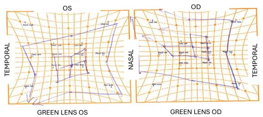
- Testing Process: Red lights are illuminated on the screen, and the patient uses a green pointer to align a green light with each red light. This is done first with one set of goggles and then with the goggles reversed. The test identifies the fixating and non-fixating eyes based on the position of the red filter. The examiner then connects the points where the patient projected the green light, creating the final graph. Left eye movements are charted on the left side of the graph, and right eye movements on the right.
Advancements in Technology[3]
Recent technological advancements offer alternatives that are less dependent on patient and examiner factors, such as eye tracker devices, for more accurate assessments.
Interpretation
Interpretation of Hess Chart Findings[2][4]
The Hess chart provides a graphical representation of ocular muscle function and deviations in different gaze positions. Understanding the key elements of interpretation is crucial for accurate diagnosis and management of incomitant strabismus.
Starting with the Primary Position
Begin by observing the deviation in the primary position, which is the central point on the chart. This provides an initial assessment of the type and magnitude of strabismus.
Identifying the Affected Eye and Muscle
- Smaller Chart: Indicates the eye with the paretic muscle.
- Larger Chart: Indicates the eye with the overacting yoke muscle. The yoke muscle is the contralateral synergist, which overacts in response to the primary muscle's underaction.
Analyzing the Charts
- Maximum Restriction: The smaller chart shows the greatest restriction in the direction of action of the paretic muscle.
- Maximal Expansion: The larger chart shows the greatest expansion in the direction of action of the overacting yoke muscle.
- Inner Field: A smaller than normal inner field indicates underaction of a muscle.
- Outer Field: A larger than normal outer field suggests overaction of a muscle.
- V- and A-Patterns: The inward or outward slope of the fields on the chart reveals V- and A-patterns in strabismus.
Additional Considerations
- Homolateral Antagonist: In the eye with the paretic muscle, the homolateral antagonist (the muscle that opposes the action of the paretic muscle) may show overaction. This occurs due to the unopposed action of the antagonist in the absence of normal function of the paretic muscle.
- Contralateral Antagonist: In the eye with the overacting yoke muscle, the contralateral antagonist (the muscle that opposes the action of the yoke muscle) may show underaction. This is a secondary effect, as the overaction of the yoke muscle inhibits the action of its contralateral antagonist. When this interaction exists, it implies a long-standing palsy.
Muscle Sequelae
Muscle sequelae are a pattern of muscle overaction and underaction seen in incomitant strabismus, which sequentially develops after the onset of the condition. These sequelae follow Sherrington's law of reciprocal innervation, which states that when a muscle receives increased innervation, decreased innervation is directed to its ipsilateral antagonist.
The order of muscle sequelae development is as follows:
- Overaction of the contralateral synergist (Hering's law)
- Overaction of ipsilateral antagonist (Sherrington's law)
- Underaction of contralateral antagonist (Hering's law and Sherrington's law)
The absence of muscle sequelae suggests a recent palsy, while their presence indicates a long-standing condition. The greater the extent of muscle sequelae, the more chronic the palsy is likely to be.
By carefully analyzing the size, shape, and slope of the Hess charts, along with considering the interplay between agonist and antagonist muscles in both eyes, ophthalmologists can understand the complex nature of incomitant strabismus.
Limitations
While the Hess test is a valuable tool in strabismus evaluation, it's essential to be aware of its limitations:[2][4]
- Patient Collaboration and Attention: The test's accuracy depends heavily on the patient's ability to understand and follow instructions, maintain concentration, and provide reliable responses.
- Time-Consuming: Administering the Hess test can be time-consuming, requiring patience from both the examiner and the patient.
- Requirement for Normal Binocular Vision: The Hess test is most informative in patients with binocular vision. It may not be suitable for those with suppression (where the brain ignores one eye's image) or abnormal retinal correspondence (where non-corresponding retinal points are interpreted as a single image).
- Color Vision: Patients with red-green color blindness cannot perform the test accurately.
- Limitations with Large Deviations: In cases of very large deviations, the perceived point may fall outside the Hess chart's field, limiting the assessment's accuracy.
- Difficulty Assessing Torsional Deviations: The Hess chart's gridded scale primarily measures horizontal and vertical deviations, making it challenging to evaluate torsional deviations (rotations of the eye around its visual axis).
- Old Paralysis Complications: In long-standing paralysis cases, secondary motor adaptations can develop, potentially obscuring the initial diagnosis.
- Manifest vs. Latent Deviations: The Hess test does not differentiate between manifest deviations (tropias, always present) and latent deviations (phorias, only present when binocular fusion is disrupted).
Understanding these limitations helps clinicians interpret Hess test results accurately and use the test appropriately in conjunction with other diagnostic methods for comprehensive strabismus evaluation.
Example

- ↑ Jump up to: 1.0 1.1 American Academy of Ophtalmology, Basic and Clinical Science Course, section 6 – Pediatric Ophtalmology and strabismus, Chapter 7.
- ↑ Jump up to: 2.0 2.1 2.2 Sociedade Portuguesa de Oftalmologia, Meios complementares de diagnóstico, Chapter 2.6, Avaliação da visão binocular
- ↑ Orduna-Hospital E, Maurain-Orera L, Lopez-de-la-Fuente C, Sanchez-Cano A. Hess Lancaster Screen Test with Eye Tracker: An Objective Method for the Measurement of Binocular Gaze Direction. Life. 2023; 13(3):668. https://doi.org/10.3390/life13030668
- ↑ Jump up to: 4.0 4.1 Von Noorden, G.K. and Campos, E.C. (2002) Binocular vision and ocular motility. 6th Edition, CV Mosby, St. Louis

