Stereopsis and Tests for Stereopsis
All content on Eyewiki is protected by copyright law and the Terms of Service. This content may not be reproduced, copied, or put into any artificial intelligence program, including large language and generative AI models, without permission from the Academy.
Introduction
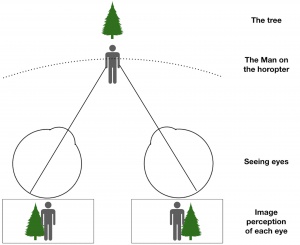
For as long as living beings have inhabited our planet, the visual system has faced a fundamental problem – reconstruction of a three-dimensional picture from two-dimensional retinal images[1][2]. When considering animals and the mechanism by which they process information to carry out their specific tasks, it has been found that the most precise information stems from the difference in vantage points of the two eyes[1]. Having different vantage points implies that the two images received by the retinae are not the same. How is it then, that we are able to reconstruct a single three-dimensional picture from different two-dimensional retinal images? The answer lies in combining information received from the two eyes[2]. Charles Wheatstone suggested in the late 1830s that fixation of the two eyes on some point in space leads to objects, near or far, casting images onto different locations on the retinae[3]. The brain is then able to detect the binocular disparity, stimulating disparity-selective neurons to fire an increased number of action potentials and encode the relationship between the two images. As a result, depth can be perceived in a phenomenon termed “stereopsis"[1][2].
Depth perception
Definition
What is depth perception? It is, plainly, the ability to see in three dimensions as well as to judge the distance of objects[4]. In a realistic sense, only one eye is needed to perceive depth. Monocular cues used to sense the presence of depth include perspective, size, order, and other movement-related cues. However, binocular depth perception is important not only for redundancy, but also to allow a symbiosis between the two eyes in extracting information from the environment[5]. An inherent dissimilarity exists between the two eyes. For example, if one were to close one eye and observe the image then close the other and observe that image, the two images would be different. However, when one uses both eyes, only one image is seen. This occurs because the two eyes are able to fixate on one point in space, and as a result, any object nearer or farther relative to that one point will cast images onto separate locations on the bilateral retinae. A binocular disparity is then detected by the brain, resulting in a sensation of depth[2].
Basis of stereopsis
Stereopsis is a word derived from Greek language meaning “solid” and “power of sight.” The phenomenon of stereopsis is important for several reasons. In the animal kingdom, having a spare eye if one is damaged is necessary for survival. In addition, certain animals have eyes on opposite sides of the head, allowing a 360° visual field from the combined fields of view and protection from potential predators. In general, horizontal separation between the eyes allows the eyes to see the same scene from two distinct vantage points. When one ‘looks’ at an object in space, one aims the fovea, the region of the retina of highest visual acuity, of both eyes at the object[6]. This is referred to as the “plane of fixation.” To understand this fully, one must consider the concept of horopters. Two types of horopters exist – geometric and empirical. The geometric horopter is a collection of points in an image projecting to corresponding points on the retina which lie at approximately the same depth as the point of fixation. On the other hand, the empirical horopter is the collection of points which lie at exactly the same depth and appear slightly flatter than the geometric horopter[6]. If an object lies in front of or behind the plane of fixation, its features will not project to corresponding points on the retinae. Therefore, the retinal distance from the focal length to the focal height on one eye’s retina will not be equal to that of the other eye’s[6]. This difference in distances is known as “binocular disparity,” and it serves as the basis of stereopsis.
Defective stereopsis and strabismus
Stereopsis is not present at birth and develops in a child at 3-6 months of age[7][8]. Any interference in the process of perceiving depth results in defective stereopsis, which can occur at any point from the infantile period to the elderly period of life. These interferences are known as binocular vision impairments. For example, a child with a congenital cataract will not be able to develop simultaneous perception if the cataract is not corrected early enough. Another possibility is a newborn with a unilateral refractive error, known as anisometropia, which can lead to anisometric amblyopia. As a result of anisometropia, the newborn cannot fuse the images from both eyes but rather produces two separate images in the brain. Depending on the difference in visual acuity, the brain can then either suppress visual capacity in the eye with greater refractive error or maintain stereopsis. Yet another common visual impediment affecting children is strabismus. Patients with strabismus exhibit tropias, which manifest as binocular misalignments. If these tropias are present from an early age, stereopsis would be expected, only shifted and with receptive fields of visual neurons offset on the retinae by the degree of shift. With misalignment, however, normal stereopsis is not possible, as the images on the retinae are too far apart for the brain to fuse. In addition, not only is eye position misaligned in strabismus, but it may also be variable[9]. When the difference in visual acuity between the two eyes is greater than the brain can overcome or when the visual fields are dissimilar, the brain chooses to suppress the bad eye, resulting in amblyopia. Although stereopsis is lost, amblyopia serves to protect the eyes from diplopia. Any interruption in vision, no matter the severity or duration, in the first 8 years of life can hinder the development of visual perception. If interruptions in vision occur after this time, stereopsis is not lost, but adaptive changes occur. Adults can acquire strabismus as part of these adaptive changes. In order to avoid diplopia, the brain deviates one eye so that the visual fields are not overlapping.
Grades of binocular vision
Three grades of binocular vision exist: 1) macular perception, the most basic grade; 2) fusion; and 3) stereopsis, the highest grade. Binocular vision can be tested for via several means. In a young child who cannot yet speak, external examination suffices, and simple assessment of head posture may point to defects in binocular vision. Abnormal head posture is a sign that binocularity is being maintained through adaptation to incomitant deviation. Another test is the cover uncover test, which should be performed in all patients to rule out strabismus and confirm binocular function[10]. In older children, the synoptophore, an instrument that measures angles of deviation, can be utilized to assess fusional ability. Additionally, Bagolini lenses may be used to evaluate binocular vision through sensory function in older children. This method involves the patient viewing a fixation light through lenses with striations at different angles. If the patient’s eyes show normal retinal correspondence, one light will be seen along with symmetrical streaks of light in a cross-like formation. The Worth Four Dot test can also be used to evaluate binocular vision. The stereopsis grade of binocular vision is assessed by various charts, including the titmus fly test and the random dot stereo acuity chart[10].
Monocular cues
The brain can achieve depth perception with a single eye through simulated stereopsis and the use of monocular cues, including texture variations and gradients, defocus, color, haze, and relative size[11][12]. These simple characteristics of an image enable the cortex to estimate the distance and depth of the object. The use of monocular cues to sense depth can be compared to sensing depth from a painting. For example, if a painting shows a tree covering part of a house, it is interpreted that the tree is located in front of the house. Though monocular cues can be helpful, they are more susceptible to optical illusions.
Quantification of stereopsis
Stereopsis, the third grade of binocular vision, is quantified by a unit known as seconds of arc. When one thinks of a circle, which consists of 360 degrees, each degree is divided into 60 minutes of arc, and each minute is divided into 60 seconds of arc. The seconds of arc represent an angle through which stereopsis is quantified and demonstrate the minute difference in depth one can perceive. If the angle represented by the seconds of arc is small, the objects being compared are very close together. This can be visualized through the following example: if one sits in the fifth row on an airplane, he or she can see not only the fourth row but also the first row in his or her field of view, as both foveae connect with both rows at separate lines of sight. Each eye has a different line of sight for each seat. The point of fixation is where the lines from each eye meet, creating the visual angle represented by the seconds of arc. Stereopsis is calculated by taking the least difference in seconds of arc that the individual can perceive binocularly. This value changes as the object’s distance from the eyes changes. Stereopsis improves at as distance from the eyes decreases.

Tests for stereopsis
The basis of testing for stereopsis resides in delivering dissimilar images to the eyes in a phenomenon called methodology of dissociation, and these tests utilize various methodologies of dissociation.
Haploscope
A haploscope is a device that delivers different images to each eye simultaneously. This optical device in its earliest stages consisted of a pair of mirrors with the shiny surfaces facing away from each other and placed at 45-degree angles. The mirror on the right corresponded to images on the absolute right, and the mirror on the left corresponded to those on the absolute left. Depending on the difference between the images perceived, the brain will attempt to fuse the images. If the images are extremely dissimilar, confusion results. If the images are only slightly dissimilar, the brain will be able to process them. When the brain is able to fuse the images, stereopsis results and depth and distance can both be perceived. The modern haploscope, also known as a stereoscope, is composed of prisms instead of mirrors, and in order to downscale on its size, lenses as eye pieces were eventually introduced[13].
Anaglyph
As the haploscope is not used often for diagnostic purposes, newer techniques were sought out to test for stereopsis while maintaining the line of sight. The anaglyph utilizes different colors, such as red and cyan, while presenting the two images to the eyes. When the images are viewed, the visual cortex of the brain fuses the images to produce one integrated stereoscopic image[14]. Wilhelm Rollmann was the first to develop a method to view anaglyph images in 1852. Using camera filters, two images from the left-sided and right-sided perspectives were projected as a single image through a red filter on one side and a contrasting color on the other side, such as blue or green. Now, however, image-processing computer programs can simulate the effect of color filters.

Vectograph
Another tool to measure stereopsis is the vectograph, which uses polarized 3D glasses. The polarized filters allow different images to be presented to each eye. To fully understand the mechanism underlying the vectograph, one must first understand polarization of light. When light travels in space, its waves normally vibrate in more than one plane. These waves, as seen from the sun or a lamp light, are produced by electric charges vibrating in all directions. Polarized light waves vibrate in just one plane. Polarization of light can occur by reflection, refraction, or use of a Polaroid filter. The axis of polarization in polarizing glasses are at right angles relative to each other. If the right eye is vertically polarizing, the left eye will be horizontally polarizing[15]. Because of this, it is possible to project different images to each eye while maintaining illumination and line of sight. From the vectograph, the titmus fly test was developed, which involves the use of an image of a large housefly portrayed at different depths[16].
Stereopsis in daily life
Not only is stereopsis relevant in the clinical setting, but it is also relevant in our everyday lives. For example, viewing devices for 3-D movies utilize two different techniques – the vectograph and the active shutter 3-D display technique. In the latter, two dissimilar images are alternated between at a very high frequency of 60 Hz or greater, meaning that each eye sees 30 images every second. The active shutter technique requires viewers to wear bulkier 3-D glasses with a built-in battery. Since the glasses have the ability to turn opaque at the same frequency, when one eye’s image is on the screen, the other eye’s glass turns opaque. By doing so, the brightness and contrast of the movie is preserved and not lost by polarization, as polarization blocks the majority of light. On the other hand, however, this technique is costlier.

Stereo-Sue
Of notable mention is Susan R. Barry, a professor of neurobiology from Massachusetts, who did not attain stereovision until adulthood. She describes her miraculous journey in a book titled Fixing My Gaze: A Scientist’s Journey into Seeing in Three Dimensions.’
Conclusion
The importance of two eyes lies in the concept of stereopsis, and developing an adequate foundation early on in training within the field of ophthalmology is essential to best clinical practice.
Additional Resources
- Hirshfield GS. What is the difference between depth perception and stereopsis? American Academy of Ophthalmology. EyeSmart Ask an Ophthalmologist. https://www.aao.org/eye-health/ask-ophthalmologist-q/what-is-difference-between-depth-perception-stereo. Accessed May 05, 2020.
References
- ↑ Jump up to: 1.0 1.1 1.2 Cumming BG, Deangelis GC. The physiology of stereopsis. Annu Rev Neurosci. 2001;24:203-38.
- ↑ Jump up to: 2.0 2.1 2.2 2.3 Cumming B. Stereopsis: how the brain sees depth. Curr Biol. 1997;7(10):R645-7.
- ↑ Wheatstone Charles XVIII. Contributions to the physiology of vision. —Part the first. On some remarkable, and hitherto unobserved, phenomena of binocular vision128Phil. Trans. R. Soc.
- ↑ Sharma P. The pursuit of stereopsis. J AAPOS. 2018;22(1):2.e1-2.e5.
- ↑ O'connor AR, Tidbury LP. Stereopsis: are we assessing it in enough depth?. Clin Exp Optom. 2018;101(4):485-494.
- ↑ Jump up to: 6.0 6.1 6.2 Ponce CR, Born RT. Stereopsis. Curr Biol. 2008;18(18):R845-50.
- ↑ Birch E, Petrig B. FPL and VEP measures of fusion, stereopsis and stereoacuity in normal infants. Vision Res. 1996;36(9):1321-7.
- ↑ Giaschi D, Lo R, Narasimhan S, Lyons C, Wilcox LM. Sparing of coarse stereopsis in stereodeficient children with a history of amblyopia. J Vis. 2013;13(10).
- ↑ Read JC. Stereo vision and strabismus. Eye (Lond). 2015;29(2):214-24.
- ↑ Jump up to: 10.0 10.1 Lee J, Mcintyre A. Clinical tests for binocular vision. Eye (Lond). 1996;10 ( Pt 2):282-5.
- ↑ Depth Estimation Using Monocular and Stereo Cues. A Saxena, J Schulte, AY Ng. IJCAI 7, 2197-2203. https://www.aaai.org/Papers/IJCAI/2007/IJCAI07-354.pdf
- ↑ Kalloniatis M, Luu C. The Perception of Depth. 2005 May 1 [Updated 2007 Jun 6]. In: Kolb H, Fernandez E, Nelson R, editors. Webvision: The Organization of the Retina and Visual System [Internet]. Salt Lake City (UT): University of Utah Health Sciences Center; 1995-.
- ↑ Krimsky E. The Stereoscope in theory and practice, also a knew precision type stereoscop. Br J Ophthalmol.1937;21(4):161-197.
- ↑ Rubino PA, Bottan JS, Houssay A, et al. Three-dimensional imaging as a teaching method in anterior circulation aneurysm surgery. World Neurosurg. 2014;82(3-4):e467-74.
- ↑ Stereoscopy.com – FAQ; www.stereoscopy.com/faq/vectographs.html
- ↑ Arnoldi K, Frenkel A. Modification of the titmus fly test to improve accuracy. Am Orthopt J. 2014;64:64-70.


