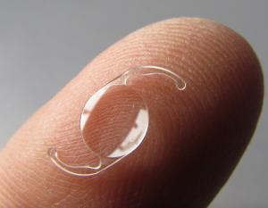
Single Piece Intraocular Lenses
All content on Eyewiki is protected by copyright law and the Terms of Service. This content may not be reproduced, copied, or put into any artificial intelligence program, including large language and generative AI models, without permission from the Academy.
Single-Piece Intraocular Lenses
Introduction
Intraocular lenses (IOLs) are used in ophthalmic surgery to replace the lens of the eye and rectify refractive errors.[1] IOLs are composed of two elements: an optic and haptics. The optic is the central area responsible for refraction, and the haptics are the appendages from the center optic that hold it in place.[2] (Figure 1) In cataract surgery, generally two haptic designs of IOLs are used: single-piece and three-piece. Single-piece IOLs (also referred to as one-piece) are named as such due to both elements being composed of the same material ( acrylic, silicone or PMMA).[3]
History
Prior to the introduction of modern lens designs, cataract surgical technique and location of lens placement differed greatly and evolved over time. The first IOL implants are credited to the British ophthalmologist, Harold Ridley, in 1949.[4] These lenses were implanted into the posterior chamber of the eye. This technique soon evolved into anterior chamber placement and iris-supported lenses, and ultimately, within the capsule placement.[5] Today, the ideal surgical technique is preservation of the lens capsule and placement within the capsule or “in the bag”.[2] Modern “in the bag” placement is superior to prior techniques, as well as made possible by, newer lens technology. Modern IOLs can be inserted through smaller incisions by nature of their more flexible designs.[2][5] This technique has been used since the 1980s.[5]
Premium IOLs
Premium IOLs have additional features in comparison to traditional monofocal IOLs and have unique materials, refractive qualities, and aspheric designs.[6] These premium designs are available to correct presbyopia and astigmatism. Regardless of IOL type, centration is key to visual acuity outcomes post-surgery. Generally, the center of the undilated pupil is used as the axial center of for IOLs.[7]
Multifocal IOLs can be used to provide correction of both near and distance visual acuity. In comparison to monofocal lenses, which have fixed focus for one distance, multifocal lenses distribute light penetrating the lens to different foci.[8] These lenses are generally classified by the physical principles used in the IOL: either refractive, or the more common diffractive.[6][9] Refractive IOLs create multiple focal points with concentric zones of different dioptric power.[9] Diffractive IOLs have multiple diffractive zones on the posterior lens surface. Multifocal IOLs can also be classified as either bifocal or trifocal. Bifocal lenses have both a near and far focus, while trifocal ones include an intermediate focus.[6]Toric IOLs can be used in patients with corneal astigmatism to provide correction of distance vision without the use of glasses. Calculation of preoperative astigmatism must be accurately obtained prior to IOL implantation for favorable results.[10] In addition to this, as well as proper centration, toric IOLs also require accurate rotational orientation. As a result, optimal axis for centration may be slightly superior and nasal from the pupil center.[7]
Toric IOLs are also available in multifocal variations to provide patients with astigmatism a multifocal option.[6] Accommodative and extended depth of focus (EDF) IOLs are two new categories of premium IOLs. Accommodative IOLs provide a dynamic refractive power with contraction and relaxation of the ciliary muscles.[6] There is one FDA approved accommodative IOL, the Crystalens by Bausch and Lomb.[11] EDF IOLs provide an extended far-distance focus through various techniques, which can improve intermediate vision.[6] Two EDOF IOLs approved by the FDA are the Symfony IOL by Johnson and Johnson that uses echelette technology and the Vivity IOL by Alcon.[12]
Single-Piece vs Three-Piece IOLs
There are certain advantages and disadvantages to single-piece IOLs when compared to three-piece IOLs. Many of these may pertain to surgeon preference or situational limitations. Three-piece IOLs are more versatile and can be placed in the capsule or in the ciliary sulcus in the event that the posterior capsule ruptures.[2] Single-piece IOLs should never be placed in the ciliary sulcus.[2][6][13] Several issues may arise with placement of this lens design in the sulcus ranging from UGH (uveitis, glaucoma, hyphema) syndrome, to shifting of the lens over time.[13] In general, the advantages of single-piece IOLs come from its less rigid haptics. The softer, longer haptics unfold in a more evenly distributed manner, causing relatively less trauma to the capsule and more uniform contact holding it in place, and reducing wrinkling.[3] Therefore, single-piece IOLs are considered superior by some surgeons for patients with competent lens capsules and inferior for those at risk of capsule rupture.[2][14] Studies comparing single and three-piece IOLs have shown mixed results in various outcome measures including stability, refractive outcomes, and more.[3][15]
- ↑ Habhab, S., & Hwang, F. S., MD. (2020, July 5). Presbyopia-correcting IOLs. Retrieved August 08, 2020, from https://eyewiki.org/Presbyopia-correcting_IOLs
- ↑ Jump up to: 2.0 2.1 2.2 2.3 2.4 2.5 Salmon, J. F., & Kanski, J. J. (2020). Kanski's Clinical ophthalmology: A systematic approach (9th ed.). Edinburgh: Elsevier Limited.
- ↑ Jump up to: 3.0 3.1 3.2 Zhong, X., Long, E., Chen, W. et al. Comparisons of the in-the-bag stabilities of single-piece and three-piece intraocular lenses for age-related cataract patients: a randomized controlled trial. BMC Ophthalmol 16, 100 (2016). https://doi.org/10.1186/s12886-016-0283-4
- ↑ Apple, D. J., & Sims, J. (1996). Harold Ridley and the invention of the intraocular lens. Survey of Ophthalmology, 40(4), 279-292. doi:10.1016/s0039-6257(96)82003-0
- ↑ Jump up to: 5.0 5.1 5.2 Werner, L., Izak, A. M., Pandey, S. K., & Apple, D. J. (2019). Evolution of Intraocular Lens Implantation. In M. Yanoff & J. S. Duker (Authors), Ophthalmology (5th ed.). Edinburgh: Elsevier. doi:9780323528214
- ↑ Jump up to: 6.0 6.1 6.2 6.3 6.4 6.5 6.6 Zvorničanin, J., & Zvorničanin, E. (2018). Premium intraocular lenses: The past, present and future. Journal of Current Ophthalmology, 30(4), 287-296. doi:10.1016/j.joco.2018.04.003
- ↑ Jump up to: 7.0 7.1 Roach, L. (2016, May 05). Centration of IOLs: Challenges, Variables, and Advice for Optimal Outcomes. Retrieved August 08, 2020, from https://www.aao.org/eyenet/article/centration-of-iols-challenges-variables-advice-opt
- ↑ Werner, L. (2020). Intraocular Lenses. Ophthalmology. doi:10.1016/j.ophtha.2020.06.055
- ↑ Jump up to: 9.0 9.1 Fragoso, V. V.& Alió, J. L. (2019). Surgical Correction of Presbyopia. In M. Yanoff & J. S. Duker (Authors), Ophthalmology (5th ed.). Edinburgh: Elsevier. doi:9780323528214
- ↑ Wang, L., Houser, K., Koch, D. D. (2019). Intraocular Lens Power Calculations. In M. Yanoff & J. S. Duker (Authors), Ophthalmology (5th ed.). Edinburgh: Elsevier.
- ↑ Crystalens AO Lens. (n.d.). Retrieved August 08, 2020, from https://www.bausch.com/our-products/surgical-products/cataract-surgery/crystalens-ao-lens
- ↑ Rocha, K. M. (2017). Extended Depth of Focus IOLs: The Next Chapter in Refractive Technology? Journal of Refractive Surgery, 33(3), 146-149. doi:10.3928/1081597x-20170217-01
- ↑ Jump up to: 13.0 13.1 Kent, C. (2018, July 11). The IOL in the Sulcus: When, Why & How. Retrieved August 08, 2020, from https://www.reviewofophthalmology.com/article/the-iol-in-the-sulcus-when-why-and-how
- ↑ Rosenthal, K. J., MD, FACS. (2011, March). CATARACT SURGERY: Optimizing IOL Selection: One-Piece vs Three-piece. Retrieved August 08, 2020, from http://epubs.democratprinting.com/article/CATARACT_SURGERY:_Optimizing_IOL_Selection:_One-Piece_vs_Three-piece/680057/64262/article.html
- ↑ Savini, G., Barboni, P., Ducoli, P., Borrelli, E., & Hoffer, K. J. (2014). Influence of intraocular lens haptic design on refractive error. Journal of Cataract & Refractive Surgery, 40(9), 1473-1478. doi:10.1016/j.jcrs.2013.12.018


