Pinhole Intraocular Lenses
All content on Eyewiki is protected by copyright law and the Terms of Service. This content may not be reproduced, copied, or put into any artificial intelligence program, including large language and generative AI models, without permission from the Academy.
Introduction
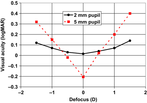
The pinhole affect is based on the stenopeic principle, which allows small central rays to enter the eye and eliminates diverging rays, thus reducing the circle of blur on the retina[1]. As a result, the depth of focus is increased, providing patients with good distance and near vision. This concept has been routinely used in ophthalmology in the form of pinhole occluders and glasses and even through the surgical constriction of the iris, however this concept has recently been introduced to the IOL plane. Although viewing through a small aperture increases depth of focus, it comes with the disadvantage of decreased brightness, decreased visual field and decreased optimal visual acuity,[2][3] as depicted in the chart below[4] .
Caption: Visual Acuity as compared with pupil size after correction of aberrations in 13 patients[4]
Depth of Focus IOL
Currently two types of pinhole IOLs exist, the IC-8 IOL (AcuFocus) and the Xtrafocus IOL (Morcher), both serving two distinct functions.
IC-8 IOL (AcuFocus)
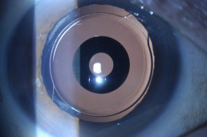
The IC-8 monfocal IOL uses similar principles to the Kamra corneal inlay[5]. The IOL is intended for monocular placement in the non-dominant eye, in an effort to provide presbyopic correction while maintains good range of distance vision.
The IOL is a foldable single piece, hydrophobic, Acrylic lens that has an embedded annular ring composed of polyvinylidene fluoride and carbon nanoparticles. It is 5um thick and the aperture measures 1.36mm, while the outer diameter measures 3.2mm, an overal 15% decrease in size from the Kamra inlay to adjust for a more posterior position.

Source: https://www.eurotimes.org/pinhole-iol/
Xtrafocus IOL (Morcher)
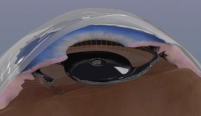
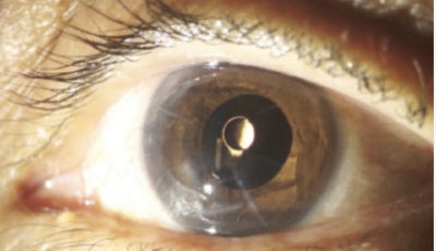
The Xtrafocus IOL is a small-aperture sulcus IOL designed for implantation as a piggy back lens for patients with a lens already implanted in the capsular bag. This lens is designed to reduce irregular astigmatism or light sensitivity in patients who have undergone PKP or who have other corneal irregularities. The IOL was given a CE mark in 2016 and is currently pending an FDA trial by Morcher. Dr. Trindade, who holds a patent for the design, implanted the IOL in a patient with Urrets-Zavala’s syndrome, suffering from intractable light sensitivity with marked improvement[6].
The IOL is composed of a foldable acrylic infra-red transparent material but otherwise appears black, opaque. The IOL measures 14mm in diameter with a 6mm optic and a 1.3mm central aperture. The lens has a concave-convex design to prevent contact with the lens already implanted in the capsular bag. It has soft, thin (250um) angulated haptics at 14 degrees that help to prevent uveitis-glaucoma-hyphema syndrome. It is placed in the ciliary sulcus in a piggy-back fashion in pseudophakic eyes. Funduscopic imaging can be obtained with an infra-red OCT imaging system.


Caption: Xtrafocus IOL with soft angulated haptics allow for ciliary sulcus placement in a piggy-back fashion in pseudophakic eyes
Source: https://crstodayeurope.com/articles/2016-feb/development-of-a-pinhole-implant-xtrafocus/
Outcomes
Accufocus IC-8 IOL
The IC-8 IOL is marketed by AcuFocus to provide 3D of functional range of vision when placed at a target of -0.75D in the non-dominant eye with an accompanying monofocal IOL in the dominant eye, targeted for emmetropia[7].
Visual Acuity
Data from AcuFocus suggests patients achieve a near visual acuity of 20/30 at 40cm and 20/40 at 33cm. Other Studies show that mean visual acuity remains 20/40 (0.3 logMAR) or better over a range of ±2 D of defocus[8][9]. The lens also corrects astigmatism up to 1.5D without needing to be placed on a certain axis.[10][11][12][13].
Higher Order Aberrations
Given the IOL is placed closer to the eye’s nodal point, there are less problems noted with slight decentration from the visual axis as compared to the corneal inlays. The small aperture helps to reduce coma as peripheral light rays are blocked.
Morcher Xtrafocus IOL
A study comprised of 21 patients with high irregular astigmatism from RK, KCN and PKP showed the median CDVA improved from 20/200 to 20/50 following IOL implantation[14].
Patient Selection
Accufocus IC-8 IOL
The IC-8 IOL is for patients undergoing cataract surgery who want a good range of vision without spectacle dependence.
Patients who are good candidates for an IC-8 IOL include:
- Patients with low degree of astigmatism, measuring <1.5D.
- Less glare and haloes compared to MFIOL due to absence of diffractive rings
- Patients w/ HOA > 0.5 who maybe unsuitable for MFIOLs
- Provides presbyopic correction in patients who cannot tolerate anisometropia.
Patients who are not good candidates include:
- Patients who want excellent near vision without glasses (trifocal IOL or a MFIOL with +3.0 add may be better)
- Patients with central corneal scarring
- Mesopic pupils >6mm, patients
- Macular pathology
- Severe glaucoma
Patients have to be counseled regarding decreased brightness and possible need for glasses during low light conditions[15].
Morcher Xtrafocus IOL
The Xtrafocus IOL is currently intended for patients with moderate-severe astigmatism resulting from KCN, trauma, PKP or post-RK. Due to its small aperture and opaque nature, peripheral vision may be significantly limited.
Retina Surgery with Pinhole Optics
Although only anecdotal reports are available, retinal surgery following implantation of an intraocular lens with a pinhole optic seems to be possible without significant obstacle[16].
References
- ↑ Lay M, Wickware E & Rosenfield M. Visual acuity and contrast sensitivity. In: Optometry: Science, Techniques and Clinical Management, 2nd edn, Rosenfield M, Logan N & Edwards K(eds), Butterworth‐Heinemann: Oxford, 2009; pp. 178–179.
- ↑ Charman WN & Tucker J. The depth‐of‐focus of the human eye for Snellen letters. Am J Optom Physiol Opt 1975; 52: 3–21.
- ↑ Kim WS, Park IK & Chun YS. Quantitative analysis of functional changes caused by pinhole glasses. Invest Ophthalmol Vis Sci 2014; 55: 6679–6685
- ↑ Jump up to: 4.0 4.1 Hickenbotham A, Tiruveedhula P & Roorda A. Comparison of spherical aberration and small‐pupil profiles in improving depth of focus for presbyopic corrections. J Cataract Refract Surg 2012; 38: 2071–2079.
- ↑ Tabernero J & Artal P. Optical modeling of a corneal inlay in real eyes to increase depth of focus: optimum centration and residual defocus. J Cataract Refract Surg 2012; 348: 270–277.
- ↑ Trindade CLC & Trindade BLC. Novel pinhole intraocular implant for the treatment of irregular astigmatism and severe light sensitivity after penetrating keratoplasty. JCRS Online Case Reports 2015; 3: 4–7
- ↑ Read L. The IC-8 IOL: Big Advantages Through Small Apertures. The Ophthalmologist. Sep 2019. https://theophthalmologist.com/subspecialties/the-ic-8-iol-big-advantages-through-small-apertures
- ↑ The small‐aperture IC‐8 intraocular lens: a new concept for added depth of focus in cataract patients. Am J Ophthalmol2015; 160: 1176–1184.
- ↑ Dick HB, Piovella M, Vukich J, Vilupuru S & Lin L. Prospective multicenter trial of a small‐aperture intraocular lens in cataract surgery. J Cataract Refract Surg 2017; 43: 956–968.
- ↑ RE Ang, “Small-aperture intraocular lens tolerance to induced astigmatism”, Clin Ophthalmol, 12, 1659 (2018). PMID: 30233128.
- ↑ Burkhard Dick et al. Prospective multicenter trial of a small-aperture intraocular lens. Journal of Cataract and Refractive Surgery 2017: Vol. 43, Issue 7; 956-968.
- ↑ Oheineachiain R. Pinhole IOL: A New IOL that uses Pinhole effect Provides Increased Depth of Field. ESCRS Eurotimes. 6 Jul 2018. https://www.eurotimes.org/pinhole-iol/
- ↑ Hilman Liz. Bringing Small Aperture Optics to the IOL Plane. ASCRS EyeWorld. April 2019. https://www.eyeworld.org/bringing-small-aperture-optics-iol-plane
- ↑ Trindade CC, Trindade BC Trindade FC, Werner L, Osher R & Santhiago MR. New pinhole sulcus implant for correction of irregular astigmatism. J Cataract Refract Surg 2017; 43: 1297–1308.
- ↑ S Manzanera et al., “Adaptation to brightness perception in patients implanted with a small aperture”, Am J Ophthalmol, 197, 36 (2019). PMID 30236772.
- ↑ Dick HB, Gerste RD. Future Intraocular Lens Technologies. Ophthalmology 2021;128:e206-e213.


