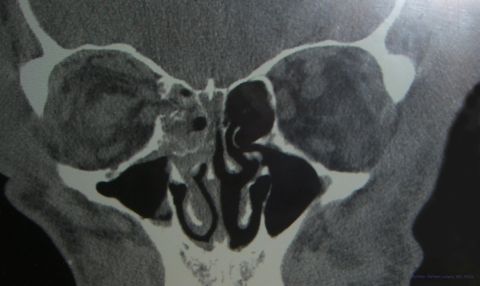
Orbital Medial Wall Fractures
All content on Eyewiki is protected by copyright law and the Terms of Service. This content may not be reproduced, copied, or put into any artificial intelligence program, including large language and generative AI models, without permission from the Academy.
Disease Entity
Orbital Medial Wall Fractures
Etiology
Fractures of the medial orbital wall most often result from blunt trauma to the periorbital area. The condition can result from traffic accidents, sports activities, violence or fall injuries. Medial wall fractures are less common in isolation and more often occur with a floor fracture or as part of a complex fracture. In addition, there may also be associated frontal, nasoethmoidal and maxillary fractures.
Risk Factors
People with injuries to the head, eye and/or periorbital area may suffer from an orbital fracture. As with most trauma, males outnumber females. High risk activities include contact sports. Some hypothesize that some ethnic groups may be anatomically predisposed to these fractures and fracture patterns differ by age due to the facial development and pneumatization of the sinuses.
General Pathology
Medial wall fractures are usually caused by blunt injury at mid face to the orbital rim and/or eye. In comminuted fractures, the most common type, bone fragments and intraorbital contents (e.g.extraocular muscle, fat or soft tissue) may herniate into the ethmoidal sinus. Others types of fractures include the hinge fracture, blow in fracture, and the trapdoor fracture. Medial orbital wall blow out fractures, by definition is a pure internal fracture confined to the orbital wall without involvement of orbital rim. Two theories have been proposed to explain how these fractures occur, the hydraulic or buckling mechanisms. Most likely it is a combination of these two mechanisms in the majority of cases.
Pathophysiology
The hydraulic hypothesis states that the orbital floor and/or medial orbital walls are fractured when there is an increase in orbital pressure with an external impact which leads to increased orbital pressure resulting in a fracture. The lateral wall and roof are usually thick enough to withstand such a trauma.
The buckling theory states that force exerted on the orbital rim transmitted the force to the weaker orbital floor or medial wall causing fracture.
The medial orbit is the thinnest of the orbital walls and is thus rendered more susceptible to fracture.
Primary Prevention
Avoiding trauma to face or periorbital area, e.g. wearing protective face and/or eyewear in contact sports and preventing slip and fall injuries, may lower the risk of medial orbital wall fractures.
Diagnosis
Radiologic studies, generally a CT, would confirm the presence of a medial orbital fracture. Plain films are not recommended for medial wall fractures as they detect less than 50% of the fractures, give less detail than CT and still utilize radiation. MRI is never the first line imaging after trauma (given concern for an unknown metallic intraocular foreign body) and provides less bone detail than CT. The CT should be evaluated in detail to rule out a concomitant Naso-Orbito-Ethmoid (NOE) fracture.
History
The patient may note recent trauma to face or present with orbital emphysema after nose blowing without prior knowledge of the fracture. If the patient has an increase in periorbital swelling after nose-blowing, this can be indicative of the open communication between the nasal cavity and the orbit causing emphysema around the eye. The medial wall injury can also be iatrogentic, due to other facial/sinus surgery. Some patients presenting with delayed enophthalmos warrant further assessment for an old fracture or other pathology.
Symptoms
Patients may be asymptomatic with an isolated medial orbital fracture. Epistaxis, soft tissue signs, and subcutaneous emphysema may be present. In cases with medial rectus muscle or associated soft tissue entrapment, patients may complain of diplopia in horizontal gaze or pain on eye movement. Children and young adults with entrapment may experience nausea, vomiting, bradycardia, dizziness because of the oculocardiac reflex. Sometimes, even without muscle entrapment, diplopia or limitation in extraocular movement is present due to soft tissue swelling and/or muscle contusion.
Physical examination
Complete eye examination is essential in all trauma cases, including measurement of visual acuity, intraocular pressure, careful pupil evaluation, red color desaturation, and dilated fundus examination. Any other trauma, particularly head trauma, should also be ruled out.
On palpation, there may be crepitus, which indicates emphysema, and palpation for step off deformity and facial sensation should be evaluation and may be present with concomitant orbital floor fracture.
Patients can have impaired horizontal eye movement in both adduction and abduction and they may complain of diplopia in such gazes.
Enophthalmos can be measured with exophthalmometer. In the early stage, enophthalmos may be masked due to soft tissue swelling. Repeat exophthalmometry in subsequent follow-up examinations may be appropriate to reveal the presence of enophthalmos. Sometimes there is proptosis early after trauma due to severe periorbital tissue edema. Although not common, retrobulbar hemorrhage can occur. In severe cases a hemorrhage can lead to an orbital compartment syndrome and can present with painful proptosis, raised intraocular pressure and a decrease in vision due to optic nerve compression. In such cases, urgent intervention may potentially be sight saving.
Besides the orbit, the trauma may cause periocular soft tissue injuries, such as eyelid laceration. Potential ophthalmic injuries include globe rupture (corneal or sclera laceration), iridodialysis, lens instability, traumatic cataract, vitreous hemorrhage, commotio retinae, retinal hemorrhage or detachment, choroidal rupture and traumatic optic neuropathy.
Apart from ophthalmic examination, one has to assess for any signs of the oculocardiac reflex including bradycardia, nausea, vomiting, syncope or even heartblock, which may happen when there is extraocular muscle entrapment, especially in children or young adults.
Clinical diagnosis
We should suspect patients having orbital fracture if there is history of trauma to the periorbital area or face. Additional signs including horizontal ocular dysmotility, emphysema and enophthalmos may hint at the presence of medial orbital wall fracture. Radiological investigation can confirm the diagnosis.
Diagnostic procedures
Computer tomography (CT) is the primary diagnostic tool in evaluating for orbital fractures, muscle entrapment or retrobublar hemorrhage. In addition, CT can help in assessing the size of fracture and plan subsequent surgical repair. While plain film x-rays of the orbit may reveal an ethmoidal sinus opacity, the test is insensitive in detecting medial wall fractures.
Differential diagnosis
Periorbital ecchymosis without fracture, in which there may be limitation in extraocular movement, not limiting to horizontal gaze. Muscle paresis, hemorrhage or contusion after trauma can also simulate entrapment.
Management
Medical therapy
When there is a medial wall fracture, patients should avoid nose-blowing to prevent the development of orbital emphysema and non-sterile tissue in sinuses getting into orbital cavity. The role of antibiotics is controversial, particularly without concominant sinus disease.
Surgery
This is one of the controversies in the management of orbital fractures. A literature review from 1983 to 2002 and published the clinical recommendation of repair of orbital floor fracture in 2002 based on 31 reviewed articles (Burnstine MA, Ophthalmology 2002). The guideline can be adopted in medial wall fracture as well.
It is recommended to repair the fracture immediately if there is non-resolving oculocardiac reflex, marked extraocular motility, entrapment of extraocular muscle evidenced in CT or early enophthalmos. However, enophthalmos is less common with an isolated medial wall fracture.
Surgical repair was traditionally ideally performed urgently when there was symptomatic diplopia and positive force duction test or within two weeks with large fractures causing enophthalmos. This data is primarily from orbital floor fractures and recent data suggests repair can be delayed even past two weeks unless there is entrapment. Surgical delay can also be necessary due to medical comorbidities or concurrent ruptured globe or intraocular injuries.
When there is good extraocular motility, minimal diplopia and no significant enophthalmos (i.e. <2mm and cosmetically acceptable), conservative management is adequate and diplopia without entrapment may resolve.
Last but not least, we also need to take into account patients’ age, pre-morbid condition and preference as operation is not without risks.
Surgical approach
Fracture can be repaired either by open or endoscopic approach. In the open approach, accessing the inferior aspect of the medial wall is performed by a subciliary incision or transconjunctival incision. The approach can also be used for combined medial and inferior orbital wall fractures. For isolated medial wall fracture, a transcaruncular incision is an option.
In the endoscopic approach, the uncinate process is cut and ethmoidectomy is performed to delineate the fracture site at medial orbital wall. Herniated orbital tissue is reduced and an implant such as silastic, merocel or medpor tailored slightly larger than the defect is inserted. Care must be taken to not oversize the implant, which can lead to higher rates of complications including extrusion. Forced duction testing and pulse test (in which globe is gently pushed externally and pulsation is observed through endoscope) are performed to confirm full ocular motility and proper placement of the implant. Post-operative nasal packing is necessary.
| Open | Endoscopic | |
|---|---|---|
| Fracture type/site | Can be use in different types of fractures | May be used in medial wall fracture and trap door fracture |
| Limitations | Relatively limited posterior orbit visibility | Can’t be utilized in large defect |
| Complications | Lid malposition |
|
| Advantages | Slight advantageous with respect to the operation time, the length of hospital stay, and cost. |
|
Complications
Fracture repair can potentially cause a drop in vision or even blindness if the optic nerve is injured during operation or if there is retrobulbar hemorrhage/hematoma that causes optic nerve compression and raised intraocular pressure. Persistent or worsening diplopia, implant-related complications such as infection or migration of implant, or absorption of bone graft may occur after the operation. Lid malposition such as cicatricial ectropion is a known complication with subciliary incisions and is reported to be less common with a transconjunctival approach. However, some suggest careful tissue dissection can render similar rates of ectropion between the 2 methods. Even with repair orbital fat atrophy can lead to enophthalmos [2]
Additional Resources
- Boyd K, Rizzuto PR. Orbital Fracture. American Academy of Ophthalmology. EyeSmart/Eye health. https://www.aao.org/eye-health/diseases/orbital-fracture. Accessed March 21, 2019.
References
- Clinical Recommendations for Repair of Isolated Orbital Floor Fractures, An Evidence-based Analysis; Michael A. Burnstine; Ophthalmology 2002; 109: 1207-1213
- Endoscopic Repair of Orbital Floor Fracutre; D. Gregory Farwell; Facial Plast Surg Clin N Am 14 (2006) 11-16
- Epidemiology and Management of Orbital fractures; Antonio Augusto V. Cruz; Current Opinion in Ophthalmology 2004, 15:416-421
- Orbital Bow-out Fractures: Surgical Timing and technique; GJ Harris; Eye (2006) 20, 1207-1212
- Update on Orbital Reconstruction; Chien-Tzung Chen; Current Opinion in Otolaryngology & Head and Neck Surgery 2010; 18:311-316
- Endoscopic Orbital Fracture Repair; Yasaman Mhadjer et al; Otolaryngologic Clinic of North America 39 (2006) 1049-1057
- Management of Orbital Fractures; Risto Kontio; Oral Maxillofacial Surg Clin N Am 21 (2009) 209-220
- An Analysis of 733 Surgically Treated Blowout Fractures; Mi Jung Chi; Ophthalmologica 2010;224:167-175
- Comparison of Endoscopic Endonasal Reduction and Transcaruncular Reduction for the treatment of Medical Orbital Wall Fractures; Kihwan Han; Ann Plastic Surg 2009;62: 258-264
- Endoscopic Approach to Medial Orbital Wall Fractures; John S. Rhee; Facial Plastic Surg Clin N Am 14 (2006) 17-23
- Modified Technique for Endoscopic Endonasal Reduction of Medial Orbital Wall Fracture Using a Resorbable Panel; Jaewoon We; Ophthal Plast Reconstr Surg 2009;25:303-305
- Treatment of Orbital Fractures : Evaluation of Surgical Techniques and Materials for Reconstruction; Daniel Nowinski; J Craniofac Surg 2010;21: 1033Y1037
- Medial orbital wall fractures: classification and clinical profile; Nolasco FP, Mathog RH; Otolaryngol Head Neck Surg. 1995 Apr; 112(4):549-56.
- Medial wall fracture: An update. Thiagarajah C, Kersten RC; Craniomaxillofac Trauma Recontr. 2009 Oct;2(3):135-139.
- ↑ American Academy of Ophthalmology. Right orbit medial wall fracture. https://www.aao.org/image/right-orbit-medial-wall-fracture-2 Accessed July 26, 2019.
- ↑ Cohen LM, Habib LA, Yoon MK. Post-traumatic enophthalmos secondary to orbital fat atrophy: a volumetric analysis. Orbit. 2020 Oct;39(5):319-324.


