Ophthalmoscopy for Medical Students and Primary Care Physicians
All content on Eyewiki is protected by copyright law and the Terms of Service. This content may not be reproduced, copied, or put into any artificial intelligence program, including large language and generative AI models, without permission from the Academy.
This article provides a summary of tips for teaching the ophthalmoscope exam to medical students and primary care physicians.
Diagnostic Intervention
Description/Overview
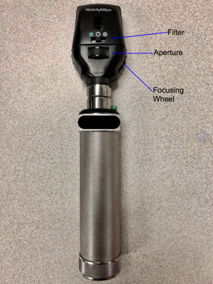
The ophthalmoscope (Figure 1) was invented in 1850 by Hermann von Helmholtz, a then 30-year-old German physicist. Helmholtz’s contribution to medicine is one of the few medical instruments so omnipresent yet so underutilized as a diagnostic tool. It is invaluable for viewing the retina, retinal blood vessels and optic nerve, and very useful in identifying ocular as well as systemic pathology. As medicine becomes increasingly specialized, it is challenging for medical schools to teach both depth and breadth in the traditional undergraduate medical curriculum. Surveys of primary care physicians have shown that training on the ophthalmoscope is often inadequate.[1] The following contains tips for teaching the ophthalmoscope exam.
Tips for Teaching Ophthalmoscopy to Trainees
Find willing patients who can have their eyes dilated
In a similar manner to detecting organomegaly by palpation or cardiac murmurs by auscultation, optimization of the learning condition by viewing the fundus through dilated pupils will help immensely in identifying normal anatomy as well as pathology. Using the ophthalmoscope through an undilated pupil requires more skill, so finding willing dilated patients will help to foster comfort and then confidence. Dilating eye drops such as 1% tropicamide or 2.5% phenylephrine are readily available in most eye clinics and one drop of each will dilate a pupil.
Ensure comfort for the patient as well as the learner
While not every clinic or school may have access to eye exam rooms, a comfortable learning environment can be created in various types of spaces. The learner should be able to hold the ophthalmoscope at a comfortable height, so he or she is not hunched over and the patient not sitting too long without back support. While most typical for a patient to sit while the examiner stands, it is equally acceptable for both to sit. If the patient and or examiner are not comfortable, appreciation for subtle findings will likely not occur.
Understand the ophthalmoscope and techniques for proper ophthalmoscopy
It is important to understand the ophthalmoscope with its capabilities and limitations. The instrument does specifically magnify images, as this is done naturally by the combination of the optical system of the patient and learner. Teaching learners that starting with the medium sized white light and the focusing wheel at the number zero is a good starting point. This wheel accounts for refractive errors (nearsighted/myopia and farsightedness/hyperopia) in the patient or observer so it should vary with each patient. The index finger should be kept on the focusing wheel and turned up or down in order to improve focus on an image such as the optic nerve (Figure 1). As the light illuminates structures in the fundus, moving the light around is the best way to visualize the retinal vessels and optic nerve as only illuminated structures will be visible, analogous to a spotlight. The ophthalmoscope is not able to visualize the entire fundus at one time, just as the stethoscope is not capable of listening to an entire lung at one time.
Right-right-right, left-left-left
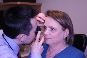
In the majority of situations, the learner should start by holding the ophthalmoscope in the right hand, while using the right eye to visualize the fundus of the patient's right eye. After the right eye is examined, the learner should hold the ophthalmoscope in the left hand, and use his/her left eye to examine the patient's left eye. These techniques help to improve viewing, increase comfort level with using both hands, and hopefully minimize uncomfortable face to face positioning between individuals. There may be patients who may prefer to have only one eye dilated or learners who may not be able to use one hand or one eye for examination, so accommodations should be allowed. The thumb of the examiner’s other hand should also be used to lift the eyelid or eyebrow to also improve viewing (Figure 2).
Nerve towards the nose
Remind examiners who may have challenges finding the optic nerve that following bifurcations of the retinal vessels should lead the path toward the optic nerve. The learner will realize that the optic nerve is located towards the nose and should naturally direct his/her position to view the optic nerve from a 45 degree angle. This position is ideal as the optic nerves attach to the eyes at an angle in the orbit. Directing the patient to look straight ahead will also improve visualization of the optic nerve, typically the most exciting moment for any observer! Once this structure is illuminated, encourage learners to adjust the focusing wheel to maximize image quality.
Open eyed test
Prior studies of medical students have shown that they may prefer learning subtle details through fundus photography rather than with the ophthalmoscope.[2] While visualizing fundus photos allows for appreciation of anatomic subtleties, medical students and primary care providers may not have photos of their patients available at the time of clinical examination. Hence, the ideal situation should involve simultaneous live fundus examination with fundus photos of that same patient. This viewing of the patient's fundus with his or her fundus photo will help enforce nuances of a patient's findings and also serve as a teaching moment for the patient. If such photos are not available, discussing the learner's findings or having him/her draw what was viewed, will help to ensure the structures such as the optic nerve were properly identified.
This manuscript is dedicated Dr. David Waters, Clinical Associate Professor of Surgery, University of Hawaii John A. Burns School of Medicine. Dr. Waters is responsible for teaching Direct Ophthalmoscopy to hundreds of medical students and Resident Physicians including one of the authors.


