Oncocytoma
All content on Eyewiki is protected by copyright law and the Terms of Service. This content may not be reproduced, copied, or put into any artificial intelligence program, including large language and generative AI models, without permission from the Academy.
Disease Entity
Oncocytoma (ICD-10 D31)
Background
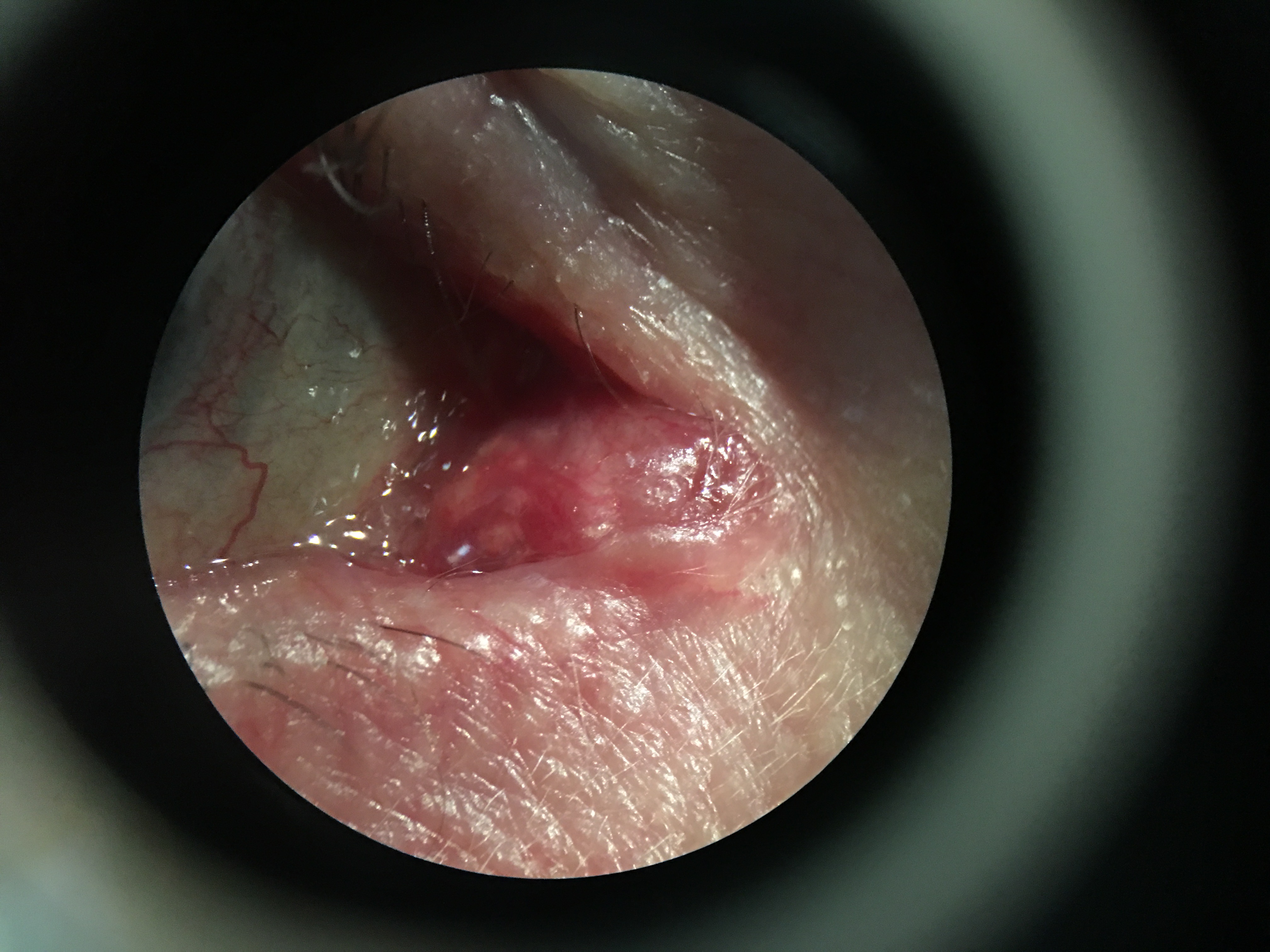
Oncocytoma of the caruncle was first described in the literature in 1941. Oncocytomas are also seen in the thyroid, parathyroid, salivary glands, and kidneys.[1] The incidence of ocular oncocytoma has been estimated to be 0.3 per million/year.[2]
Presentation
From a literature review of 212 cases of oncocytic lesions (oncocytoma) of the ocular adnexa and orbit, the most common presentation was a mass with possible symptoms of discomfort and/or epiphora. Of the 212 cases reviewed, the distribution of tissue involvement was 127 (60%) caruncle, 20 (10%) conjunctiva, 15 (6%) eyelid, 10 (5%) lacrimal gland, and 40 (19%) lacrimal sac. Only 3 (lacrimal gland) and 11 (lacrimal sac) were oncocytic carcinoma. Fifteen of the oncocytoma cases underwent ultrasound biomicroscopy and anterior segment optical coherence tomography which showed mixed solid and cystic components without evidence of scleral invasion.[1]
Pathophysiology
The mechanism for oncocytoma development is thought to be associated with mitochondrial and somatic gene mutations, resulting in oxidative phosphorylation deficiency. This results in reduced ATP in the mitochondria and therefore a reduction in energy production, which indicates that the increased mitochondrial content may be compensatory.[3]
General Pathology
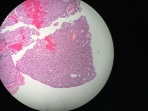
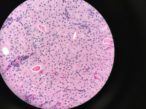
Enlarged, polygonal to square-shaped, modified, bland-appearing epithelial cells with voluminous, and intensely eosinophilic cytoplasm due to the accumulation of mitochondria that were classically considered “burnt out” mitochondria.[1][3][4]
Diagnosis
Differential Diagnosis
A pathology review of 112 lesions of the caruncle showed that 43% were nevi (pigmented from brown to black), 13% squamous papilloma, 9% Sebaceous gland hyperplasia, 5% chronic inflammation, 4% oncocytoma, 4% epithelial inclusion cyst, 3% foreign body granuloma, 3% pyogenic granuloma, 2% malignant melanoma, 2% capillary hemangioma.[5] Other less common lesions include lymphoma, basal cell carcinoma, and squamous cell carcinoma.[6]
Management
Oncocytomas have excellent prognosis. Recurrence after total resection in the locations of the caruncle and conjunctiva has never been reported.[1] However, although very rare, recurrence of the eyelid, lacrimal sac, and lacrimal gland have been reported.[7][8] Fifteen cases of ocular oncocytic adenocarcinomas have been described in the literature which underwent more aggressive management with radiation therapy and chemotherapy.[1][9]
References
- ↑ Jump up to: 1.0 1.1 1.2 1.3 1.4 Say EA, Shields CL, Bianciotto C, et al. Oncocytic lesions (oncocytoma) of the ocular adnexa: report of 15 cases and review of literature. Ophthal Plast Reconstr Surg 2012;28(1):14-21.
- ↑ Ostergaard J, Prause JU,Heegaard S. Oncocytic lesions of the ophthalmic region: a clinicopathological study with emphasis on cytokeratin expression. Acta Ophthalmol 2011;89(3):263-7.
- ↑ Jump up to: 3.0 3.1 Mete O, Asa SL. Oncocytes, oxyphils, Hurthle, and Askanazy cells: morphological and molecular features of oncocytic thyroid nodules. Endocr Pathol 2010;21(1):16-24.
- ↑ Hamperl H. Benign and malignant oncocytoma. Cancer 1962;15:1019-27.
- ↑ Luthra CL, Doxanas MT, Green WR. Lesions of the caruncle: a clinicohistopathologic study. Surv Ophthalmol1978;23(3):183-95.
- ↑ Kapil JP, Proia AD, Puri PK. Lesions of the lacrimal caruncle with an emphasis on oncocytoma. Am J Dermatopathol 2011;33(3):227-35.
- ↑ Lamping KA, Albert DM, Ni C, Fournier G. Oxyphil cell adenomas. Three case reports. Arch Ophthalmol 1984;102(2):263-5
- ↑ Morand B, Bettega G, Bland V, et al. Oncocytoma of the eyelid: an aggressive benign tumor. Ophthalmology 1998;105(12):2220-4.
- ↑ Gonnering RS, Sonneland PR. Oncocytic carcinoma of the plica semilunaris with orbital extension. Ophthalmic Surg 1987;18(8):604-7.


