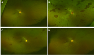
Ocular Decompression Retinopathy
All content on Eyewiki is protected by copyright law and the Terms of Service. This content may not be reproduced, copied, or put into any artificial intelligence program, including large language and generative AI models, without permission from the Academy.
Disease Entity
Disease
Ocular decompression retinopathy (ODR) is an rare postoperative complication that occurs as a result of rapid intraocular pressure lowering interventions. The clinical appearance is characterized by multiple round intraretinal hemorrhages, some with white centers, located in the postequatorial retina[1].
One case of medically treated acute primary angle closure (APAC) resulting in severe visual loss owing to retinal haemorrhage has been report in literature.
The term ocular decompression retinopathy was coined by Fechner et al (1992) to describe retinal changes subsequent to iatrogenic lowering of intraocular pressure after glaucoma filtering surgery[2][3]
Etiology
Glaucoma drainage implant insertion and trabeculectomy but may also occur after anterior chamber paracentesis, cataract surgery, vitrectomy, silicone oil removal, and pars plana sclerotomies. Despite the small sclerotomy caliber, it has been demonstrated that some patients develop transient hypotony and even choroidal detachment in the immediate postoperative period.[4]
Primary Prevention
Care should be taken to prevent ocular hypotony at the end of the procedure, especially in eyes with high preoperative IOP[1].
Diagnosis
The diagnosis of ocular decompression retinopathy is mainly clinically where about 80% of patients are asymptomatic
Symptoms
Although the fundus findings in ODR can be striking, about 80% of patients remain asymptomatic. When symptomatic, patients present with of decreased vision, a central scotoma, or floaters[4]. In the early postoperative period. Other contributing factors, such as corneal edema, may affect vision, and it may be difficult to determine what portion of the visual loss is due to the retinal hemorrhages[4].
Signs
Fundoscopic findings most commonly include scattered superficial and deep blot intraretinal hemorrhages, with some white-centered hemorrhages, in the posterior pole and equatorial retina[1]. Subhyaloid and vitreous hemorrhages are less common, but may also be present. Retinal vessels usually have a normal appearance, whereas retinal venous tortuosity and dilation are typically not observed. Choroidal detachment and macular edema are very rare in ODR.
Retinal venous tortuosity and dilation are typically not observed.
Management
Surgery
A small percentage of patients may require vitrectomy for non-resolving vitreous hemorrhage[5].
Prognosis
In most of the cases the disease follows a benign course with visual acuity usually returning to preoperative levels without treatment[5].

References
- ↑ Jump up to: 1.0 1.1 1.2 Rezende FA, Regis LGT, Kickinger M, Alcântara S. Decompression Retinopathy After 25-Gauge Transconjunctival Sutureless Vitrectomy: Report of 2 Cases. Arch Ophthalmol. 2007;125(5):699–700. doi:10.1001/archopht.125.5.699
- ↑ Jea SY, Jung JH. Decompression retinopathy after trabeculectomy. Korean J Ophthalmol. 2005 Jun;19(2):128-31. doi: 10.3341/kjo.2005.19.2.128. PMID: 15988929.
- ↑ Fechtner RD, Minckler D, Weinreb RN, Frangei G, Jampol LM. Complications of glaucoma surgery. Ocular decompression retinopathy. Arch Ophthalmol. 1992;110(7):965-968. doi:10.1001/archopht.1992.01080190071032
- ↑ Jump up to: 4.0 4.1 4.2 Mukkamala, Sri Krishna; Patel, Amar; Dorairaj, Syril; McGlynn, Robert; Sidoti, Paul A.; Weinreb, Robert N.; Rusoff, Jade; Rao, Sunil; Gentile, Ronald C. (2013). Ocular decompression retinopathy: A review. Survey of Ophthalmology, 58(6), 505–512. doi:10.1016/j.survophthal.2012.11.001
- ↑ Jump up to: 5.0 5.1 Reddy, Shubakar; Doshi, Shreyansh; Pathengay, Avinash; Panchal, Bhavik Ocular decompression retinopathy following intracameral bevacizumab injection in a case of proliferative diabetic retinopathy with neovascular glaucoma, Indian Journal of Ophthalmology: June 2020 - Volume 68 - Issue 6 - p 1206-1209 doi: 10.4103/ijo.IJO_1401_19
- ↑ 6. Weber P, Amin S, Qiu M. Decompression Retinopathy after Ahmed Glaucoma Valve in a Patient with Neurofibromatosis Type 1 and Moyamoya Syndrome. Ophthalmology. 2022 Jul;129(7):802. doi: 10.1016/j.ophtha.2022.01.006. PMID: 35738703.

