Microperimetry
All content on Eyewiki is protected by copyright law and the Terms of Service. This content may not be reproduced, copied, or put into any artificial intelligence program, including large language and generative AI models, without permission from the Academy.
Description/Overview
Microperimetry is a visual field test that incorporates perimetry and retinal imaging. It allows for the direct mapping of the stimulus in the region of interest, thereby correlating functional information (visual field testing) with structural/anatomical data (retinal imaging). Other designations for microperimetry include fundus-related perimetry or macular perimetry.
Indications
Microperimeters are the method of choice for residual visual function assessment. They have been used for the study of retinal macular disorders (age-related macular degeneration [AMD], diabetic macular edema (DME), macular dystrophies). Low vision training also benefits from microperimetry. The identification of eccentric fixation locations, know as preferred retinal loci (PRL), and fixation-stability estimates at those sites allows the practitioner to use the best residual visual function available for rehabilitation.
Procedure
As in conventional perimetry, visual field testing starts by determining the patient’s light sensitivity by finding the minimal light intensity stimulus that the patient can recognize when spots of light stimulate specific locations of the retina. Each software has a standart exam, that can be adapted to the patient (variations of diameter area and number of measurement points). Of note, care is needed when comparing results from different devices. Maximum luminance may vary between devices and the Decibel scale is relative to that value.
Constant tracking of the eye during microperimetry allows for real-time correction of retinal movement and evaluation of fixation. Data regarding PRL may be presented in two ways: as the percentage of fixation points inside circles centered by retinography or by mathematical calculation of the area and orientation of the best elipse that describes such points, the bivariate contour elipse area (BCEA). The first method can be used to produce a clinical classification, as suggested by Fuji et al. (table 1 and 2)[1]. Whilst not providing a classification, the second technique has the advantage of giving a more accurate and reproducible fixation stability measurement[2].
Table 1 - Classification of location of fixation, by Fuji et al.[1]
| Classification of location of fixation | Percentage of preferred fixation points located within 2º-diameter fovea centered circle |
| Predominantly central fixation | >50% |
| Poor central fixation | 25-50% |
| Predominantly eccentric fixation | <25% |
Table 2 - Classification of fixation stability, by Fuji et al.[1]
| Classification of fixation stability | Percentage of preferred fixation points located within 2º- and 4º-diameter circles centered in the gravitational center of all fixation points |
| Stable fixation | >75% within 2°-diameter circle |
| Relatively unstable fixation | < 75% within 2°-diameter circle, but > 75% within 4°-diameter circle |
| Unstable fixation | < 75% within 4°-diameter circle |
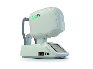
The retina may be imaged through an integrated color fundus camera (e.g., MP-3) or scanning laser ophthalmoscopy (SLO) (e.g., MAIA and Optos OCT-SLO), depending on the choice of device.
After completion of measurements, the perimetry grid is superimposed on the retinal image facilitating evaluation of a specific region of interest. Frequently, the Decibel scale is color coded according to normative studies and summarized indices that describe the mean macular sensitivity data, adjusted for age and/or gender normative data (e.g., Macular Integrity Index from MAIA) are displayed to aid with interpretation.
Devices
The first microperimeter (SLO101) was manufactured by Rodenstock Instruments (Munich, Germany) in 1982. Fundus mapping was based on SLO technology and a modulated helium-neon laser (633nm) was used to manually generate background and stimulus illumination in the 33x21º central field[3]. It lacked an eye-tracking capability and the stimulus were presented in a semi-automated fashion[4].
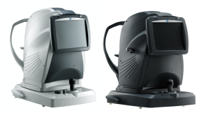
To date, there are three microperimeters available on the market: the MP-3 by Nidek Technologies (Padua, Italy), the OCT-SLO from Optos (Marlborough, USA) and the MAIA microperimeter from CenterVue (Padua, Italy). Albeit different technical specificities, their functions are similar[5]. Relevant particularities include that Optos OCT-SLO offers the possibility of superimposing functional deficits with cross-sectional OCT retinal images (besides en face images) and MP-3 and MAIA allow for scotopic microperimetry, performed under scotopic luminance conditions[6].
Benefits
When compared to conventional perimetry, microperimetry offers the following advantages:
- Real-time detection and correction of testing in a given retinal location (stimuli are directly projected onto the retina with eye-tracking feedback, allowing for testing-retesting).
- Direct correlation between fundus morphology and light sensibility, by presenting the light stimulus in a particular region of interest (e.g., small scotoma created by a macular scar).
- Continuous eye-tracking makes it the preferred exam when foveal function or fixation stability is compromised (it does not assume a stable foveal fixation, like conventional perimetry).
- Biofeedback for training in low vision patients (e.g., relocation of a PRL to a TRL [trained retinal loci] predetermined by the practitioner to improve functional visual outcomes).
Clinical applications of microperimetry include, but are not limited to:
- Atrophic and neovascular AMD: functional assessment of vision quality; identification of central scotoma; diminished macular sensitivity in AMD correlates with disease severity and progression[7]; these alterations are also observed at a microstructural level (morphofunctional correlation), with worst macular sensitivity in areas of retinal pigment epithelium-drusen complex[8][9], pigment epithelium detachment, subretinal fluid[10] and geographic atrophy[11]; anti-VEGF therapy efficacy assessment[12].
- DME: diminished macular sensitivity in DME correlates with degree of macular edema[13]; evaluation of different laser modalities on macular function[14].
- Glaucoma: although standart automated perimetry remains the gold standart for monitoring glaucoma, detection of nerve fiber layer defects and eccentric fixation in advanced glaucoma can be enhanced by microperimetry[15].
- Low-vision patients: rehabilitation with microperimetry biofeedback in patients with central scotoma has demonstrated to improve fixation stability, visual function and quality of life[16].
- Any other retinal pathologies affecting macular structure and/or function: central serous corioretinopathy, hydroxychloroquine maculopathy, rod-cone alterations, macular hole and epiretinal membrane[17]…
Limitations
As with most diagnostic tests, patient cooperation is necessary, especially since it relies on patients answers to identify light sensibility (e.g., it is subject to false negative/positive answers). Because it is a relatively recent technology it is not yet in widespread clinical use. In a study of their own experience, Romaniuk et al. pinpoint long examination time in selected strategies (fatigue effect) and cost as microperimetry’s drawbacks[18].
References
- ↑ Jump up to: 1.0 1.1 1.2 Fujii GY, de Juan E Jr, Sunness J, Humayun MS, Pieramici DJ, Chang TS. Patient selection for macular translocation surgery using the scanning laser ophthalmoscope. Ophthalmology. 2002;109(9):1737-1744.
- ↑ Crossland MD, Dunbar HM, Rubin GS. Fixation stability measurement using the MP1 microperimeter. Retina. 2009;29(5):651-656.
- ↑ Rohrschneider K, Fendrich T, Becker M, Krastel H, Kruse FE, Völcker HE. Static fundus perimetry using the scanning laser ophthalmoscope with an automated threshold strategy. Graefes Arch Clin Exp Ophthalmol. 1995;233(12):743-749.
- ↑ Laishram M, Srikanth K, Rajalakshmi AR, Nagarajan S, Ezhumalai G. Microperimetry - A New Tool for Assessing Retinal Sensitivity in Macular Diseases. J Clin Diagn Res. 2017;11(7):NC08-NC11.
- ↑ Markowitz SN, Reyes SV. Microperimetry and clinical practice: an evidence-based review. Can J Ophthalmol. 2013;48(5):350-357.
- ↑ Taylor LJ, Josan AS, Pfau M, Simunovic MP, Jolly JK. Scotopic microperimetry: evolution, applications and future directions. Clin Exp Optom. 2022;105(8):793-800.
- ↑ Vujosevic S, Smolek MK, Lebow KA, Notaroberto N, Pallikaris A, Casciano M. Detection of macular function changes in early (AREDS 2) and intermediate (AREDS 3) age-related macular degeneration. Ophthalmologica. 2011;225(3):155-160.
- ↑ Wu Z, Cunefare D, Chiu E, et al. Longitudinal Associations Between Microstructural Changes and Microperimetry in the Early Stages of Age-Related Macular Degeneration. Invest Ophthalmol Vis Sci. 2016;57(8):3714-3722.
- ↑ Iwama D, Tsujikawa A, Ojima Y, et al. Relationship between retinal sensitivity and morphologic changes in eyes with confluent soft drusen. Clin Exp Ophthalmol. 2010;38(5):483-488.
- ↑ Sulzbacher F, Kiss C, Kaider A, et al. Correlation of SD-OCT features and retinal sensitivity in neovascular age-related macular degeneration. Invest Ophthalmol Vis Sci. 2012;53(10):6448-6455.
- ↑ Pilotto E, Convento E, Guidolin F, et al. Microperimetry Features of Geographic Atrophy Identified With En Face Optical Coherence Tomography. JAMA Ophthalmol. 2016;134(8):873-879.
- ↑ Squirrell DM, Mawer NP, Mody CH, Brand CS. Visual outcome after intravitreal ranibizumab for wet age-related macular degeneration: a comparison between best-corrected visual acuity and microperimetry. Retina. 2010;30(3):436-442.
- ↑ Vujosevic S, Midena E, Pilotto E, Radin PP, Chiesa L, Cavarzeran F. Diabetic macular edema: correlation between microperimetry and optical coherence tomography findings. Invest Ophthalmol Vis Sci. 2006;47(7):3044-3051.
- ↑ Vujosevic S, Bottega E, Casciano M, Pilotto E, Convento E, Midena E. Microperimetry and fundus autofluorescence in diabetic macular edema: subthreshold micropulse diode laser versus modified early treatment diabetic retinopathy study laser photocoagulation. Retina. 2010;30(6):908-916.
- ↑ Scuderi L, Gattazzo I, de Paula A, Iodice CM, Di Tizio F, Perdicchi A. Understanding the role of microperimetry in glaucoma. Int Ophthalmol. 2022;42(7):2289-2301.
- ↑ Qian T, Xu X, Liu X, et al. Efficacy of MP-3 microperimeter biofeedback fixation training for low vision rehabilitation in patients with maculopathy. BMC Ophthalmol. 2022;22(1):197. Published 2022 Apr 28.
- ↑ Molina-Martín A, Pérez-Cambrodí RJ, Piñero DP. Current Clinical Application of Microperimetry: A Review. Semin Ophthalmol. 2018;33(5):620-628.
- ↑ Romaniuk W, Gościniewicz P, Mrukwa-Kominek E, et al. Clinical applications of microperimetry techniques – advantages and disadvantages. Own experience. Postępy Nauk Medycznych. 2013;26(12):843-846.


