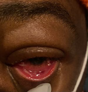
Membranous Conjunctivitis and Pseudomembranous Conjunctivitis
All content on Eyewiki is protected by copyright law and the Terms of Service. This content may not be reproduced, copied, or put into any artificial intelligence program, including large language and generative AI models, without permission from the Academy.
Membranous conjunctivitis and pseudomembranous conjunctivitis form thin-yellow sheets of fibrin and inflammatory debris on the palpebral conjunctiva. Etiologies include inflammation from bacterial or viral infections, drug reactions, or systemic and local ocular autoimmune disease. Membranous conjunctivitis differs from pseudomembranous by the formation of true membranes that bleed more significantly upon peeling, representative of a more intense inflammation. Most cases are acute and require treatment of the underlying etiology. Peeling of membranes is recommended in pseudomembranous conjunctivitis but is controversial in membranous conjunctivitis[1] . Prompt identification and treatment may result in good prognosis and prevent complications such as corneal scarring, symblepharon formation, dryness or secondary infection.
Disease Entity
- Membranous Conjunctivitis ICD -10: H10.89
- Pseudomembranous Conjunctivitis ICD-10: H10.22
Disease
Membranous conjunctivitis and Pseudomembranous conjunctivitis cause conjunctival inflammation with the formation of fibrinous sheets and inflammatory debris on the epithelial surface of the conjunctiva.[2][3]
Membranous conjunctivitis involves growth of true membranes into the conjunctival epithelial surface, resulting in significant bleeding upon peeling of membranes. Pseudomembranous conjunctivitis membranes are more superficial, with no growth into the conjunctival epithelium, and can be removed with minimal bleeding.
Etiology
Similar etiologies may cause membranous and pseudomembranous conjunctivitis.
Causes of Membranous conjunctivitis include:
- Bacterial and viral infections can cause true membrane formation. Bacterial infections include Beta-hemolytic streptococci (most common), Staphylococcus aureus, Neisseria gonorrhea, Corynebacterium diphtheria, and Chlamydia.[2][3][4] Viral causes include adenovirus (most common) with Epidemic Keratoconjunctivitis and potential for fibrosis. Herpes simplex virus (HSV) and Epstein-Barr virus (EBV) may also be implicated.[2][5]
- Drug reactions including hypersensitivity spectrum reactions, Stevens-Johnson syndrome (SJS) and toxic epidermal necrolysis (TEN), often in response to adverse drug reactions.[3][1]
- Autoimmune diseases
- Eye-limited inflammatory etiologies include ocular cicatricial pemphigoid and chemical or thermal injury.
Causes of Pseudomembranous conjunctivitis include:
- Infectious causes include Corynebacterium diphtheria, Neisseria gonorrhea, Streptococcus pyogenes, Chlamydia, and adenovirus; COVID-19 has also been implicated.[6][7]
- Inflammatory etiologies include hypersensitivity reactions, such as SJS, TEN, and erythema multiforme; graft-versus host-disease, specifically in allogeneic hematopoietic stem cell transplants; and foreign body reactions, especially in cases with difficult-to-treat pseudomembranes.[3][8][9]
- Ligneous conjunctivitis can cause chronic, recurrent pseudomembrane conjunctivitis, with fibrin deposits on the conjunctiva and systemically due to genetic plasminogen deficiency.[10]
Risk Factors
Risk factors for both Membranous and Pseudomembranous conjunctivitis are similar and relate to the etiologies of disease outlined above. Exposure to causative infections, adverse drug reactions, and autoimmune disease may lead to formation of true membranes or pseudomembranes. Genetic predisposition to ligneous conjunctivitis is a risk factor for pseudomembrane formation.
Pathophysiology
Membranes and Pseudomembranes are pale sheets of fibrin deposits and inflammatory exudates that coagulate on the conjunctival surface. True membrane formation occurs when the fibrinous deposit secreted by the invading microorganisms or ocular tissues adheres to the conjunctiva's epithelial surface due to interdigitating capillaries' growth into the membrane.[11] Peeling membranes in membranous conjunctivitis causes bleeding and leaves behind a raw surface, representing more intense inflammation.
Pseudomembranes are more superficial and can be easily removed, leaving behind an intact epithelium with minimal bleeding. However, bleeding may still be observed in pseudomembranous conjunctivitis due to severe inflammation and friability of the underlying conjunctiva.[11] The amount of bleeding due to less invasive growth and an intact epithelium can differentiate pseudomembranous conjunctivitis from membranous conjunctivitis.
Membranes and pseudomembranes arise due to an inflammatory reaction within the eye due to systemic or local inflammation. The exact pathogenesis of the formation of membranes and pseudomembranes is related to its specific etiology.
Histopathology
Histopathologic features of Membranous and Pseudomembranous conjunctivitis can be identified under the microscope (Figure 1).[12] Examination of the membranes revealed a fibrinous exudate matrix composed of fibrin, fibronectin, and tenascin, with neutrophils and necrotic epithelial cells; macrophages may be present in older membranes.[13] In membranous conjunctivitis, fibroblasts and blood vessels provide a scaffold for the highly vascularized inflammatory membrane containing granulation tissue. If membranes form due to the presence of a foreign body, foreign body giant cells surrounding birefringent foreign bodies may be seen.[9]
Diagnosis
The diagnosis of Membranous and Pseudomembranous conjunctivitis is often a clinical diagnosis based on the history and physical examination.
History
The onset and timing of Membranous and Pseudomembranous conjunctivitis are related to the underlying etiology. Viral and bacterial conjunctivitis present acutely, whereas SJS and TEN arise sub-acutely after exposure to an inciting medication within three to four weeks. Ligneous conjunctivitis presents with a chronic and recurrent course of pseudomembrane formation.[10]
Physical examination and Clinical Presentation
Membranous and Pseudomembranous conjunctivitis both present with thin-yellow membranes in the fornices and palpebral conjunctiva. The membranes may be patchy or cover the entire palpebral conjunctiva, starting at the lid edge, but rarely involve the bulbar conjunctiva.[14]
Fluorescein staining in Pseudomembranous conjunctivitis will reveal bright green staining of the membranes, and corneal epithelial defects may be present in some instances.[9][15] Visual acuity may be decreased in the affected eyes. Pre-auricular lymphadenopathy may be present with suppuration.[14]
Differentiation of true membranous conjunctivitis and pseudomembranous conjunctivitis:
- True membranes are more adherent to the conjunctival surface and result in more significant tearing and bleeding when peeled with a raw surface uncovered
- Formation of a free discharge and necrosis of conjunctiva and cornea leading to membranes that separate less readily and bleeding of the underlying surface.[14]
- Pseudomembranes can be readily scraped away from the conjunctival surface with minimal bleeding
Signs and Symptoms
Patients usually report symptoms of eye redness, eyelid swelling, tearing, watery discharge, and visual disturbances. In addition, a foreign body sensation may be present due to irritation from the membranes, resulting in ocular discomfort. Symptoms may be unilateral or bilateral, depending on the underlying etiology of inflammation.
Signs include conjunctival inflammation with conjunctival hyperemia, chemosis, eyelid edema, corneal edema, and mucopurulent discharge.
Diagnostic procedures
Fluorescein staining is essential in making the diagnosis to visualize the membranes and detect any corneal epithelial defects. Removal of membranes and pathologic examination can help diagnose Membranous and Pseudomembranous conjunctivitis with the histopathologic features described above if the diagnosis is unclear from the clinical examination. Gram stain, culture, and PCR of the membranes may help identify organisms involved in infectious forms of conjunctivitis. Systemic signs and symptoms of infection or inflammatory disease are supportive findings in determining the etiology of the formation of conjunctival membranes.
Differential diagnosis
The differential diagnosis includes any infectious or inflammatory etiology of membranous or pseudomembranous conjunctivitis as well as other diseases that resemble this process.
- Bacterial conjunctivitis (beta-hemolytic Streptococcus, S. aureus. N. gonorrhea, C. diphtheriae)
- Viral conjunctivitis (adenovirus, HSV, EBV)
- Inclusion conjunctivitis
- Ligneous conjunctivitis
- Toxic conjunctivitis
- Chemical conjunctivitis
- Hypersensitivity reaction (SJS, TEN)
Management
Treatment of Membranous and Pseudomembranous conjunctivitis is highly dependent on the underlying etiology of the inflammation resulting in the membrane formation.
Medical therapy and follow
Management of Membranous conjunctivitis is similar to that of Pseudomembranous conjunctivitis with the judicious use of topical steroids and antibiotics as necessary. However, membranes can dissolve on their own, allowing the cornea to repair.[1] Lubricants assist in rehydrating the eye and improving comfort with a moist ocular surface and can be accomplished with gels, ointments, and preservative-free artificial tears.[1]
Management of Pseudomembranous conjunctivitis involves peeling the membranes and topical steroid use, in addition to removing any foreign bodies if identified.[9] Topical steroids can reduce ocular inflammation.[16] Washing of the ocular surface to remove chemical irritants is also crucial in preventing the reformation of membranes.[15] Use of topical antibiotics to treat infections is warranted, and prophylactic use of antibiotics to prevent secondary infection, especially if corneal defects are noted, may be reasonable.[9][16]
Systemic treatment of conditions such as SJS, TEN, and infection may be warranted if inflammation is severe. Treatment of SJS-related membrane formation involves cessation of any offending or high-risk medications and preventing the use of similar drugs. In addition, adequate lid hygiene and lubrication are essential in reducing discomfort and preventing sequelae.
Close follow-up for resolution and complications is recommended. Patients should be followed up on in 3-7 days due to the risks for secondary corneal infection or symblepharon formation. Specific follow-up for the inciting etiology is also recommended.
Surgery and follow up
Debridement of true membranes is controversial as removal will result in a raw exposed surface and can increase the risk of conjunctival scarring and chronic ocular complications[1]
Surgery may be used to treat complications of disease. If symblepharon are present acutely, daily lysis of adhesions with sweeping of the fornices by a glass rod or similar device is recommended.[1] Patients should be followed closely after removal of adhesion to monitor for re-development of symblepharon or secondary complications of intervention.
Complications
Prompt identification and adequate treatment of conjunctival membrane formation are necessary to prevent sight-threatening complications. Healing of ocular inflammation and ulceration may result in scarring and contraction of the conjunctiva and symblepharon formation.[1] Other ocular complications include keratitis or uveitis.[16] Corneal epithelial defects can result in secondary ocular infection.
Prognosis
Prognosis of Membranous or Pseudomembranous conjunctivitis is generally good with treatment of the underlying disease and any related complications to the eye. Prompt identification and management of the etiology will help prevent complications, such as corneal infection or scarring.
References
- ↑ Jump up to: 1.0 1.1 1.2 1.3 1.4 1.5 1.6 Kirkwood BJ, Billing KJ. Membranous conjunctivitis from adverse drug reaction: Stevens-Johnson syndrome. Clin Exp Optom. 2011;94(2):236-239. doi:10.1111/j.1444-0938.2010.00548.xa
- ↑ Jump up to: 2.0 2.1 2.2 Grayson M. Diseases of the Cornea. Mosby; 1979.
- ↑ Jump up to: 3.0 3.1 3.2 3.3 Bhat A, Jhanji V, Ung L, Rajaiya J, Chodosh J. Infections of the Cornea and Conjunctiva. (Das S, Jhanji V, eds.). Springer Singapore; 2019. doi:10.1007/978-981-15-8811-2
- ↑ Lindquist T. Cornea and External Disease: Clinical Diagnosis and Management. Vol II. (Krachmer J, Mannis M, Holland E, eds.). Mosby; 1997.
- ↑ Slobod KS, Sandlund JT, Spiegel PH, et al. Molecular evidence of ocular Epstein-Barr virus infection. Clin Infect Dis. 2000;31(1):184-188. doi:10.1086/313932
- ↑ Sahay P, Nair S, Maharana PK, Sharma N. Pseudomembranous conjunctivitis: Unveil the curtain. BMJ Case Rep. 2019;12(3). doi:10.1136/bcr-2018-228538
- ↑ Navel V, Chiambaretta F, Dutheil F. Haemorrhagic conjunctivitis with pseudomembranous related to SARS-CoV-2. Am J Ophthalmol Case Rep. 2020;19. doi:10.1016/j.ajoc.2020.100735
- ↑ Liu YC, Gau JP, Lin PY, et al. Conjunctival acute graft-versus-host disease in adult patients receiving allogeneic hematopoietic stem cell transplantation: A cohort study. PLoS One. 2016;11(11). doi:10.1371/journal.pone.0167129
- ↑ Jump up to: 9.0 9.1 9.2 9.3 9.4 Ho D, Lim S, Kim Teck Y. Pseudomembranous Conjunctivitis: A Possible Conjunctival Foreign Body Aetiology. Cureus. Published online May 18, 2020. doi:10.7759/cureus.8176 11. Schuster V, Seregard S. Ligneous conjunctivitis. Surv Ophthalmol. 2003;48(4):369-388. doi:10.1016/S0039-6257(03)00056-0
- ↑ Jump up to: 10.0 10.1 10.2 Schuster V, Seregard S. Ligneous conjunctivitis. Surv Ophthalmol. 2003;48(4):369-388. doi:10.1016/S0039-6257(03)00056-0
- ↑ Jump up to: 11.0 11.1 de Cock R. Membranous, Pseudomembranous and Ligneous Conjunctivitis. In: Cicatrising Conjunctivitis. KARGER; 1997:32-45. doi:10.1159/000060697
- ↑ Chintakuntlawar A v., Chodosh J. Cellular and Tissue Architecture of Conjunctival Membranes in Epidemic Keratoconjunctivitis. Ocul Immunol Inflamm. 2010;18(5):341-345. doi:10.3109/09273948.2010.498658
- ↑ Kivelä T, Tervo K, Ravila E, Tarkkanen A, Virtanen I, Tervo T. Pseudomembranous and membranous conjunctivitis Immunohistochemical Features. Acta Ophthalmol. 1992;70(4):534-542. doi:10.1111/j.1755-3768.1992.tb02128.
- ↑ Jump up to: 14.0 14.1 14.2 14.3 Sihota R, Tandon R. Parsons’ Diseases of the Eye. 23rd ed. Elsevier; 2019
- ↑ Jump up to: 15.0 15.1 Ono T, Nejima R, Kinoshita K, Mori Y, Iwasaki T, Miyata K. Pseudomembranous Conjunctivitis following Exposure to Arisaema ringens Sap: A Case Report. Case Rep Ophthalmol. Published online May 10, 2022:350-354. doi:10.1159/000524726
- ↑ Jump up to: 16.0 16.1 16.2 Maden CL, Ah-Kye L, Alfallouji Y, Kulakov E, Ellery P, Papamichael E. Erythema multiforme major with ocular involvement following COVID-19 infection. Oxf Med Case Reports. 2021;2021(11-12). doi:10.1093/omcr/omab120


